
Abstract
Abstract
Introduction
Case
A 60-year-old postmenopausal woman, para 5, presented to the emergency room (ER) at the Post Graduate Institute of Medical Sciences, Rohtak, Haryana, India, with fever and abdominal pain for 10 days prior to presentation. Her pain was intermittent, nonradiating, and noncolicky. Her fever was associated with rigor and chills and a Widal test was positive (1:160) that was performed at a private clinic showed positive results for antibodies against Salmonella typhi and Salmonella paratyphi. She also had a history of mild abdominal distension for 3 days prior to the current ER visit. She had no history of IUCD insertion, dilatation and curettage, postmenopausal bleeding, or discharge or significant medical or surgical illness. She did not have diabetes mellitus and had never had sexually transmitted diseases.
An examination revealed that she was dehydrated with a pulse rate of 110 beats per minute, blood pressure of 90/60 mm Hg, and a respiratory rate of 20 inspirations per minute. Blood testing revealed mild anemia with leucocytosis. The findings on chest examination were normal, as was her cardiovascular system. Abdominal examination revealed slight distension with diffuse tenderness, guarding, and rigidity. Bowel sounds were absent.
A speculum examination revealed that her ectocervix appeared normal and a vaginal examination revealed a soft uterus of normal size with slight tenderness in the fornices. Sonography showed her abdomen was flatulent with free fluid, and her uterus had a minimal amount fluid in the endometrial cavity. An X-ray of the abdomen did not show flatulence under her diaphragm. Because of the absence of flatulence under her diaphragm, perforation of her uterus was ruled out. Within 6 hours of admission, this patient's general condition deteriorated, and she started gasping for breath. She was admitted to the RICU for stabilization. However, her condition continued to deteriorate. Computed tomography (CT) scanning of her abdomen and pelvis were done, which revealed a perforation in her uterus with multiple pelvic and intra-abdominal collections of flakes (Fig. 1). A laparotomy was planned immediately. Operative findings revealed a hole measuring 1×1 cm on the posterior side of the uterine fundus sealed by pus flakes, and multiple pelvic and abdominal adhesions (Fig. 2). A total abdominal hysterectomy with a bilateral salpingo-oopherectomy was performed, along with peritoneal lavage. Cut section specimens of her, uterus and cervix did not reveal any abnormalities (Fig. 3). Histopathologic examination of the surgical specimens revealed features of necrotic endometrium with no evidence of malignancy.
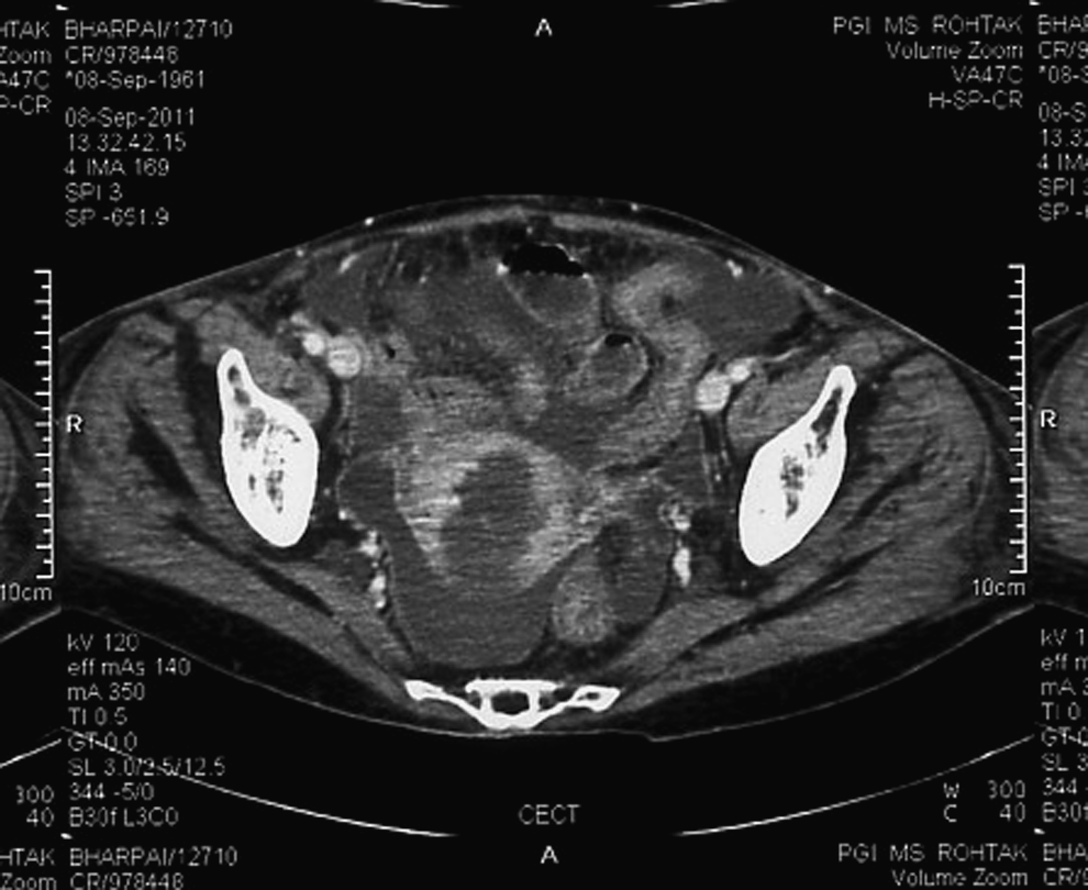
Computed tomography scan showing perforation on the posterior aspect of the fundus.

Perforation of the uterus on posterior aspect of fundus (which was sutured during the hysterectomy).

Cut open section of the uterus showing no growths on the uterus and cervix.
Results
This patient was readmitted to the RICU but she died on the third postoperative day.
Discussion
Pyometra is an uncommon condition occurring mainly in elderly postmenopausal females, and results when natural drainage of the uterine cavity is compromised. Pyometra is a serious medical condition because of its association with malignant disease and danger of spontaneous perforation of the uterus that carries significant morbidity and mortality. 3 Spontaneous perforation is thought to occur at a site of degenerative or necrotic change after pyometra develops as a result of blockage of natural drainage caused by a stenotic cervix. Cervical occlusion may be caused by malignant or benign tumors, radiation cervicitis, atrophic cervicitis, infection, or congenital anomalies. 4 In this case, it was probably because of stenosis of the postmenopausal cervix. The risk factors may include a decline in activity, incontinence, diabetes, and long-term use of an IUCD.
The classical triad of symptoms consists of purulent vaginal discharge, postmenopausal bleeding, and lower abdominal pain. 5 In the current, case these symptoms were absent, and this patient presented to the ER with an acute abdomen. When symptoms are nonspecific, the diagnosis of pyometra is difficult to make unless it is already suspected and looked for specifically. Typically, surgeons will perform an exploratory laparotomy with a provisional diagnosis other than pyometra perforation.
The diagnosis of pyometra perforation is usually one of exclusion. Abdominal X-ray may show flatulence under the diaphragm. In the current case, there was no flatulence under this patient's diaphragm, probably because pus flakes had sealed the perforation. Confirmation is aided by ultrasonography and CT abdomen. Sonography plays a limited role in the diagnosis of ruptured pyometra because of its inability to demonstrate the uterine breech and the limited Sonographic window available due to perforation. Sagittal and coronal reformats in multidetector CT is very helpful for depicting the site and size of uterine breach, showing the resultant intra-abdominal collections of flakes, and staging of cervical cancer. The site of a uterine breach can be missed on axial images, as it is usually seen on the fundus; hence, in elderly postmenopausal females with peritonitis, sagittal and coronal reformats of the uterus should be carefully evaluated.
The correct diagnosis has rarely been made preoperatively, because the most common presenting symptoms are abdominal pain, vomiting, nausea, and fever of short duration. The most common preoperative diagnoses are generalized peritonitis, pneumoperitoneum, and perforation of the gastrointestinal (GI) tract. Yildizhan et al. concluded that most common preoperative diagnoses of acute pain in the abdomen in postmenopausal females were generalized peritonitis (47.4%), pneumoperitoneum (47.4%), and perforation of the GI tract (36.8%)—and often more than one symptom is present)—while pyometra perforation was suspected only in 15.8%. 6
The indexed English literature contained articles with 28 reported cases of spontaneous pyometra rupture. Table 1 decscribes these cases, grouped according to the etiology along with presenting symptoms and their outcomes. All of the cases were postmenopausal elderly females, mostly in the sixth-to-tenth decade of life. The most common universally present symptom was abdominal pain. Moreover, some of the patients had fever at the time of presentation. An analysis of preoperative status in the above cases hardly shows spontaneous perforation of pyometra in carcinoma of the cervix. The common preoperative diagnosis was generalized peritonitis (50%), perforation of the GI tract (40%), and pneumoperitoneum (30%). Laparotomy was performed in all of these cases Only 6 of the 28 cases had untreated carcinoma of the cervix as the cause of spontaneous pyometra perforation. Five of the cases did not have a preoperative diagnosis of malignancy and were missed cases of carcinoma cervix. 7
Case in current article.
GP, generalized peritonitis; PP, pneumoperitoneum; Nm, not mentioned, Panhysterectomy, TAH and bilateral salpingo-oopherectomy; PPU, perforated peptic ulcer; PIT, perforated gastrointestinal tract.
Total hysterectomy along with bilateral salpingo-oopherectomy and thorough drainage and irrigation of the abdominal cavity is the preferred treatment. Postoperatively, broad-spectrum antibiotics and intensive care can facilitate good recovery followed by specific management according to the etiology. Cancer is managed according to cancer stage in consultation with a gynecologist–oncolgist. Radiation in combination with cisplatin-based chemotherapy has become the standard of care for patients with locally advanced cervical cancer.
Conclusions
Pyometra is a serious medical condition, because of its association with malignant diseases and the danger of spontaneous perforation. Although rare, ruptured pyometra should be considered in the differential diagnosis of acute abdomen in elderly women, especially those with malignant disorders of the genital tract. The treatment of pyometra rupture is immediate peritoneal lavage and drainage, or simple hysterectomy. Intensive care with strict management of respiration and circulation is essential. A histopathologic diagnosis should always be established.
Footnotes
Disclosure Statement
No competing financial conflicts exist.