
Abstract
Abstract
Introduction
Case
A 61-year-old female presented with clinical features of subacute intestinal obstruction of 3 weeks' duration. Her past history revealed that she had carcinoma of the cervix, stage IV, 2 years prior, which was managed by radiotherapy. She was totally asymptomatic after radiotherapy until 3 weeks prior to her current presentation. At that time she started experienced a bloating sensation, and distension and pain in her abdomen that was associated with vomiting. She did not have a history of vaginal bleeding, vaginal discharge, hematuria, or bone pain. Her clinical workup confirmed the presence of a distal large-bowel obstruction, and a computed tomography scan revealed an infiltrating cervical mass in her rectal area, with narrowing of her rectum and signs of colonic obstruction. A colonoscopy showed normal rectal mucosa with overlying extrinsic compression and proximal dilatation. She was diagnosed as having recurrent cervical tumors with extrinsic rectal compression causing obstruction. She was evaluated for surgery, placement of a rectal SEMS was chosen instead. Under fluoroscopy and colonoscopy guidance, a 12-mm Wilson cook stent was placed at the level of her obstruction (Fig. 1).
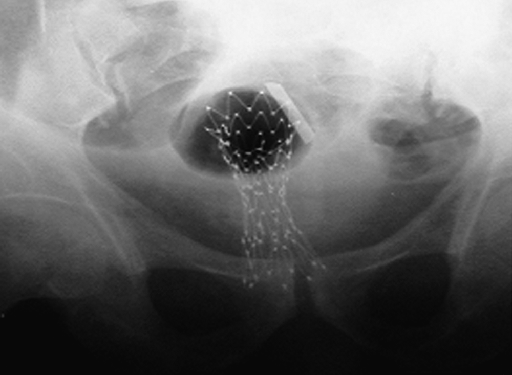
X-ray of successful placement of rectal stent.
Results
The patient's symptoms were reduced immediately, except for mild discomfort in her lower abdomen. She was followed for 11 months and remained free of obstruction.
Discussion
Cervical cancer is one of the most prevalent malignancies in the world, especially in developing nations. Unfortunately, colonic obstruction is usually a late stage of presentation of this disease and is more typical of an advanced and noncurable lesion. Surgical therapy has been the standard approach in this situation, with patients generally undergoing temporary or permanent colostomy. The surgical approach, however, is associated with significant risks and morbidity. Typically, these patients are not optimal surgical candidates because of the advanced stage of the tumor and their associated poor nutritional status. A palliative approach is standard treatment for some of these patients, with the intent being to relieve the obstruction and thereby treat the obstructive symptoms. In addition to surgical strategies for relief of colon-cancer obstruction, a variety of endoscopic interventions have been tried. These include palliative dilatation and ablative techniques, using laser or argon-plasma coagulation. These approaches, however, have been associated with risk for perforation as well as with rapid recurrence of obstruction. Rectal obstruction in cervical growths is rare and occurs when the tumor infiltrates or causes an extrinsic mass effect on the bowel wall—obstruction. The treatment in these cases is mainly surgery to bypass the obstruction. However considering the morbidity and mortality outcomes of surgery, a better option for these patients is nonoperative restoration of luminal patency.
SEMS has been used increasingly for palliation of malignant large-bowel obstruction or as a bridge to surgery. Patients with inoperable malignancies with either obstruction or with narrowed lumen can benefit greatly from placement of SEMS. In this case, the patient's symptomatic rectal obstruction had been diagnosed as resulting from advanced cervical carcinoma. The obstruction was been relieved effectively with the placement of a SEMS across the obstruction. The primary indications for expandable metal stent placement in the colon and rectum is preoperative decompression of inoperable malignant obstruction to allow bowel cleansing for a single-stage surgical resection and long-term colonic decompression for patients with unresectable malignant obstruction.1,2
Up to 30% of patients experience obstructive symptoms with colorectal malignancy at initial clinical presentation.3–6 Emergent surgical resection for acute malignant colonic obstruction has a mortality rate ranging from 7.2% to 22.4%.5,7 Placement of a colorectal stent enables bowel preparation, stabilization of the patient, and an elective one-stage surgical procedure. 8 The Wallstent Enteral was first approved for use in malignant colorectal obstruction in 1996. 3 Although esophageal stents have been used in the left colon and rectum, these stents can be problematic for navigating the tortuous distal colon.4,8,9 With its through-the-scope delivery option, the Wallstent Enteral has been the only metal stent placed in the transverse and right colon. 10 An Nd:YAG laser can be used to increase luminal diameter prior to stent placement, 11 but the added efficacy of this approach is unproven, and the procedure certainly increases risk. Neither balloon dilation nor laser therapy is universally recommended prior to colorectal stenting. 8
The efficacy of SEMS for palliating or allowing preoperative preparation in patients with malignant colorectal obstruction approaches 90%.4,8,9 Stents can also be placed in patients with benign (e.g., diverticular) obstruction to reestablish luminal patency and allow bowel cleansing prior to surgical resection. Stenting of known benign strictures is otherwise discouraged. Earlier, uncovered (bare metal) designs allowed ingrowth of tumors through wire mesh, causing the stents to anchor in place. In contrast, covered versions prevent such tumor ingrowth effectively but have a higher rate of migration.
Complications associated with stent placement include perforation, bleeding, stent migration, tumor ingrowth/overgrowth, and fistula formation. The decreasing diameters of delivery systems make perforation a rare occurrence and are generally related to prestent dilation. Tumor ingrowth rates are obviously higher with use of uncovered stents, but overgrowth at the distal or proximal ends can occur with covered versions. Tumor ingrowth and/or overgrowth is a function of tumor proliferation, length of follow-up, stent design, and possibly prior therapy. 3 This complication may be treated by coaxial placement of another stent,12,13 laser therapy, argon plasma coagulation, 14 photodynamic therapy, 15 and dilatation. 12 Placement of a colonic stent has a 5% perforation rate. 8 Dilation of the malignant colorectal stricture is not recommended because this action can increase risk of perforation. Stent migration is reported to be higher in the colon (up to 40%) secondary to increased peristalsis. Stents may generally be retrieved or passed in the rectum without sequelae. 4 The goal of SEMS is to maintain luminal patency; there must be a favorable balance between migration rates and prevention of tumor ingrowth. Current designs generally incorporate a covered midsection with uncovered flared ends that provide an anchoring mechanism.
Conclusions
Nonsurgical relief of malignant colonic obstruction is a great therapeutic challenge. The advent of self-expanding metal stent placement has been revolutionary for reducing surgical morbidity and mortality in patients with certain slow-growing tumors Although obstructive symptoms are rare, when this occurs, relief of symptoms can be obtained by a surgical approach with considerable morbidity or can be managed with placement of a SEMS, with a high success rate of relief of symptoms or as a bridge for elective surgery. The current patient had cervical growth and had successful placement of a SEMS that relieved her obstruction completely, avoiding the need for a major surgical procedure and improving her QoL.
Footnotes
Disclosure Statement
No competing financial conflicts exist.