
Abstract
Abstract
Introduction
Case
A healthy 26-year-old female, primipara presented to a labor room with excessive bleeding after normal delivery. She had delivered an average-sized full-term neonate 4 hours back at home. On examination, the patient was noted to be significantly pale with a pulse rate of 150 beats/minute, a systolic blood pressure of 60 mm Hg, and a respiratory rate of 30 breaths/minute. Her abdomen was soft and her uterus was “flabby.” A vaginal examination, revealed that was profuse bleeding. Pieces of placental tissue and blood clots were felt on bimanual examination and the tissue and clots were removed. Cervicovaginal exploration was performed, and no cervical tear or vaginal laceration occurred. Uterotonic agents were administered along with bimanual compression and abdominal aortic compression, but the patient continued to bleed profusely. A decision was made to perform a laparotomy. Modified B-Lynch sutures were applied along with uterine artery and ovarian artery ligation. The patient stayed in the intensive care unit for 2 days and was then shifted to the regular ward after her condition stabilized. Her postoperative period was uneventful. She was discharged to go home on the tenth postoperative day. After 8 weeks, the patient reported in the outpatient department with complaints of a mass being expelled from her vagina. On examination, this mass appeared to be part of her gut (Fig. 1).
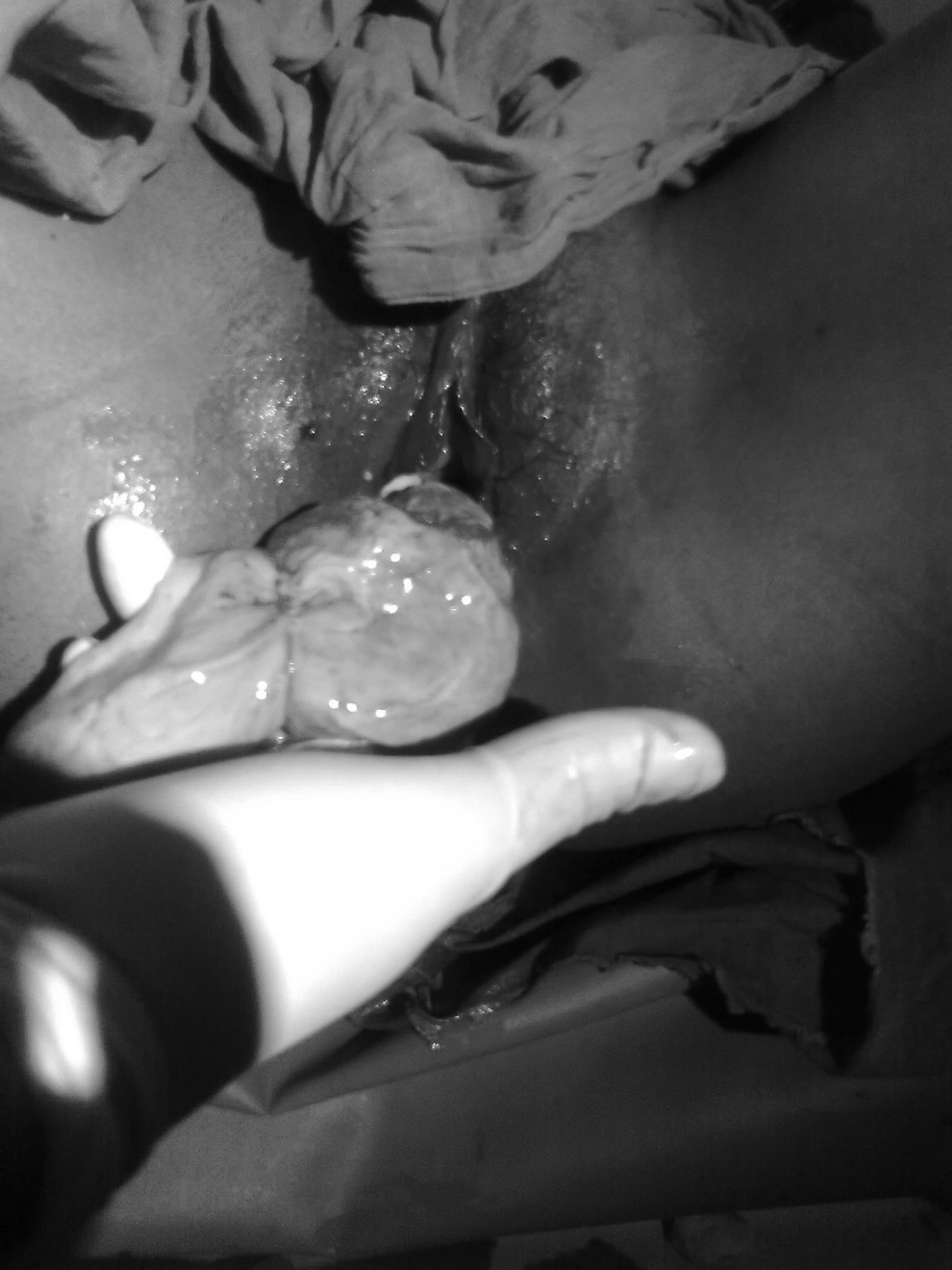
Decidual cast being expelled through the vagina.
Results
An examination was performed under anesthesia, and the mass was removed without any resistance. The mass had sutures embedded in it (Fig. 2). A provisional diagnosis of a decidual cast was made, and the tissue was sent for histopathologic examination, which confirmed it to be a necrosed decidua. After 6 months, a hysteroscopy was performed, which revealed that her endometrium was now normal.
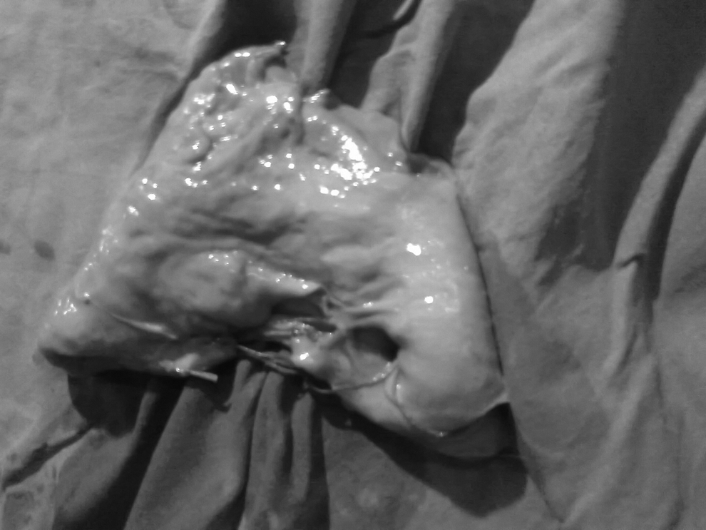
Decidual cast with sutures embedded in it.
Discussion
PPH typically occurs unpredictably, and no woman is exempt from the risk of PPH. Known risk factors for PPH arising from uterine atony include nulliparity, uterine overdistension (from fetal macrosomia, multiple pregnancy, hydramnios), prolonged or augmented labor, precipitate labor, maternal obesity, operative delivery, chorioamnionitis, and a history of PPH. Prompt diagnosis, rapid action, and a multidisciplinary approach are crucial for preventing and managing PPH.
Compression suture techniques have shown great promise in decreasing maternal morbidity and mortality associated with uterine atony, while maintaining fertility. In 1997, B-Lynch et al. described a novel technique of a uterine compression suture placed vertically as “belt and braces”; this suturing method was applied successfully in 5 patients as an alternative to cesarean hysterectomy. 2 In 2000, Cho et al. published an article about a case series of 23 patients with primary PPH who were treated by multiple square sutures compressing the anterior and the posterior walls of their uteri. 3 In 2002, Hayman placed two vertical sutures on each side of the uterus in each of 3 patients who had PPH. 4
The first rationale for all the improved methods is based on hemodynamic studies, which have shown that uterine compression sutures reduced pelvic blood flow and pulse pressure, resulting in venous pressures in the arterial circuit and thus promoted hemostasis. The second rationale is based on the uterine compression suture rapidly reducing the surface of the uterine wall from which the placenta has detached. The third rationale is based on the lack of blood to stimulate uterine contractions, which should be studied further and clarified. 5
Most uterine compression sutures attempted have been successful in controlling PPH, thus avoiding hysterectomy, but all of these situations need careful evaluation. The reported complications resulting from these sutures are pyometra, uterine synechiae, uterine necrosis, partial ischemic necrosis, and, in a few of the procedures, hysterectomy was not averted because the sutures “slid off” at the uterine fundus, and uterine avulsion occurred after the knots were tied too tightly. 6
A review by Grotegut et al. 7 reported 3 failures in 35 patients who received B-Lynch suturing, a success rate of 91.4%. These researchers have reported identifying erosion of B-Lynch sutures at the 6-week postpartum examination of an asymptomatic woman. Ultrasound examination confirmed the presence of a defect over her lower uterine segment. The researchers attributed this minor complication to the use of a suture with delayed absorption for controlling uterine bleeding and recommended the use of rapidly absorbed sutures. 7
In a 7-year prospective case series of 28 patients, Baskett reported a success rate of 82%. 8 He observed the presence of grooves over the uterine fundus at the site of previous B-Lynch sutures in 3 of seven patients who had subsequent cesarean sections after the initial compression sutures. In 2 of these 3 cases, there were additional signs of minor ischemic necrosis: a whitened myometrium between the grooves at the fundus and puckering at the posterior entry and exit sites of the previous B-Lynch suture. 8
Joshi and Srivastava described a case of severe postpartum hemorrhage in a 26-year-old primigravida who developed disseminated intravascular coagulopathy in the immediate postoperative period. After partial correction of the disseminated intravascular coagulopathy, the patient underwent a laparotomy. The surgeons found that the B-Lynch suture had cut through and become embedded in the patient's uterine wall, causing ischemic congestion and distension of the mid-section of her uterus between the B-Lynch vertical braces, giving her uterus a lobulated appearance. 9
Akoury and Sherman are the first authors to describe focal myometrial necrosis following the use of simultaneous B-Lynch and Cho-square sutures. During a subsequent pregnancy in the same patient, a large triangular myometrial defect was identified in her mid-anterior uterine wall and two smaller defects were noted in her posterior wall. 10
Treloar et al. reported the use of the B-Lynch suture to control severe PPH in a 33-year-old multigravida. Over the following 3 postpartum weeks, the woman experienced persistent vaginal bleeding and was found to have a tender subinvoluted uterus of 18-weeks' size. Multiplanar multisequence magnetic resonance imaging, with and without intravenous gadolinium contrasting, revealed that her uterus was avascular, except at the peripheral rim of the uterine wall. 11
The term “decidual cast” is used because the area of decidua that is shed is in the shape of the uterine cavity. Decidual casts have a well-known association with ectopic pregnancies. In nonpregnant women, decidual casts has been reported in patients taking human menopausal gonadotropin, human chorionic gonadotropin, and progestogens.
Unpredictable effects on endometrial and myometrial vasculature could be produced by compressive forces of uterine compression sutures. Enough external pressure to interrupt the blood supply of the outer myometrium may cause pressure necrosis, whereas if the blood supply of the inner myometrium and endometrium is disrupted, then decidua may be shed in form of a cast, as occurred in the current case.
Conclusions
Passage of a decidual cast after application of B-Lynch suturing, is reported for the first time. Women requiring uterine compression sutures should be informed about the possible complications of this procedure. Postoperative follow-up for these women should include pelvic examination, pelvic ultrasound, and sonohysterography to identify any decidual casts, uterine-wall defects, or uterine-cavity adhesions.
Footnotes
Disclosure Statement
No competing financial conflicts exist.