
Abstract
Abstract
Introduction
Case
A 61-year-old woman, gravida 6, para 6, presented to the emergency department with nausea, vomiting, and decreased appetite on postoperative day 7 after having undergone VH with BSO. The surgery was uncomplicated and was performed because of persistent cervical intraepithelial neoplasia grade 3 on the last two loop electrosurgical excision procedures. It was performed by a fourth year obstetrics and gynecology resident assisted by a gynecologic oncologist surgeon. The patient's past medical history was significant for hypertension and arthritis. Her past surgical history was significant for cesarean delivery. She was a smoker, but rarely drank alcohol. Her only medication was tramadol for joint pain.
On the day of presentation to the emergency department, the patient had increased nausea and vomiting, was passing flatus, and had one episode of loose stool. She denied any chest pain, shortness of breath, fever, or chills. She had no difficulties with urination. Physical examination demonstrated mild tachycardia and a distended, appropriately tender abdomen, with hypoactive bowel sounds. There was no rebound or guarding and no costovertebral angle tenderness. An abdominal series revealed numerous dilated loops of small bowel with air-fluid levels present (Fig. 1). Electrolyte levels were notable for mild dehydration. The patient was diagnosed with a small bowel obstruction, and she was readmitted to the hospital for conservative management that included nasogastric decompression. On hospital day 3, after persistent symptoms and bowel distension, she was taken to the operating room.
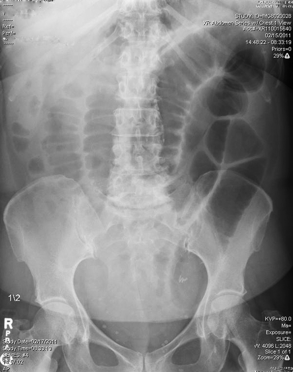
Upright abdominal radiograph demonstrating a small bowel obstruction.
During the second operation, the surgeon noted a hematoma present at the right pelvic side wall, measuring ∼2 cm×2 cm where the ovarian vessels had been ligated during the previous surgery that did not require evacuation. The ligation technique of the infundibulopelvic ligament used in the first surgery included two sutures. The first suture had been placed as a free tie behind the clamp. The second suture was a transfixation stitch, placing the suture in the midportion of the pedicle and then passing it around the tip and behind the heel of the clamp and tying again. The exposure of the infundibulopelvic ligaments was facilitated by transecting the round ligament on each side as a separate pedicle. Adhesions of the near terminal ileum to the vascular pedicles were observed. The appendix was also adherent to the hematoma and was secondarily swollen. The majority of the small bowel was distended except for the terminal ileum. The small bowel stuck to the hematoma was released, and the patient underwent an appendectomy. The dilated small bowel was emptied into the stomach and aspirated via nasogastric tube. The patient tolerated the procedure and had an uneventful postoperative course.
Discussion
Small bowel obstruction after VH and BSO is a very rare surgical complication. A PubMed search using the relevant key words
The complications of VH are well known and include a hematoma above the vaginal vault that might cause fever, infection, bleeding, bladder injury, nerve injury, and need for conversion to laparotomy/laparoscopy. 3 Long-term complications are vault prolapse or evisceration and formation of a fistula. 3 The incidence of bowel injury is very low with VH, but might include injury to the rectum, or laceration of a small bowel. 4 Ureteric injury is also rare. 4
Removal of adnexa at the time of VH is a feasible procedure and was shown to be successful in many studies.2,5 Prerequisites for vaginal salpingo-oophorectomy as described by Sheth are normal tubes and ovaries, easy access to the tube, mobile ovaries with free infundibuloplevic ligament, and an experienced vaginal surgeon. 6
Transvaginal removal of adnexa was not found to be associated with increased rate of complications.2,3,5,7 Neither was it shown to significantly increase the operative time. 8 The main complication that was cited in the literature was incomplete removal of the ovarian tissue.4,8 Sheth and Malpani 2 reported technical difficulties at the time of vaginal oophorectomy during which the tube or the ovary were torn; however, it did not cause any problems during the surgery. Small hematomas formed at the site of infundibulopelvic ligament were reported as well. 4 They were self limited, did not require laparotomy or transfusion, and were not associated with small bowel obstruction. 4
In the present case, bowel obstruction occurred secondary to adhesions created between a loop of a small bowel and a right pelvic side wall hematoma, at the site of the ovarian vessel pedicle. This is a very rare complication during VH with or without salpingo-oophorectomy, as there is much less bowel handling, and fewer adhesions are associated with this procedure. It is assumed that the pelvic side wall hematoma predisposed the tissue to adhesion formation and subsequent small bowel obstruction. In our first surgery, Trendelenburg position was used at the time of infundibulopelvic ligament ligation, to elevate the bowel out of the surgical field. In retrospect, adding a moist roller gauze to pack the bowel out of pelvis might have been helpful as well.
Conclusions
In conclusion, bowel obstruction is rare complication of VH with salpingo-oophorectomy. It can occur in association with hematoma formation on the pelvic side wall at the time of the surgery. Although rare, it should be considered in the differential diagnosis of a postoperative patient with nausea and distension after VH.
Footnotes
Disclosure Statement
No competing financial conflicts exist.