




Abstract
Abstract
Introduction
Case
Miss PO was a 20-year-old nullipara whose last menstrual period had been on August 4, 2010. She presented to the gynecology clinic on September 3, 2010 with a 7 month history of a rapidly growing abdominal mass and associated mild, intermittent abdominal pains. She had no history of vomiting or change in bowel habits. Her appetite was good. There was no history of weight loss. She had no significant urinary or respiratory symptoms.
She had attained menarche at the age of 13 years. She menstruated 3–5 days in a normal, regular, menstrual cycle of 28–30 days. There was associated dysmenorrhea, but no menorrhagia. The patient was aware of contraceptives, but had never used any method in the past. She had not had surgery in the past; and had neither diabetes nor hypertension. There was no family history of similar illness. She was the first of two children in a polygamous setting. She neither drank alcoholic beverages nor smoked cigarette. She had just completed her senior secondary education.
Health-seeking attitudes and the myth of the African tradition
The patient was taken to a rural private health care center where treatment with injectable medication was administered to her. She was later referred to a diagnostic center in the nearby town for ultrasonography. The utrasonographer was said to have diagnosed uterine fibroids. The patient was subsequently counseled for surgery, but her relatives later declined this modality of treatment and instead opted for traditional treatment. The patient was given traditional herbs to drink, with no effect.
Her relations later decided to seek divine intervention through the services of a traditional priest, in the belief that some evil powers were responsible for her illness. A consultation fee of $7.60 was paid to the traditional priest. The patient was expected to pay $276.10 later, for prayers to be offered to the gods for her deliverance. She was, however, unable to make this payment. The priest had warned her never to contemplate surgery on the grounds that she will die if operated upon. An enlightened relation of hers eventually decided to disregard this advice by taking her to The Holy Family Hospital, Ikom, Cross-Rivers State, Nigeria for proper treatment.
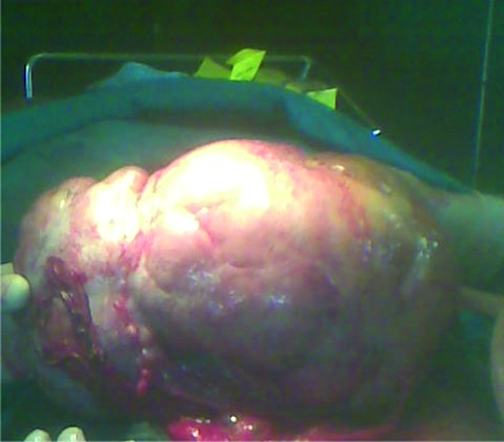
The tumor being removed at laparatomy. It weighed 3.4 kg. Color images available online at www.liebertonline.com/gyn
Physical examination
On examination at the gynecology clinic, the patient was found to be an attractive young woman, anicteric, afebrile, and not pale. She had no pedal edema. There was no peripheral lymphadenopathy. She weighed 41 kg with a height of 1.48 m. Her body mass index (BMI) was 18.7 kg/m2. Her lung fields were clinically clear. Her pulse rate was 80 beats per minute and blood pressure was 110/80 mm Hg, with only first and second heart sounds heard on auscultation of the chest. The abdomen was uniformly enlarged with a 36 weeks sized, firm to hard, and nontender, immobile, irregular-surfaced mass. It was, however, possible for the examining finger to get above and below the mass. Neither differential warmth nor ascites were demonstrated. The vagina and vulva were grossly normal. The cervix was healthy looking with a closed external os. It was difficult to perform a bimanual palpation for the body of the uterus. The pouch of Douglas was free. Cervical motion tenderness was negative. An impression of a huge right adnexal mass to rule out ovarian tumor was made.
An abdominopelvic ultrasound revealed a normal sized, empty uterus with a visible endometrial plate. The left ovary appeared grossly normal. There was a huge right adnexal hyperechoic mass measuring 24 cm×15 cm. There was no significant fluid collection in the pouch of Douglass. The patient's hemogram, liver function test (LFT), and urea and electrolytes (U/E) urinalysis tests all showed normal results. Her blood group was O+ and she was HIV negative. Her hemoglobin (Hb) electrophoresis result was AA.
The patient was counseled for exploratory laparotomy, for which she consented. Three units of blood were grouped and cross-matched. The anesthetist also reviewed the patient. She was placed in the supine position under general anesthesia. After she was cleaned and draped, a midline supra-umbilical incision was used to gain access to the abdominopelvic cavity.
The intraoperative findings at surgery were as follows: a huge solid, irregular surfaced, right ovarian mass measuring 26 cm×20 cm and weighing 3.4 kg. The right fallopian tube was attached to the mass. The left ovary and fallopian tube appeared grossly normal. The uterus was normal sized. There were mild filmy pelvic adhesions, but no peritoneal implants or omental deposits. There was no ascitic fluid in the peritoneal cavity or paracolic gutters.
The patient had a unilateral salpingo-oophorectomy (USO) and omental biopsy. The wound was closed in layers using the appropriate sutures. Estimated blood loss was ∼400 mL. The mass was sent for histopathologic assessment. The patient had initial postoperative treatment with intravenous fluids, analgesics, and antibiotics.
Histology report
Gross
There was an irregularly shaped tan tissue with nodular surface and attached Fallopian tube as well as omentum. The mass weighed 3.4 kg and measured 26 cm×19 cm×14 cm.
Cut section
The cut section showed a lobulated greenish-white surface with cavitary degeneration, filled grayish-tan gel. The fallopian tube measured 9 cm in length, and 0.7 cm in widest diameter. The cut surface showed a central empty lumen.
Microscopy
A section of tissue showed a tumor consisting of sheets, cold and trabecular cohesive polygonal cells with vesicular nuclei, prominent nucleoli, abundant clear cytoplasm, and distinct cell membrane. The cellular clusters were separated by variable fibrotic bands harboring moderate lymphocytes cells. Also seen were sections of fallopian tube with liberal folds of mucosa lined by columnar cells and bounded by smooth muscular wall. Section of omental tissue showed matured adiposite and fibrocollagenous tissue.
Diagnosis
The patient was diagnosed with ovarian dysgerminoma.
Treatment
Six weeks after surgery, the patient was administered six cycles of adjuvant chemotherapy with bleomycin, etoposide and cisplatinum as treatment for International Federation of Gynecology and Obstetrics (FIGO) stage 1C disease. She did well and was subsequently placed on monthly follow-up visits.
Discussion
Dysgerminomas are a subtype of MOGCT. They are found in young girls, adolescents, and young women such as the patient presented here. The clinical symptoms of chronic abdominal pains and a rapidly growing abdominal mass were typical in this patient. Unfortunately, prompt diagnosis and appropriate treatment was delayed because of the myths of the African tradition, and patronage of poorly equipped, peripheral health facilities, staffed by lower cadre health personnel. Public health education campaigns, and reduction of extreme poverty and cost of medical services may encourage the use of orthodox healthcare services (where care is delivered by skilled medical professionals) particularly by rural dwellers in sub-Saharan Africa.
The main conditions for obtaining a good result in MOGCT is a quick diagnosis and appropriate treatment, usually surgery associated with multidrug chemotherapy. 4 The stage of the disease is also a good prognostic factor. 4 In young patients suspected of having an OGCT, laparatomy is initially indicated both for diagnosis and treatment. For most patients, unilateral salpingo-oophorectomy with preservation of the contralateral ovary and the uterus is appropriate. 2 This was the treatment given to the patient described here.
Fertility-conserving surgery was performed on the basis of patient's age, parity, stage of tumor, and reproductive wishes. This form of surgical treatment has been shown to be effective in retaining patient's fertility potential.5,6 For all OGCT patients, except those with well- documented stage 1A grade 1 pure immature teratoma or stage 1A pure dysgerminoma, postoperative chemotherapy is indicated. 2 The patient described here was treated as FIGO stage 1C. She therefore had further treatment with adjuvant chemotherapy; the current regime of which is bleomycin, etoposide, and cisplatinum, a combination that appears to result in at least a 95% cure rate for stage 1 disease and at least a 75% cure rate for advanced stage disease.2,3 The patient did well and is on monthly follow-up visits.
Conclusions
Early diagnosis and treatment of dysgerminoma ensures good prognosis. Fertility- conserving surgery and adjuvant chemotherapy are appropriate for young, nulliparous women who wish to retain their fertility potential. Education, public health enlightenment, and reduction of extreme poverty and cost of hospital services will promote better health-seeking behavior and use of quality orthodox health facility services in sub-Saharan Africa.
Footnotes
Acknowledgments
I appreciate the assistance and cooperation of Ojo Solomon, Friday Ashikabe, and Christiana Obasi.
Disclosure Statement
No competing financial interests exist.