
Abstract
Abstract
Introduction
Case
An 29-year-old primigravida without a prior appointment was admitted in early labor to the obstetric department of the Postgraduate Institute of Medical Sciences, in Rohtak, Haryana, India, at 39 weeks of gestation. She had been married for 11 years and had taken some indigenous medicines for infertility. There was no significant family or past history. There was no history of dyspareunia. Her vital signs were stable. A consultant was called by the residents, because the woman's cervix was not visible. Abdominal examination revealed a term-sized uterus with the fetus in cephalic presentation with a regular fetal heart rate, and regular uterine contractions of mild intensity. Speculum examination revealed a transverse vaginal septum with a pinpoint septal opening. Vaginal examination confirmed the diagnosis of transverse vaginal septum with a pinpoint opening. Neither the woman's cervix nor fetal head could be felt. A pediatric Foley's catheter was inserted freely up to 3–4 cm through the hole in the septum, suggestive of the length of the vagina above the septum (Fig. 1). Emergency cesarean section was performed, and a healthy male baby weighing 3150 g was delivered. The woman's internal os admitted one finger and this os was dilated (Fig. 2). To prevent hematometra, the small opening in the vaginal septum was dilated after the cesarean section (Fig. 3).
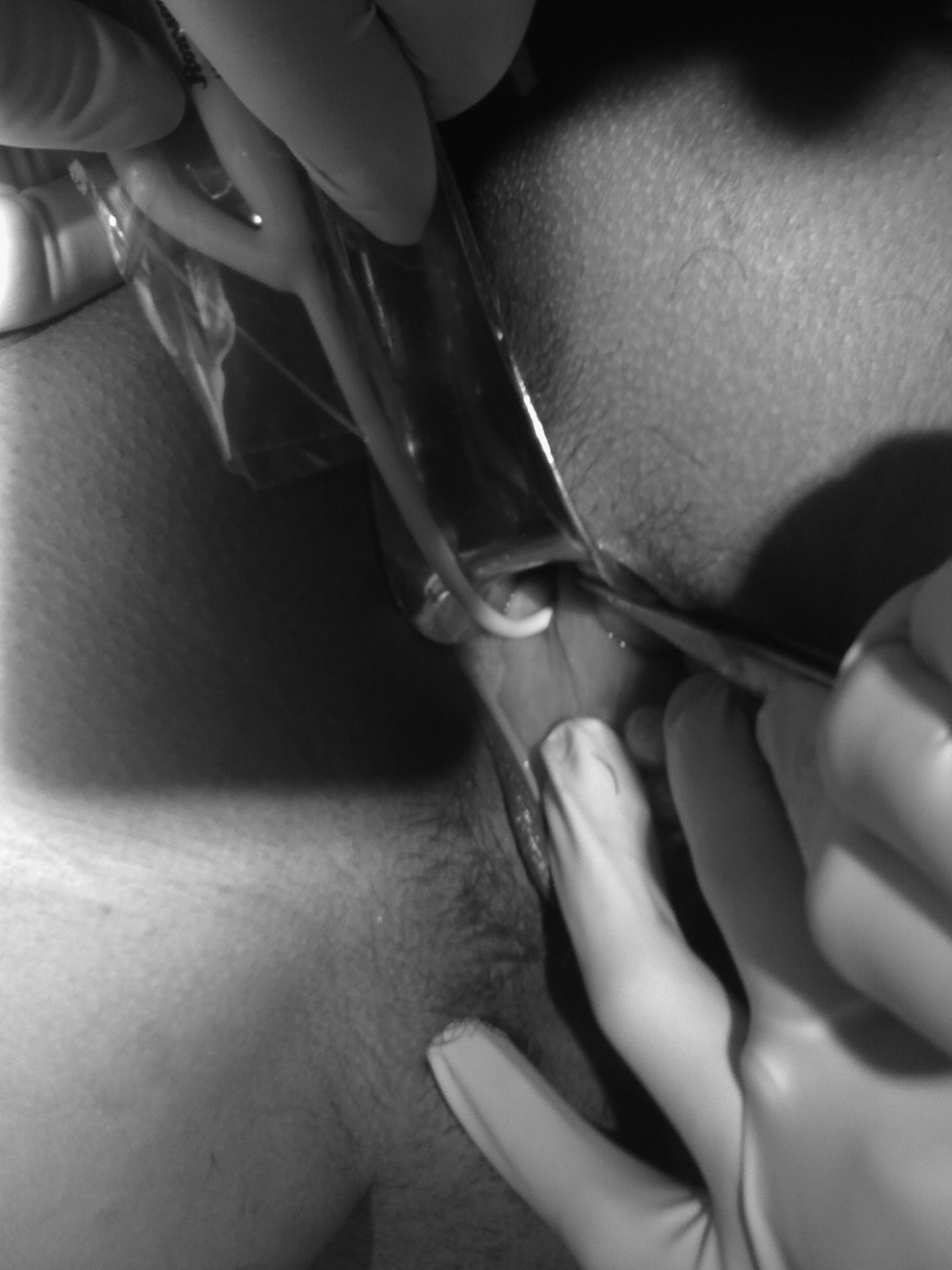
Pediatric Foley`s catheter passed through septal opening in transverse vaginal septum.
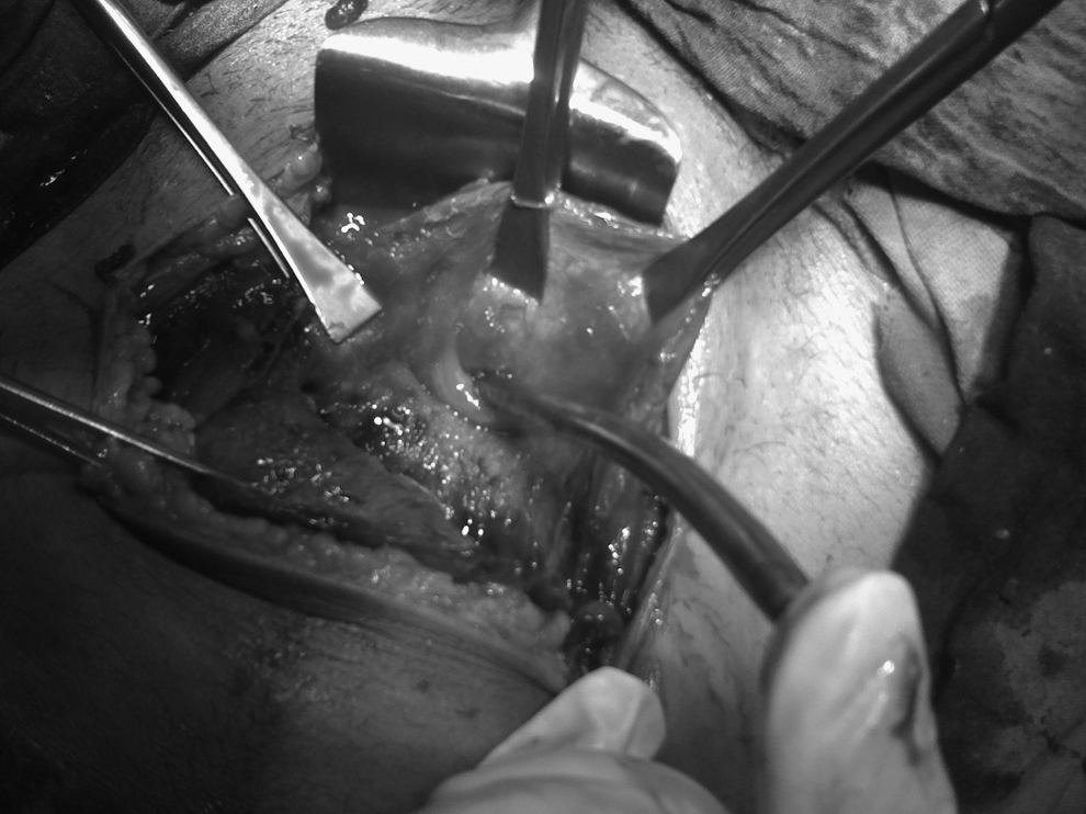
Internal os being dilated during cesarean section.
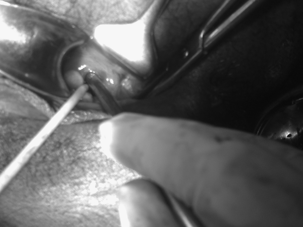
Dilatation of septum at time of vaginal toileting.
Results
Ultrasonography of the abdomen was done on the fourth postoperative day. No associated genitourinary malformations were present. The patient's postoperative period was uneventful.
Discussion
Transverse vaginal septum is a rare congenital anomaly of the reproductive tract. The estimated incidence is 1/30,000–84,000 women. 2 These defects are much less common than midline longitudinal defects.3–8
Any defect in the normal organogenesis involving the urogenital sinus or the Müllerian duct can result in genital-tract anomalies. Embryologically, the lower two thirds of the vagina develop from the urogenital sinus. The upper vagina, cervix, uterus, and Fallopian tubes develop from the Müllerian duct system. Failure of vertical fusion or canalization of the two systems in utero may result in cervical stenosis or atresia, vaginal atresia, or transverse vaginal septa.
There are two kinds of vaginal septa: transverse and longitudinal. Transverse vaginal septum is thought to be the result of faulty canalization of the embryonic vagina. A longitudinal vaginal septum occurs when the distal ends of the Müllerian ducts fail to fuse properly.
Transverse septa may be complete, resulting in cryptomenorrhea and hematocolpometra, or partial, with pinpoint openings allowing menstrual flow. These septa are usually found in the midvagina, but may occur at any level. When the septum is in the upper vagina the septa is more likely to be incomplete. If it is located in the lower part of the vagina, the septa is more likely to be complete.
Diagnosis of complete transverse vaginal septum is often delayed until after menarche, when menstrual blood is trapped behind an obstructing membrane. 9 Ahmed et al. described a 12-year-old girl with distal mucocolpos and proximal hematocolpos secondary to a concurrent imperforate hymen and a transverse vaginal septum. 10
An incomplete septum is usually asymptomatic and, therefore, does not require correction during childhood and early adolescence. The central aperture allows for vaginal secretions and menstrual flow from the vagina. 1
The presence of a septum can cause dyspareunia and infertility. A persistent transverse vaginal septum does not appear to be linked to obstetric complications such as pregnancy loss, fetal anomalies, or premature delivery, or to urinary-tract malformations similar to those associated with longitudinal defects.3–8 During labor, a transverse vaginal septum may result in significant vaginal lacerations during vaginal delivery or obstructed labor resulting in rupture uterus.
Cesarean section in early labor appears to be the best option for these patients. Fenton and Singh have also advocated cesarean section for them. 7 However, other options suggested in world literature include: (1) expectant management with a plan of either allowing spontaneous dissection of the septum as a result of dilatation of cervix and descent of the fetal head, or incision late in labor, if needed, after the septum has been thinned and pressure from head can provide hemostasis; or (2) incision of the septum before labor.
Blanton and Rouse have also reported 2 patients with transverse vaginal septa who were allowed a trial of labor. Their septa were incised in active labor, resulting in vaginal delivery with no related complications. 11
Ustun et al. described a case of an 18-year-old nullipara with transverse vaginal septum, at 28 weeks of gestation. Pelvic and vaginal ultrasonography with gynecologic examination established a diagnosis of transverse vaginal septum in the midvagina with a fully dilated cervix behind the septum. The woman's septum was incised with the patient under epidural analgesia, followed by delivery of a preterm infant.
However, trial of labor is not recommended, as the progress of labor cannot be assessed, and internal monitoring is also not possible. Levin et al. opined that incision before labor was likely not to be optimal because this protocol creates scarring, resulting in obstructed labor, and, therefore, requiring two separate operative interventions. 8 Cesarean section is the best choice for patients with transverse vaginal septum diagnosed during pregnancy and labor. The ideal time for correction of this disorder is incision immediately before a woman becomes sexually active, to avoid dyspareunia and improve reproductive performance.
Conclusions
To avoid obstructed labor and injuries to the patient, prophylactic cesarean section is recommended for patients presenting in labor with transverse vaginal septum.
Footnotes
Disclosure Statement
No competing financial interests exist.