
Abstract
Abstract
Introduction
Case
A 67-year-old Greek female patient presented to the University General Hospital of Alexandroupolis with significant abdominal distension and complaints of nonspecific abdominal pain and heaviness. Her past medical and family history were unremarkable, as was her gynecologic history, with menarche at the age of 15 and menopause at the age of 52. Her general condition and vital signs were normal. During the physical examination, the abdomen was found to be distended, and a large mass was palpable in the inferior abdomen and in the lesser pelvis. An ultrasound (US) examination of the abdomen was performed and showed a huge abdominal thin-walled cyst spanning the abdomen and pelvis with no evidence of any solid element or any papillary projections arising from either the internal or external wall surfaces. The liver, spleen, and kidneys were normal, and the pancreas was not observed. A computerized tomography (CT) scan was immediately performed and revealed a large (13.5 cm×11.4 cm×19.5 cm) cystic mass spreading from the right ovary and the right fallopian tube to the inferior abdomen and the whole lesser pelvis (Fig. 1). Because of the huge size of the cyst, its origin could not be defined. Abnormalities in the uterus and left ovary were not detected, and the CT scan excluded the dilatation of the inguinal or cervical lymph nodes, the infiltration of adipose tissue, and ascites. The overall morphologic view did not indicate malignancy. The patient's laboratory results were in the normal range except for her C-reactive protein (CRP) level, which measured 3.27 mg/dL (normal range 0–0.5 mg/dL). In addition, her CA-125 level measured 20 U/mL, which is in the normal range (0–30 U/mL). On the day of admission, after a preoperative consultation and considering the low risk of malignancy, the patient and her family opted for a laparoscopic cystectomy. A wedge resection of the ovary and a right salpingo-o ophorectomy were performed.
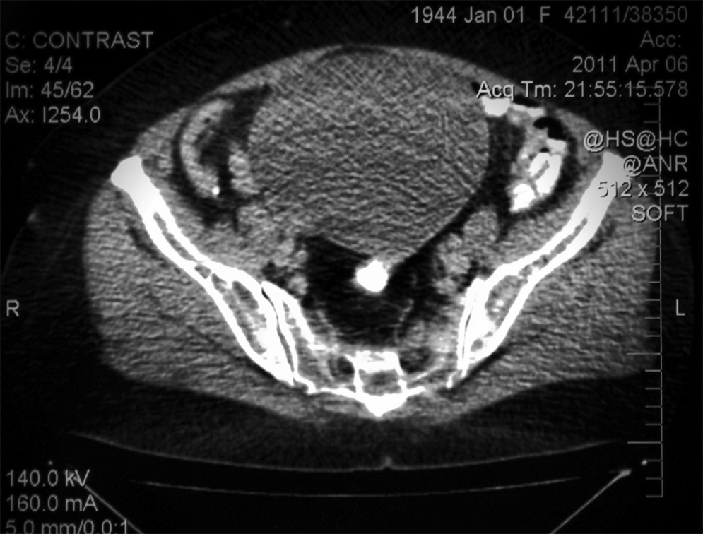
Computerized tomography (CT) scan revealing a sizable cyst 13.5 cm×11.4 cm×19.5 cm originating from the right ovary and extending from the lesser pelvic to the umbilicus. There were no signs of malignancy.
Consent
Written informed consent was obtained from the patient for the publication of this case report and the accompanying images.
Surgical details
After appropriate preoperative care, laparoscopic surgery was performed with the patient under general anesthesia with endotracheal intubation. The patient was placed supine in the Trendelenburg position (20 degrees), after insufflation of CO2 (2.5 L), at a pressure of 12 mm Hg to create pneumoperitoneum. Afterwards, a camera (0°) was inserted through the first 10-mm trocar, above the umbilicus, into the peritoneal cavity, to visualize the exterior of the cyst, and the whole peritoneal cavity was assessed. The second 10-mm trocar was placed, laterally, to the right outer edge of the rectus abdominis muscle and another 5 mm to the left side. The cyst appeared to arise from the right paraovarian region (Fig. 2). Ligature systems were used to separate the cyst from the connective tissue and adhesions. After this procedure was complete, grasping forceps were used to close the puncture site of the cyst. The first task was to place the whole cyst into an Endobag® without rupturing the cyst wall. However, it was not possible to use an Endobag because of the large size of the cyst, and, therefore, an alternative, uncommon approach was tried. After the cyst was grabbed, it was moved to just below the umbilical portside. Then, the contents of the cyst were aspirated by placing a 5-mm trocar in combination with an aspiration system into the cyst, to prevent leakage, and the cyst was removed through the umbilical incision for the first trocar (Fig. 3). This new technique of aspirating outside of the peritoneal cavity was effective, and it was possible to excise the whole serous cystadenoma without causing any rupture or intraperitoneal diffusion of the serous fluid. The whole procedure lasted ∼55 minutes. The whole procedure was documented in a short video file.
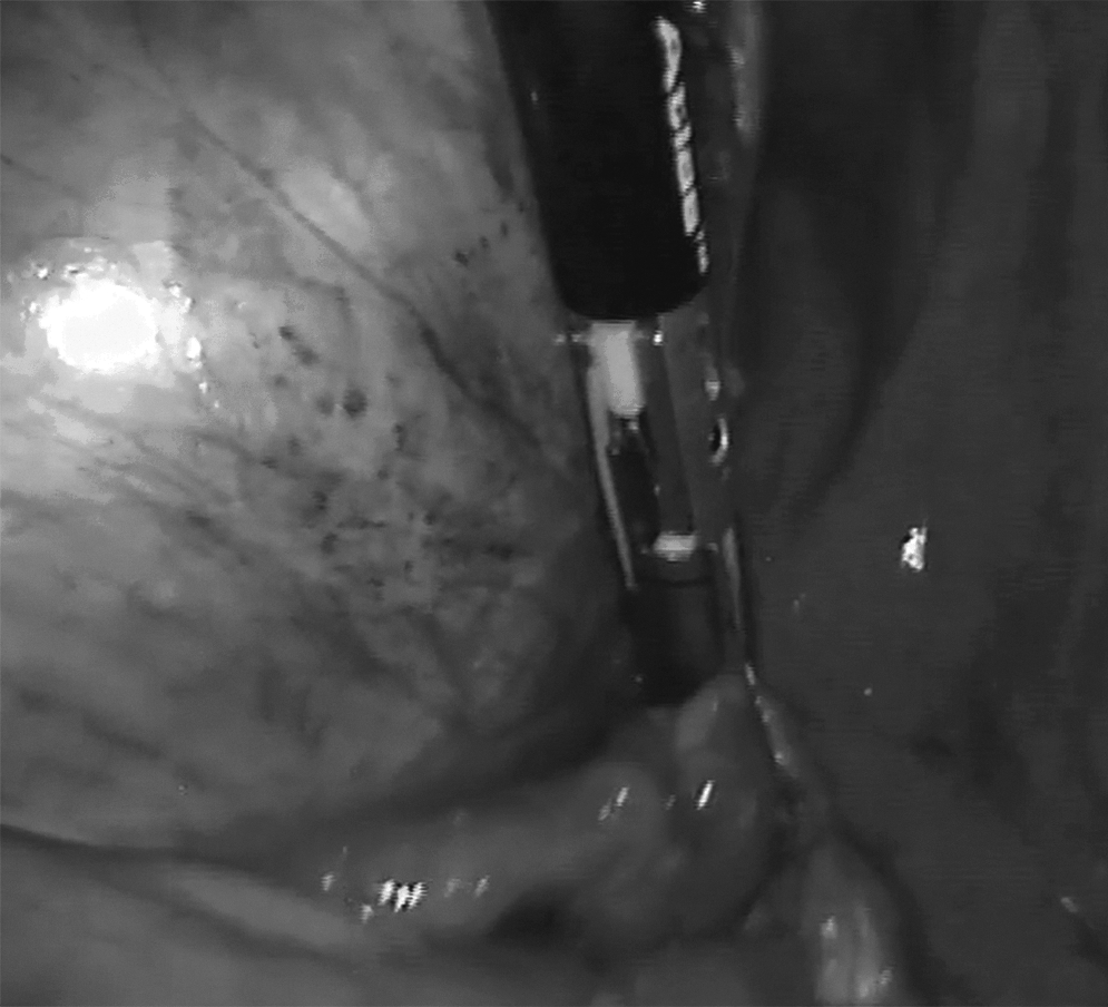
Intraoperative view through a laparoscopic endoscope of the huge serous cystadenoma extending into the abdomen and pelvis.
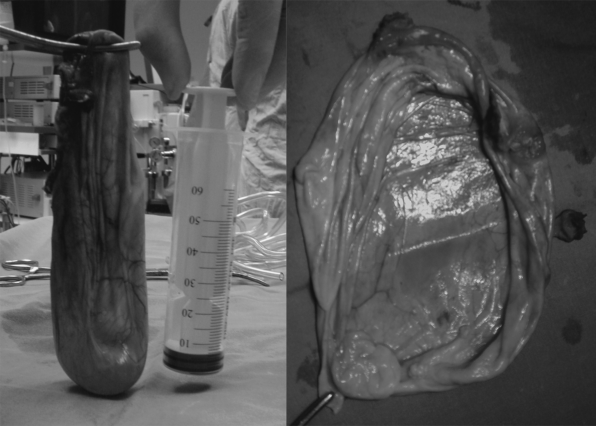
Macroscopic view of the excised serous cystadenoma.
In addition, a drain was placed, and specimens and the cystic fluid were sent for histopathology and cytologic examination.
Results
Postoperatively, the patient had an uneventful recovery, and she did not experience any complications. Her recovery was quick, and she was discharged on postoperative day 3.
The pathology report revealed a unilocular cyst with no solid areas, and the cyst wall was lined with a single cuboidal epithelium. All the features were consistent with a benign serous cystadenoma.
At her latest follow-up appointment 6 months after surgery, she presented with no symptoms, and there has been no recurrence of the cyst.
Discussion
Cystadenomas account for ∼20% of all ovarian neoplasms. 2 These tumors originate from the mesothelium of the peritoneal cavity, and, therefore, they can be divided into serous and mucinous types according to the type of cells. 1 Furthermore, this neoplasm cannot produce ovarian hormones. As a result, it does not cause menstrual disorders.1,3
Serous cystadenomas are very common benign ovarian neoplasms that can reach very large sizes. These tumors can reach 40 cm in diameter and form spherical masses.2,4 Approximately 70% of all serous cystadenomas are benign tumors, 5%–10% have borderline malignant potential and 20%–25% are malignant. This neoplasm is most often diagnosed during menopause, during the 5th decade of life.3,4 The macroscopic morphology of a benign cystadenoma is a unilocular cyst with a thin and fibrous wall that contains serous, achyrochrous fluid.2,5 Microscopically, the wall of cyst consists of cuboidal cells, similar to those of the epithelium of fallopian tubes.1,5,6 A benign, large, unilocular cyst with dimensions of 13.5 cm×11.4 cm×19.5 cm is reported here.
Benign serous cystadenomas represent one of the greatest current challenges for medicine with respect to diagnosis and treatment. Diagnosis is difficult, especially during the primary stages and for small cysts, because the clinical presentation is not characteristic. 1 As a general rule, benign serous cystadenomas are asymptomatic or lack specific symptoms, and, thererfore, these tumors are often large by the time the patients are diagnosed. Pain is rare except when the cyst rupture, when there is inflammation, or when there is ovarian torsion. 4 Large cysts can extend from the lesser pelvis to the whole abdomen. 5 The findings of physical examinations are not identical. During gynecologic examination, a spherical, painless mass is typically revealed. A computed tomography scan and US can be performed to support the diagnosis.4,7–9
The treatment of serous cystadenomas is also complicated. All ovarian tumors with sizes >5 cm must be excised immediately because of the likelihood of malignancy and the possible complications unrelated to the histopathologic type of grade of the tumor. Currently, there are two surgical options for the treatment of serous cystadenomas: laparotomy and laparoscopy.1,2,6,8 Both of these techniques must be always followed by histologic examination of the cyst to exclude malignancy.1,3,4,9
When deciding on the treatment plan for serous cystadenomas, it is necessary to choose a plan that decreases the risk of the implantation of cancerous cells. Implantation and metastasis can occur via the consecutive tissue, through the circulation of lymph or blood, and through intraperitoneal diffusion. 8 The last mode of implantation and metastasis is the most common, and follows the flow of intraperitoneal fluid to the right side. Cancerous cells are transferred by the serous intraperitoneal fluid, especially after the rupture of the cyst wall, along the colon, where they implant, and then toward the subdiaphragmatic region and Glisson's capsule. This transfer is facilitated by the different levels of pressure that occur during respiration. 7
Since 1930, gynecologists from France and Germany have led the development and application of laparoscopic surgery and have encountered challenges for which technology should give answer within the next several decades. Laparoscopic surgery has made a significant contribution to the field of gynecology. Semm performed gynecologic operations, such as cystectomies and hysterectomies, from 1965 to 1980 using laparoscopic equipment and techniques such as suturing, aspiration, and ligation; these same techniques are used today. 10 In the last 15 years, the laparoscopic treatment of ovarian cysts has advanced greatly, although there are a lack of data to compare surgical laparoscopy with laparotomy for sizable ovarian cysts. The use of a laparoscopic approach in cases of huge ovarian cysts is controversial, and is rarely performed successfully.11,12
A search of the medical literature published since 2000 and indexed in PubMed using the key words
Idotta R. Removal of a voluminous serous papillary paraovarian cystadenoma by endoscopic surgery: A case report. Clin Exp Obstet Gynecol 2000;27:150.
Eltabbakh GH, Kaiser JR. Laparoscopic management of a large ovarian cyst in an adolescent: A case report. J Reprod Med 2000;45:231.
Postma VA, Wegdam JA, Janssen IM. Laparoscopic extirpation of a giant ovarian cyst. Surg Endosc 2002;16:361.
Lin YH, Hwang JL, Huang LW, Seow KM. Successful laparoscopic management of a huge ovarian tumor in the 27th week of pregnancy: A case report. J Reprod Med 2003:48:834.
Sagiv R, Golan A, Glezerman M. Laparoscopic management of extremely large ovarian cysts. Obste Gynecol 2005;105:1319.
Mittal S, Gupta N, Sharma AK, Dadhwal V. Laparoscopic management of a large recurrent benign mucinous cystadenoma of the ovary. Arch Gynecol Obstet 2008;277:379.
Garg P, Misra S, Thakur JD, Song J. Single incision laparoscopic surgery ovarian cystectomy in large benign ovarian cysts using conventional instruments. J Minim Access Surg 2011;7:232.
Surgical laparoscopy is a relatively minor procedure that allows the visualization of the intraperitoneal cavity and the organs therein, and several operations can be performed using laparoscopy, obviating the need for open laparotomy. There are many clear benefits of laparoscopic treatment of benign ovarian cysts. The morbidity is always less than that for similar major surgeries performed by laparotomy. In addition, the operating time is reduced, postoperative pain and other complications are rare, recovery is improved, discharge is quick, and patients return to their daily routine in a few days.2,3,6,8–10,12
However, the major problem with laparoscopy is the risk of the intraperitoneal diffusion of cancerous cells, which can lead to implantation and metastasis. According to a study performed by Dembo (1990) 13 , the factors that contributed to the recurrence of disease, in 519 patients with stage I ovarian cancer after laparoscopic surgery, were the tumor grade, the existence of fixed adhesions, and ascites. Rupture of the cyst was not one of these factors. Other serious problems, especially for huge serous cystadenomas, are the result of the limited working space, the risk of cyst rupture, and the intraoperative cardiorespiratory function. 9
Even if all the preoperative examinations indicate that the tumor is a benign cystadenoma, pathologic evaluation of frozen sections can be performed during surgery to diagnose or exclude malignancy. 14 When malignancy is suspected, laparoscopy is contraindicated, and a median laparotomy is appropriate for radical extirpative surgery. 1 Then, the external surface of ovaries should be checked, and any adhesions should be removed and evaluated. If all results indicate a benign serous cystadenoma, the cyst should be aspirated and then placed into an Endobag.2,8
The sizable serous cystadenoma that was presented in the case report is the largest benign ovarian serous cystadenoma at University General Hospital of Alexandroupolis that has been treated laparoscopically in combination with a new aspiration system outside of the peritoneal cavity. Because of the huge size of this serous cystadenoma, an Endobag could not be used. However, the extraction of the cystadenoma through the umbilical incision, after aspiration, led to the successful, complete excision of the cystadenoma. This method was based on the macroscopic and immediate extraction of the cyst to reduce the risk of the intraperitoneal diffusion of cancerous cells.
Conclusions
Laparoscopic surgery has advanced greatly over the last few years, and now plays an important role in treatment of small ovarian cysts. This case report demonstrates that laparoscopic ovarian cystectomy is safe even for huge serous cystadenomas, because intaperitoneal diffusion can be prevented. Furthermore, the justification for adopting this laparoscopic technique depends upon the perceived value of its ability to improve short-term postoperative outcomes, especially recovery time and time to discharge. The method described herein is an alternative option for the successful laparoscopic treatment of huge serous cystadenomas.
Footnotes
Disclosure Statement
No competing financial interests exist.