
Abstract
Abstract
Introduction
B
Case
A 35-year-old woman, gravida 7, para 5+1, who had 5 living children at home and a history of one first-trimester abortion, was admitted to the Women Health Hospital in Assiut University, Assuit, Egypt, with a history of 8 weeks' amenorrhoea and intermittent vaginal bleeding; she also had mild lower abdominal pain of 1 week's duration. A urine pregnancy test on the day of her admission was positive. She had been married for 13 years. Her menstrual history was not relevant. There was no past history of contraception use or previous abdominopelvic surgery. She also had no history that was suggestive induction of ovulation.
On general examination, this patient was noted to be conscious, with a pulse of 100 beats per minute, a temperature of 37°C, and blood pressure of 110/70 mm Hg; her cardiovascular and respiratory systems were normal. Her abdomen was tender on palpation with a positive rebound and guarding. On pelvic examination, it was noted that there was mild spotting, her cervical os was closed, and her cervix tender on transverse motion. Her uterus was bulky; there was fullness in all the fornices with tenderness; and the adnexae were difficult to palpate. Haematologic examination showed a white blood cell count of 8×10⁹ cells/L and a hemoglobin level of 9.5 g/dL. Pelvic ultrasound examination showed a bulky uterus, a homogenous texture, and a thick endometrium with a smooth outline. No free intraperitoneal fluid collection was detected. There was an echogenic mass lesion in the right adnexae measuring ∼4 cm×4 cm with an ovarian cyst in the left side measuring 3 cm×3 cm, suggestive of a right unruptured ectopic pregnancy with a left ovarian cyst (Fig. 1).

Right-sided echogenic mass lesion measuring ∼4 cm×4 cm, with ovarian cyst in the left side measuring 3 cm×3 cm, suggestive of right unruptured ectopic pregnancy with left ovarian cyst.
The patient was counseled concerning the possibility of an ectopic pregnancy, and informed consent for abdominal exploration with the possible need for salpingostomy or salpingectomy was obtained.
Exploratory laparotomy under general anesthesia through a Pfannenstiel incision revealed a right unruptured ampullary ectopic pregnancy (Fig. 2) with a marked hematosalpinx. The left tube showed an unruptured ampullary ectopic pregnancy 2×2 cm with no apparent hematosalpinx but with a left simple ovarian cyst of 3 cm×3 cm (Fig. 3). In view of these findings, a right salpingectomy and a left salpingostomy with cautery were carried out with removal of the products of conception.
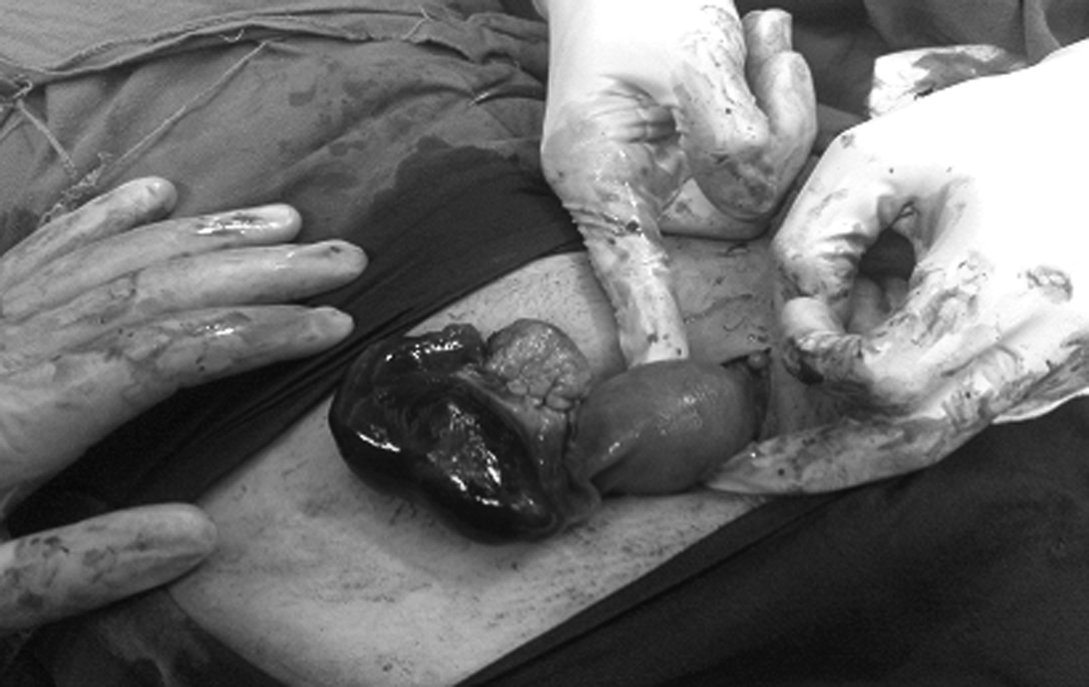
Introperative view of right unruptured ectopic pregnancy.
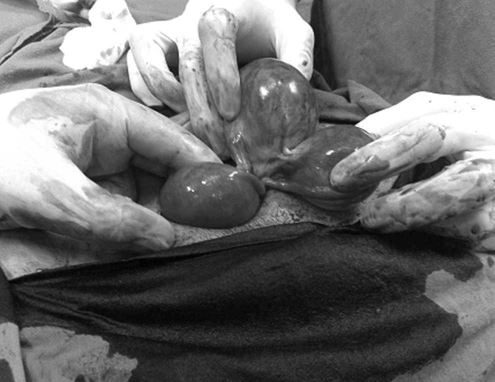
Intraoperative view of left unruptured tubal pregnancy with the ovarian cyst.
Results
This patient's postoperative follow-up was uneventful and she was discharged on the fifth postoperative day. Two weeks after surgery the level of β subunits of human chorionic gonadotropin (hCG) was zero. Histopathologic examination of the specimens, including the excised right unruptured tube and the product of conception extracted from the left tube, confirmed the diagnosis. There were tubal tissues with decidua and chorionic villi on the right unruptured tube, and inflamed decidua with chorionic villi, but no tubal tissues, was seen on the specimen from the left side.
Discussion
Spontaneous bilateral ectopic pregnancy is a rare event; therefore, preoperative diagnosis is uncommon. The frequency of bilateral ectopic pregnancy has been estimated at 1/200,000 uterine pregnancies and 1/725–1/1580 ectopic pregnancies. 4 In the past 20 years, a threefold increase in the incidence has been observed. 5 Heterotopic as well as bilateral tubal ectopic pregnancies are seen after the introduction of assisted reproductive treatment. 6 Several theories have been attempted to explain the occurrence of bilateral tubal pregnancies. Bilateral tubal gestation requires multiple ovulations to occur with implantation of the fertilized oocytes at the tubes. 7 Another possible theory is transperitoneal migration of fertilized oocytes from one tube to another. 8 Superfetation, another possible theory, implies fertilization and development of a second oocyte when a woman is already pregnant. This is considered to be an extremely rare event in humans and is difficult to prove. 9
Most patients with bilateral tubal pregnancies have similar clinical pictures and risk factors to patients who have unilateral ectopic pregnancies. The most frequent findings are amenorrhea, vaginal bleeding, and abdominal pain. Levels of serum hCG and the discriminatory zone are not reliable markers for patients with bilateral disease. 10 Consequently, the diagnosis of bilateral tubal pregnancy is usually made intraoperatively, 11 so examination of both tubes at the time of surgery is very important. This article discussed a rare case of spontaneous bilateral intact ectopic pregnancy involving the ampullary segments with an ovarian cyst. Ultrasonography in this patient's case failed to facilitate such a diagnosis, and this is in agreement with other reports (i.e, use of ultrasound is not necessary to make a diagnosis of bilateral ectopic pregnancy). 7 Therefore, diagnosis of bilateral ectopic pregnancy continues to be an important challenge facing emergency-room physicians.
Conclusions
In the current case, surgical management to preserve the left tube was by linear salpingostomy with evacuation of the hematoma; hemostasis was achieved by cauterization. Careful attention should be directed to follow-up tests. A serial measurement of serum concentrations of hCG is necessary to rule out the risk of a persistent trophoblast. 8
Footnotes
Disclosure Statement
No financial conflicts exist.