




Abstract
Abstract
Introduction
Case
A 35-year-old woman was admitted to the gynecology ward with pregnancy at 26 weeks of gestation, and a mass protruding through the vaginal introitus. She had had three full- term normal vaginal deliveries at home. She had noticed the protrusion 4 weeks prior to admission.
On general examination, her pulse rate was 88 beats per minute and her blood pressure (BP) was 110/80 mm Hg. Abdominal examination revealed a uterine height corresponding to 26 weeks of gestation. Fetal heart sound was 134 beats per minute and regular. Local examination showed a third degree cervical prolapse (International Continence Society Pelvic Organ Prolapse Quantification System [ICS-POP-Q] III C). The cervix was lying 3 cm outside the introitus, hypertrophied. and edematous with no ulceration.
The patient was put on bed rest, with the foot end of the bed raised. Acriflavin and glycerine were applied on the cervix, and the prolapse was repeatedly reduced. The patient was discharged on the 7th day when cervical edema subsided. At the time of discharge, she was advised to rest, and a plastic ring pessary (Fig. 1) was inserted in the vagina to keep the cervix in place. She was asked to come for follow-up after 2 weeks, or earlier, if she had any discomfort.
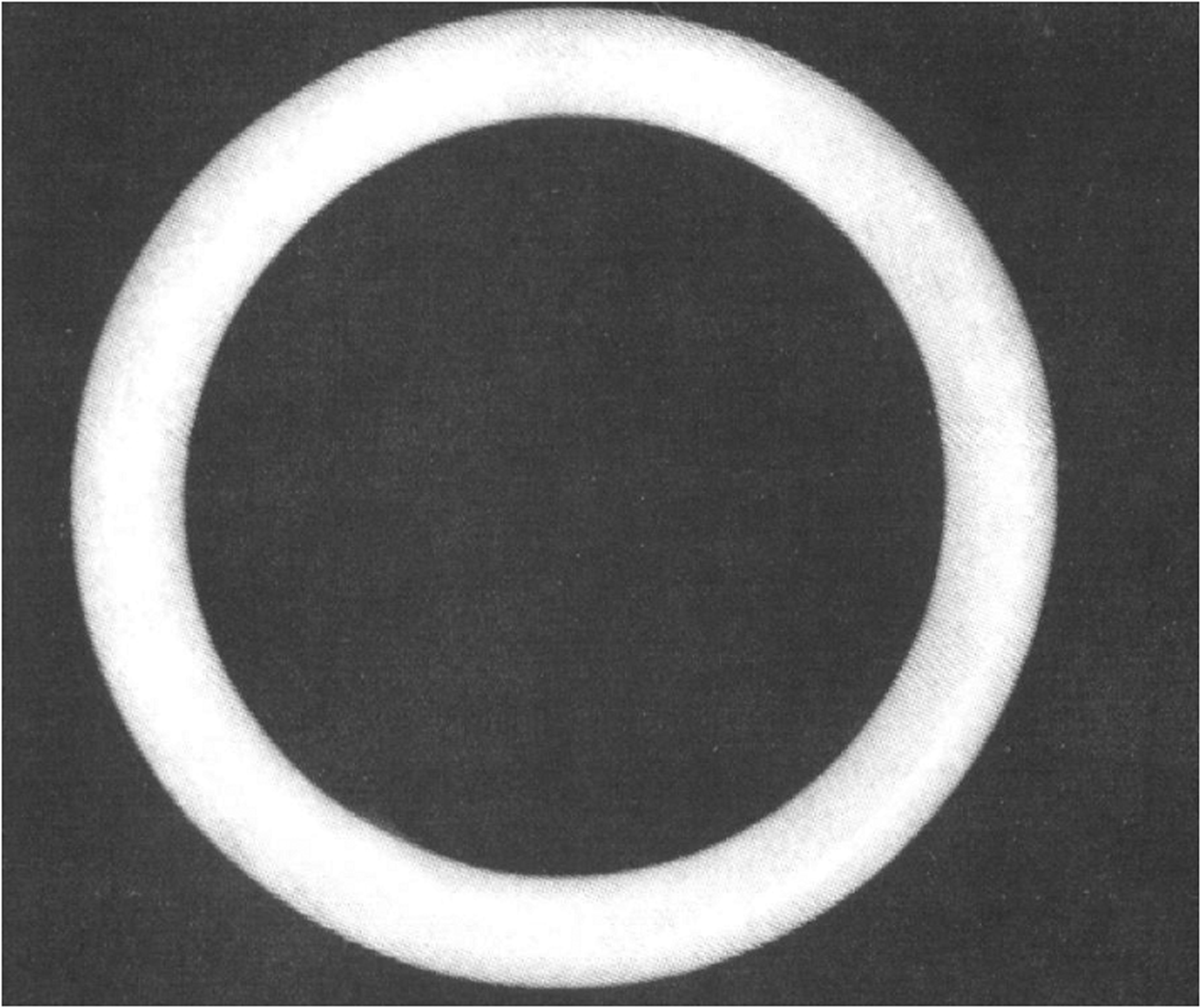
Plastic ring pessary (fenestrated).
The patient did not come for any antenatal checkup thereafter, but came as an emergency admission in late labor at 37 weeks of gestation. Her pulse rate was 100 beats per minute and her BP was 220/120 mm Hg. Her urine albumin was 2+. Abdominal examination revealed 30 weeks' uterine height, and breech presentation with nonlocalization of fetal heart sound and good uterine contractions.
On pelvic examination, the fetus's feet were lying outside the introitus, the patient's cervix was fully dilated, and the ring pessary was felt anteriorly high up. The ring pessary was greatly tightened around the fetus's abdomen and the umbilical cord. The patient was put on antihypertensive medication and a prophylactic anticonvulsant Zuspan regimen.
Results
The pessary was cut with scissors, and an assisted vaginal breech delivery was performed. A stillborn male, small for gestational age, weighing 1.6 kg, was born. There was no cervical prolapse. A follow-up examination at 6 weeks postpartum also showed no uterine prolapse.
Discussion
Uterocervical prolapse is an uncommon condition during pregnancy. In the majority of cases, pregnancy is superimposed on a preexisting prolapse. 1 In this case, the prolapse developed during pregnancy late in the second trimester, as has been reported by Brown and Toy et al.2,3
A combination of pregravid structural injury to the support system, hereditary weakness in the connective tissues, and the pressure and hormonal effects of an established pregnancy, may be responsible for symptomatic gestational decensus.
The ring pessary allowed the pregnancy to continue to term in the present case, but caused entrapment of the fetus during labor. The ring pessary has led to an unusual complication that was never anticipated.
Conclusions
Obstetricians should be aware of this condition to ensure a successful pregnancy outcome. Early recognition and close follow-up during pregnancy is essential, especially when a ring pessary is inserted for uterine prolapse.
Footnotes
Author Disclosure Statement
No competing financial interests exist.