
Abstract
Abstract
Introduction
I
Case
A 34-year-old para 3, abortus 1 married woman presented with AUB in the form of polymenorrhea and hypomenorrhea. On examination, she was lean and of slender build, with moderate pallor. Bimanual examination revealed a normal size uterus and normal adnexae. Laboratory examination revealed hemoglobin 9 g/dL. Remaining hematologic and biochemical parameters were within normal limits. Pelvic ultrasonography (USG) did not reveal any remarkable findings. Hysteroscopy revealed multiple tiny bony chips in the uterine cavity, which were removed by sharp curette and submitted for histopathologic examination. Gross examination revealed multiple small flattened bony chips each measuring 0.8 and 0.7 cm in addition to a tiny grayish-brown endometrial soft tissue piece (Fig. 1). Microscopic examination revealed an early secretory endometrium with fragmented osseous tissue bits. A thorough review of the patient's past history revealed that she had become pregnant 2 years earlier. Because of personal problems, she had undergone termination of pregnancy at 16 weeks, by dilatation and evacuation (D&E) method.
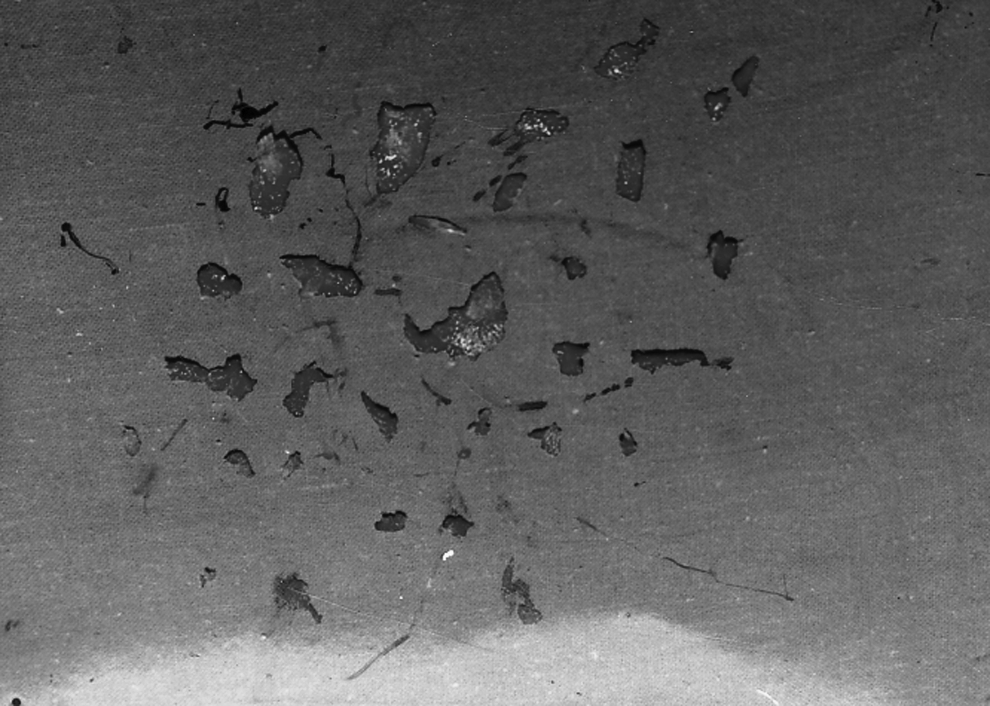
Retained fetal bones.
Results
Correlating the patient's present situation with her past history, it was concluded that she had retained fetal bony fragments during her second-trimester D&E, leading to AUB. Removal of the fragments by curettage regularized her menstrual cycle.
Discussion
This patient had complications from a D&E for a mid-trimester miscarriage. D&E is associated with high risk of excessive hemorrhage and uterine perforation. Furthermore, D&E in the mid-second trimester is highly likely to be complicated by retained products of conception including fetal bones, unless it is performed with USG guidance. Fetal bones can be retained freely in the endometrial cavity. On the other hand, they can be totally or partially embedded in the myometrium. 6 The usual symptoms of retained fetal bones are abnormal uterine bleeding, dysmenorrhea, dyspareunia, chronic pelvic pain, and secondary infertility. 7 The retention of a bony structure in the endometrial cavity or ossification of the endometrium may act as an intrauterine contraceptive device (IUCD) in preventing conception. 4
A high index of suspicion must be maintained for those patients who have a history of mid-trimester termination of pregnancy by D&E, in order not to miss the diagnosis. Apart from history and examination, imaging studies are very important in the diagnosis. Pelvic USG, especially with the vaginal probe, is particularly reliable. We can stretch the spectrum of transvaginal sonographic diagnosis further by performing a saline infusion sonohysterography (SIS). This technique allows detailed visualization of the uterine cavity in both longitudinal and transverse planes, and the endometrium can be evaluated for the presence of polyps, submucosal fibroids, intrauterine synechiae, and foreign bodies. 8 Hysterosalpingogram is useful in outlining the endometrial cavity and in determining the state of the Fallopian tubes, but its usefulness in the diagnosis of retained bones is limited. Hysteroscopy has both diagnostic and therapeutic values. Presumably, hysteroscopy should be the most accurate diagnostic tool. Hysteroscopic removal of the bony pieces should be regarded as the gold standard of treatment, as it enables a complete removal of the bones under direct vision. In this case, the bony chips could not be visualized by USG, although their size was >0.5 cm, which suggests that radiologists should also keep in mind this rare entity while evaluating USG findings in such a case. The treatment is removal of the retained bones, and the success rate in terms of resumption of normal fertility and the resolution of other symptoms can be very high.
In addition to retained intrauterine bones after second trimester D&E, endometrial calcified lesions and the presence of ectopic bones can also occur by metaplasia in association with chronic inflammation and tissue destruction, which are likely to be present after repeated spontaneous or therapeutic abortions. Osseous metaplasia usually presents as diffuse, sporadic ossifications without the tissue reaction frequently occurring around retained fetal tissue. 9
Conclusions
Today, effective and cheap prostaglandins are available for terminating mid-trimester pregnancies, 8 and when D&E is used, pelvic USG must be performed to confirm uterine emptiness. The important role of routine pelvic USG, or, where facilities exist, routine hysteroscopy for AUB, especially for those patients with a history of mid-trimester D&E, is emphasized.
Disclosure Statement
No competing financial interests exist.