
Abstract
Abstract
Introduction
I
Simulation is being increasingly incorporated into graduate medical education and the Accreditation Council for Graduate Medical Education (ACGME) will soon require that programs provide educational simulation sessions to maintain accreditation. The use of animal models, including animal tissue, is one such method for teaching surgical techniques. Use of live and freshly killed porcine bladder to simulate repair of cystotomy has been previously described as part of complications courses, but not has not been studied as a model.2,3
Fresh-frozen animal tissues are readily available from medical-education companies and are much less expensive than live animals for simulation purposes. The current authors have experience with simulation utilizing fresh-frozen porcine intestines. Based on this and the current authors' experience with live animals, it was hypothesized that fresh-frozen porcine bladder would be a low-cost, high-fidelity model for simulating cystotomy repair.
There were two objectives of this investigation: (1) subjective evaluation of the porcine bladder for simulation of cystotomy repair; and (2) assessment of the educational benefit of a didactic session and demonstration on the technical skill of obstetrics and gynecology (OB/GYN) residents in repair of cystotomy.
Materials and Methods
This study was approved by the University of South Florida (USF) Morsani College of Medicine committee on graduate medical education and institutional review board. The participants were consenting USF OB/GYN residents; 16 learners were available for this wet laboratory.
Each subject was assigned a random number via a random-number generator on a computer when they arrived for the session. These numbers were used on the surveys throughout the laboratory work to keep track of the subjects' results. The subjects were given the opportunity to opt out of any formal assessments. This assessment and wet laboratory was a learning opportunity for the residents, who were informed that no part of their formal grades or ability to move to the next level would be affected by their decisions to participate or not participate.
Immediately prior to the laboratory work, a questionnaire (Box 1) was administered to the residents in a classroom to assess previous experience with repair and fund of knowledge regarding appropriate cystotomy care. During this time, all participating faculty members were given preparatory information by the principal investigator on how to observe, administer, and evaluate the proceedings.
The training session took place at the USF Center for Advanced Medical Learning and Simulation. Use of animal tissues for this purpose was approved by the USF institution for animal care and use committee (protocol T 4164). Following completion of the questionnaire, the session started with 1 hour of laboratory work. This consisted of separate “wet” training stations, with 2 residents working at each station (Fig. 1). There was a freshly thawed large (from >50 kg animal) porcine bladder with attached urethra and ureteral segments at each station. The total cost of the porcine tissue and equipment was ∼$400. A cystotomy was created in the dome prior to the start of the laboratory work. The residents were asked to place a Foley catheter. The more junior of the 2 residents at each station was asked to identify and evaluate the injury to the dome of the bladder, note its anatomical context, and repair it. Integrity of the repair was tested by backfill through the Foley catheter with 200 cc of water (an amount determined prior to the laboratory work to fill the bladder). The more senior resident was asked to evaluate and repair an injury close to the trigone of the bladder (Fig. 2). Integrity of repair was tested as before. A pair of faculty members completed an objective standardized assessment of technical skills (OSATS) (Box 2) for each of these residents. Procedural time was assessed by comparison to the performance of the course director and rated accordingly. Immediately following this laboratory work, a 30-minute didactic session was conducted on incidental cystotomy during gynecologic surgery.
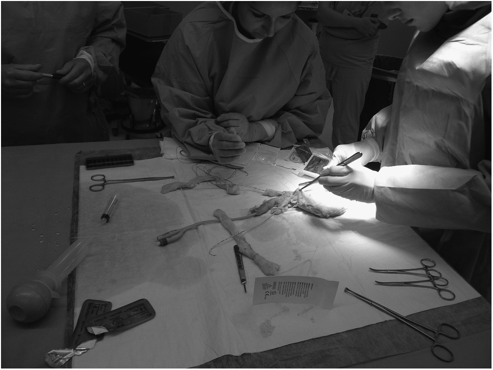
Resident closing cystotomy near trigone. Foley catheter and ureteral stents have been placed.
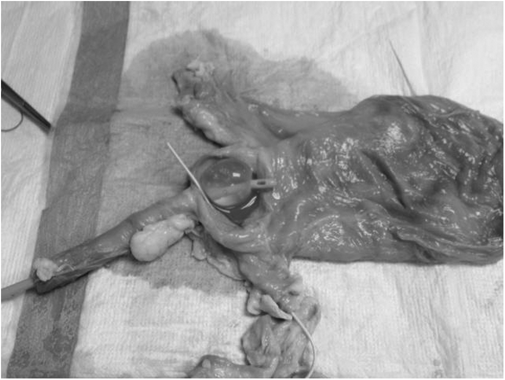
Cystotomy near trigone. Foley bulb is at urethrovesical junction and stent is seen exiting ureteral orifice.
Following the didactic session the participants returned to the laboratory to observe a demonstration of assessment and repair of a cystotomy, both at the dome of the bladder and in close proximity to the trigone. The participants then returned to the stations; each station had a new bladder with a laceration. As before, the junior resident repaired the dome injury and the senior resident repaired the injury near the trigone. The same faculty members who did the initial OSATS performed a repeat OSATS (by participant pair) based on the second laboratory session. Comparing these second OSATS results to the first attempt, was the basis for assessment of the potential effect on technical competence and educational benefit of the aforementioned “sandwich” approach to the didactic session and demonstration. Both single-inverting layer and two-layer imbricating closures (2-0 or 3-0 delayed absorbable suture) were considered to be appropriate.
Finally, a postassessment (Box 3) was administered at the conclusion of the laboratory experience to gauge new comfort levels with repair as well as fund of knowledge regarding clinical treatment decisions for a cystotomy. A post-test questionnaire was also administered to all learners and faculty members regarding subjective assessment of fidelity of the model (Box 4). The results of this questionnaire were excluded from the two first-year residents because it was felt that their experience to date would not provide an adequate basis upon which to gauge fidelity.
Descriptive statistics were presented as medians and ranges for continuous variables and as frequencies and percentages for categorical variables. The average score/rating from the two raters was calculated for each resident and the results of the pre- and postassessments were compared, using the Wilcoxon Rank Sum test. The pre- and postassessment pass–fail rate was ultimately evaluated, using an exact binomial test because of the computational difficulties associated with the 100% post-pass rate.
Inter-rater reliability was assessed using the intraclass correlation coefficient for the OSATS and the κ statistic for the pass–fail result. Results of the assessment of the contextual fidelity of the bladder models for use in further resident education are also presented in this article.
Results
Sixteen obstetrics and gynecology residents (postgraduate levels noted in tables) participated. The faculty consisted of 5 generalists, 2 female pelvic medicine subspecialties, 1 gynecologic oncologist, and 4 fellows (1 in gynecologic oncology, 2 in female pelvic medicine, and 1 in minimally invasive surgery). The fidelity of the model (faculty and residents; Table 1) received a rating of “good” by 85%, 82%, 100%, and 67% of the evaluators for anatomy, tissue handling, repair of an injury to the dome of the bladder, and repair of an injury to the trigone of the bladder, respectively. There were no “poor” ratings for any aspect of fidelity. The most common reasons cited for a “fair” rating of the trigone model were the difference in anatomy and difficulty with passing stents. The gynecologic oncologist and both urogynecologists rated the trigone model as “good.” Careful evaluation of the anatomy in all of the specimens revealed that the ureteral orifices are in the proximal urethra (Box 2). This created some initial confusion with passage of stents, which had to be passed antegrade as a matter or practicality. For the purpose of demonstrating the difficulties and anatomic hazards of trigone injury (ureteral proximity and potential entrapment), the model was rated well among subjects and faculty.
Following the didactic session and demonstration, there was significant improvement in overall technique and overall pass-rate as measured by OSATS (Table 2A). When the group was divided further into the results for dome and trigone specifically (Table 2B and C), the pattern remained the same. However, the improvement in pass-rate failed to reach statistical significance, most likely secondary to the reduced sample size. There was no significant change in time scores. The inter-rater reliability was technically statistically significant for most of the measures, but the agreement was poor for technique and overall pass-rate (Table 3). All residents were passed by all raters during the second laboratory session.
Median (range).
OSATS, objective standardized assessment of technical skills.
κ statistic p-value cannot be computed in cases of 100% agreement among all raters.
ICC, intraclass correlation coefficient.
What was notable from the prelaboratory questionnaire was that 11 of 16 residents had seen cystotomy repairs, but only 4 of 16 had performed such repairs (Table 4). Furthermore, only 1 resident rated him/herself as comfortable with repair of an injury to the dome of the bladder and all 16 rated themselves as not comfortable with repair of an injury to the bladder trigone. Following completion of the laboratory sessions, 12 of 16 rated themselves as comfortable with repair of an injury to the dome of the bladder, but only 3 rated themselves as comfortable with repair of injury to the trigone.
Discussion
The incorporation of simulation laboratory sessions offers tremendous potential for improvement of gynecologic surgical training and will soon be required by the ACGME. The extent to which simulation-based training (SBT) can reduce learning curves and make up for potential deficiencies in volume remains unclear. With respect to the present study, it is hoped that SBT can help bridge gaps in managing certain uncommon but important surgical procedures and complications.
The results of the present study suggest that the porcine bladder is a low-cost, high-fidelity model for simulation of cystotomy repair. The possible exception is the fidelity of the model for repair of injury to the bladder trigone. However, the residents' and generalists' lack of familiarity with ureteral management issues probably affected their assessments. Repair of trigonal injury is difficult and is associated with greater risk relative to repair of a dome injury. It is probably not a realistic expectation to train OB/GYN residents adequately in managing bladder trigone injury. It will be of interest to assess this model with other types of trainees, such as urology residents, and gynecologic oncology and female pelvic medicine fellows. Important factors affecting the model are ease of ex-vivo repair and the lack of attachments (which skips the critical step of bladder mobilization for repair without tension).
The improvement in technique and overall pass-rate, as measured by OSATS, is encouraging in terms of the construct validity of this model. The subjective self-assessment of comfort with repair noted in the pre- and postlaboratory questionnaires is supportive of this as well. As mentioned above, this study was not designed to measure predictive validity (how these results will translate into clinical practice). However, the current authors do plan to send 6- and 12-month follow-up questionnaires to the participants regarding the perceived impact this laboratory work may have had on their subsequent clinical experiences.
The poor inter-rater agreement for technique and overall pass-rate may have been related to utilization of less-experienced faculty members (young generalists, fellows).
Conclusions
The large porcine bladder is a useful and appropriate model for simulation of repair of a cystotomy in the dome of the bladder.
Footnotes
Acknowledgments
The authors gratefully acknowledge the participation of the following faculty and fellows, without which the study and laboratory work could not have been conducted: Shelly Holmstrom, MD, Cathy Lynch, MD, Stuart Hart, MD, Mona McCullough, MD, Hye Sook Chon, MD, Xiaomang Stickles, MD, Mark Zakaria, MD, Carol Cox, MD, Renee Bassaly, MD, and Kristie Greene MD.
Disclosure Statement
None of the authors have a conflict of interest.