
Abstract
Abstract
Introduction
C
Case
A 50-year-old multiparous female presented to the outpatient department with the complaint of a slow-growing swelling in the pubic region of 6 months' duration. It was painless, and not associated with symptoms of dysuria, dyspareunia, or pruritus vulvae. There was no history of any type of genital trauma. Her medical history was unremarkable. On examination, a painless cystic mobile mass extended from the clitoral region up to the labia minora, having a diameter of 5×6 cm in the clitoral region, with regular margins and a smooth surface (Fig. 1). There was no ulceration. An ultrasound scan of the cystic mass was performed, which revealed it to be a sebaceous cyst. The patient was scheduled for surgical enucleation of the cyst. With the patient under anesthesia, a reverse V-shaped incision was made on the swelling, and the cyst was easily dissected from surrounding structures without any damage to the neurovascular bundle of the clitoris (Fig. 2). The skin was approximated with interrupted sutures after achieving complete hemostasis. Normal cosmetic appearance of the clitoris was restored.
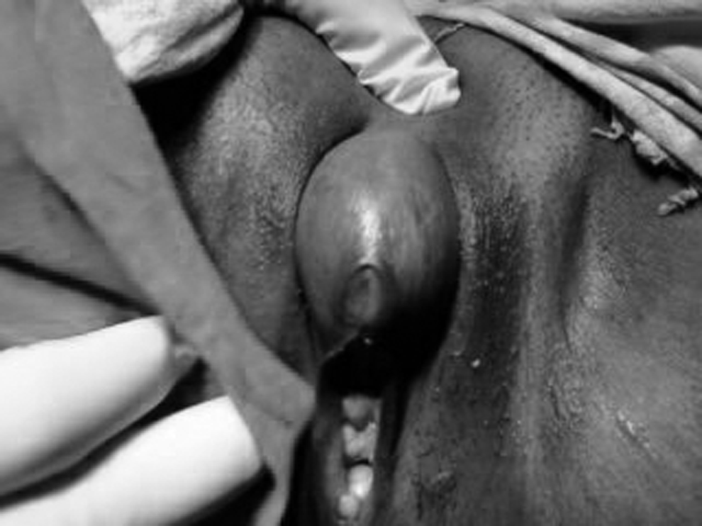
Cyst in the clitoral region.
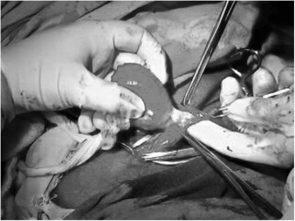
Removal of cyst after preserving the neurovascular bundle of the clitoris.
Results
On the second postoperative day, a small hematoma was observed, which was removed after cutting one stitch. The patient was discharged on the fourth postoperative day. At a follow-up visit after 2 weeks, the patient was comfortable and her stitch line was healthy. The histopathology report revealed an epidermoid cyst lined by stratified squamous epithelium filled with keratinous material (Fig. 3).

Photomicrograph showing epidermoid cyst lined by stratified squamous epithelium.
Discussion
Clitoral enlargement can be caused by a variety of conditions. Clitoromegaly can be classified into four groups: hormonal conditions; nonhormonal conditions; pseudoclitoromegaly; and idiopathic clitoromegaly (Box 1). 2
Among the nonhormonal causes of clitoromegaly, clitoral epidermoid inclusion cysts are reviewed in the literature.2–4 Epidermoid cysts are slow-growing, intradermal or subcutaneous tumors with a wall composed of true epidermis. Whereas the most common locations are the face, scalp, neck, and trunk, the external genitalia can also be involved in the form of clitoral, labial, or scrotal implantation. Those inclusion cysts arise from the invagination of keratinizing squamous epithelium within the dermis, which becomes cystic and filled with laminated keratin. 5 This implantation in the clitoris is most commonly induced by trauma, and is rarely encountered appearing spontaneously originating from dysontogenetic displacement. 6 Epidermoid cysts of the clitoris are seen commonly after type I genital mutilation/female circumcision performed in some ethnic communities in Africa and West Asia.7,8 Cases lacking any kind of trauma are scarce, and 2 of those patients had a history of oral contraceptive use, which could indicate the putative role of estrogen in stimulation of the implanted epidermis and sebaceous glands.6,9
There have only been 4 reported cases of epidermoid cysts of the clitoris not associated with female genital mutilation. 10 In this case, there was no history of female genital mutilation/trauma, and no history of oral contraceptive use or any other drug intake. Merlob et al. 11 reviewed female births in a 2-year period, and observed 19 cystic lesions on 3026 female newborns (0.6%). The cysts were readily apparent, and the majority were located in the vagina or paraurethral area. Only 1 patient had a clitoral cyst. The cysts drained spontaneously and resolved within 2 months.
The clinical presentation of this clitoral pathology usually consists of a silent course with a painless swelling gradually increasing in size. A soft, mobile, nontender mass in the clitoral region in the absence of any virilization is the typical physical finding, which is in accordance with the findings of this patient.
The preoperative investigation of the reported cases in the literature with the final diagnosis of clitoral epidermoid cyst consisted of detailed hormonal and chromosomal analysis, abdominopelvic ultrasonography, and even the sonographic and magnetic resonance imaging of the cystic lesion.5,6,9,12 In this patient, careful evaluation of the patient with history, local examination of the cyst, and ultrasound of the cyst prevented unnecessary laboratory and radiologic studies.
Incision for surgical enucleation of cyst was a reverse V-shaped incision, which was similar to the incision made by Celik et al. 1 in their case, which both omitted the necessity for skin trimming and provided a fine cosmetic result.
This case was unique among the other reported epidermoid cysts with respect to its spontaneous onset, without any history of genital mutilation or trauma, and its preoperative evaluation.
Conclusions
Immediate surgical excision, even on an outpatient basis, would be the ideal therapeutic approach for epidermoid cystic lesion of the clitoris, for which the clinical and pathologic findings are very characteristic, although the underlying etiology for the onset can differ.
Footnotes
Disclosure Statement
No competing financial interests exist.