
Abstract
Abstract
Introduction
R
Case
A 27-year-old woman with a pregnancy of 8 weeks' duration presented to the early pregnancy unit with bleeding per vaginum. Two years previously, she had undergone a surgical termination of a pregnancy during her second trimester. An ultrasound scan of the current pregnancy showed it to be an intrauterine pregnancy. However, inferior to the pregnancy, a 17 mm echogenic linear structure with strong posterior acoustic shadowing was noted in the mid/lower uterine cavity, suggesting a foreign body (Fig. 1). There was no history of the patient having used an intrauterine device in the past.
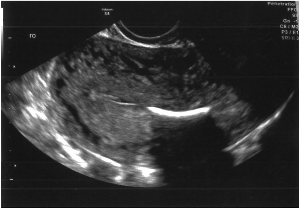
Ultrasound image displaying the echogenic linear structure, suggesting a foreign body.
The patient developed bleeding and had a repeat ultrasound scan. Appearances were of a missed miscarriage, and the previously seen foreign body was still present. The patient chose conservative management of her miscarriage.
One month later, the patient was offered a hysteroscopy to remove this foreign body. The findings included a bony tissue projecting from the right lower uterine wall into the cavity as well as costal cartilage tissues (Fig. 2). The projecting bony tissue and costal cartilages were removed with a hysteroscopic resectoscope.
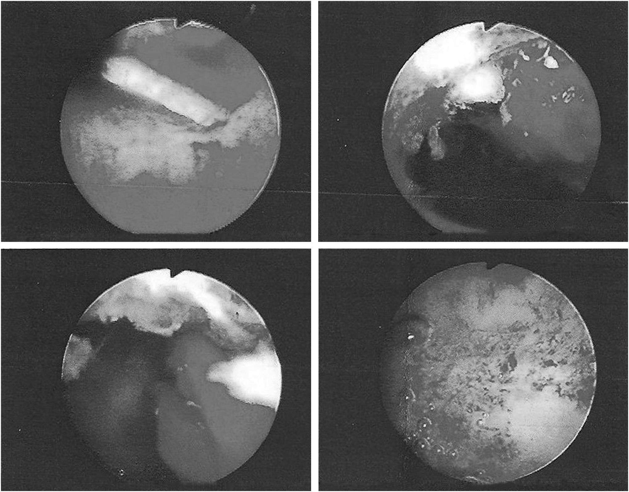
Hysteroscopic images. The top two images show the bony tissue within the uterine cavity. The bottom two images were taken after resection of the foreign body.
Histology confirmed that these were dead bony fragments related to products of conception.
Results
The patient recovered well postoperatively. At her follow-up consultation, she expressed her desire to wait for a period of time before trying to conceive. She was advised to contact her local early pregnancy unit when she becomes pregnant.
Discussion
It is known that the retention of fetal bones is a rare complication of termination of pregnancy. The majority of these cases have been identified during investigation for secondary infertility.1,2 Other symptoms that patients may present with include dysmenorrhea and chronic pelvic pain. 3 Samuel et al. 3 reported a case of chronic pelvic pain and after removal of fetal bones that were identified on an ultrasound scan, the patient's symptoms resolved. This was also seen in cases described by Graham et al. 2 and Basu et al. 1 in which fertility was restored for several patients after they underwent surgical removal of retained fetal bones.
This case study, however, highlights that the retention of fetal bones could impact the viability of early pregnancy. As has been suggested previously, fetal bones act in a similar manner to an intrauterine device, 3 thereby preventing implantation of the blastocyst. With the presence of fetal bones in close proximity to a viable pregnancy within the uterine cavity, there may be an increased risk of miscarriage in the first trimester, as described in this case report.
Conclusions
This case highlights the importance of identifying the retention of fetal bones, as this not only has an impact on future fertility but also possibly has an impact on maintaining the viability of early pregnancy. These patients should be offered surgical removal of the fetal bones to help reduce the risk of miscarriage in subsequent pregnancies.
Footnotes
Disclosure Statement
No competing financial interests exist.