




Abstract
Abstract
Introduction
M
Although rare, the most common mesenchymal neoplasm of the vagina is the leiomyoma. 4
Case
A 50-year-old multipara (para 5) female was referred from a private practitioner to the outpatient department of gynecology with complaints of menorrhagia of 4 months' duration, lower abdominal pain, and difficulty with defecation of 3 months' duration. On general physical examination, the patient was noted to be anemic. On abdominal examination, the abdomen was soft with no palpable mass. Vaginal examination revealed a firm nontender mass that was 8×10 cm, occupying the posterior upper third of the vagina. The upper limit of the mass could not be reached. The cervix could not be felt separately, and the uterus was ∼6 weeks of gestation in size.
A provisional diagnosis of cervical fibroid was made. Ultrasonography showed a large hypoechoic mass that was 8×9 cm, in the lower uterine segment, both the ovaries were normal, and the endometrium was 4 mm thick and uniform. Endometrial sampling from the patient could not be performed, because of an inaccessible cervical os. The patient's hemoglobin was 8.9 gm%. Exploratory laparotomy was planned after counseling the patient, and 1 unit of crossmatched blood was transfused prior to surgery. On laparotomy, the uterus, cervix, bilateral tubes, and ovaries were normal. A total abdominal hysterectomy was performed. There was a large myoma arising from the posterior vaginal wall; this tumor was ∼8×10 cm (Fig. 1), and a myomectomy was also performed.
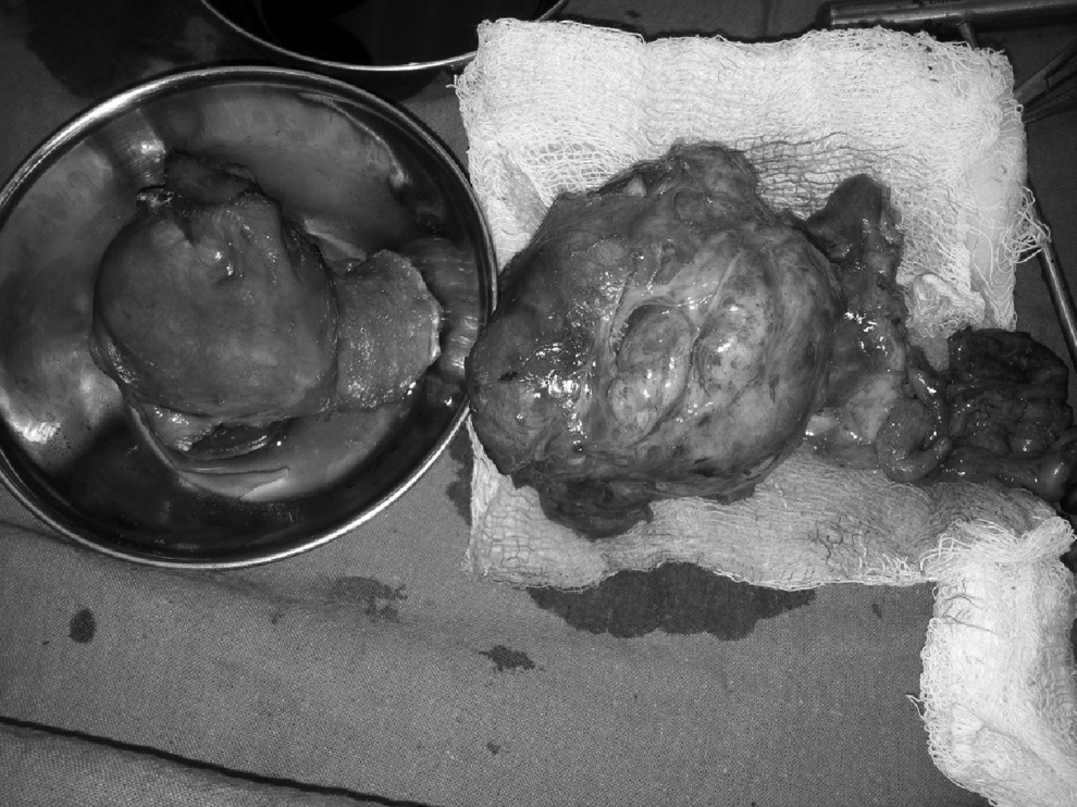
Uterus with cervix and vaginal fibroid (placed over gauze piece).
Results
This patient's postoperative period was uneventful.
Discussion
The majority of leiomyomas arise from body of the uterus, and sometimes from the cervix. The extrauterine sites of the tumor are ligament, ovary, inguinal canal, and, very rarely, the vagina and vulva. 2 Since the first report by Denys De Leyden in 1733, ∼300 cases of vaginal leiomyoma have been reported worldwide. 5 In the vagina, leiomyoma usually present as a solid single nodule, mostly from the midline anterior wall,2,5 and less commonly arise from the posterior and lateral walls. 6 In the present patient, a single vaginal leiomyoma was arising from the posterior upper third of vagina, which is a rare presentation. Young et al. reported that leiomyoma usually presents as a single solid nodule, mostly from the anterior vaginal wall in women between ages 35 and 50 years. 2
Although these tumors are often asymptomatic, larger tumors may be associated with pain, dystocia, dyspareunia, or obstructive urinary symptoms. In the present patient, vaginal leiomyoma was found on the posterior wall. Consequently, she did not have any urinary tract symptoms, but had difficulty with defecation, which may have been because of the large size of the posterior vaginal wall myoma. Shimada et al. 5 reported a case of a vaginal leiomyoma from the posterior wall, which was asymptomatic.
Conclusions
Treatment is always surgical; the only thing that must be decided is the most effective approach. Surgery is performed either via the abdominal or vaginal route, depending upon the location of the tumor. 7 The practical approach to such a vaginal mass entails careful excision. 8 Horn et al. reported that these tumors were enucleated, with an inconspicuous postoperative follow-up; however, they, too, suggested early enucleation because of the possibility of the tumor's undergoing malignant change. 9 Excision and enucleation are the treatment of choice.2,10 A vaginal approach may cause severe hemorrhage if the base of the tumor is not approachable, or if it is present in the upper part of the vaginal fornices. In this case, an abdominal route was planned, because clinically and on ultrasonography, the mass was diagnosed as a cervical fibroid. Agarwal et al. also performed excision and enucleation via a transabdominal route. 11 Hence, it is advisable to choose an abdominal route for cases in which the base of tumor is not approachable.
Footnotes
Acknowledgments
Dr. Singhal was the main operating surgeon. Dr. Katyal was the senior resident who assisted with the surgery. Dr. Singh helped in writing this article.
Disclosure Statement
No competing financial interests exist.