
Abstract
Abstract
Introduction
P
Case
A 27-year-old woman, gravida 2, para 1, living 1, was referred to an obstetric unit at 24 weeks of gestation in view of failed induction for intrauterine death. She had been married for 5 years prior. There was no history of severe dysmenorrhea or infertility. Her previous delivery was a spontaneous vaginal delivery 3 years ago at home, but no antenatal and postnatal records were available. Her current pregnancy had been supervised at a private hospital and apparently had no complications until 1 month ago, when she noticed decreased fetal movements.
Outside sonography revealed a deceased fetus of 20 weeks' gestation, following which pregnancy termination was tried with prostaglandin E2 gel, ethacrydine lactate solution, prostaglandin F2α, injection, and prostaglandin E1 tablets, but none of these agents was successful. On examination, the patient looked well and she had stable vital signs. Uterine height corresponded to 20 weeks of pregnancy. During a vaginal examination, her cervix was noted to be deviated to the left and was posterior, her uterus was also deviated to the left and was normal-size. However, this enlargement felt in her abdomen did not appear to communicate with the cervix and was felt through right fornix.
A repeat ultrasound was performed, which revealed a bulky uterus with a cavity showing a pseudogestational sac. Anteriorly and to right of the uterine cavity, there was a deceased fetus corresponding to 20 weeks of gestation with reduced liquid surrounded by a thin layer of myometrium. The gravid horn had no communication with the cervix. Thus, a sonographic diagnosis of an unruptured rudimentary horn pregnancy was made and the patient was prepared for a laparotomy. Intraoperatively a rudimentary horn on right side was seen, enlarged to 20 weeks' size with thinned out walls and was connected to the main uterine horn with a thick fibrous band (Fig. 1). There was no evidence of endometriosis or hemoperitoneum. Excision of the right horn with an ipsilateral salpingectomy was performed (Figs. 2 and 3). Both kidneys appeared and felt normal.
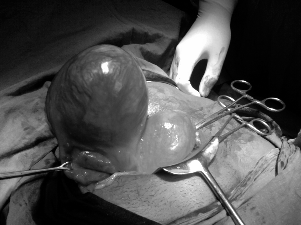
Laparotomy findings suggestive of intact rudimentary horn pregnancy.
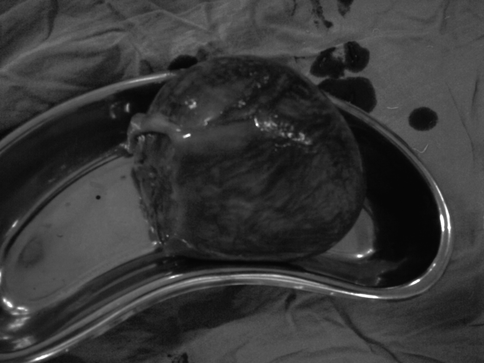
Excised intact rudimentary horn.
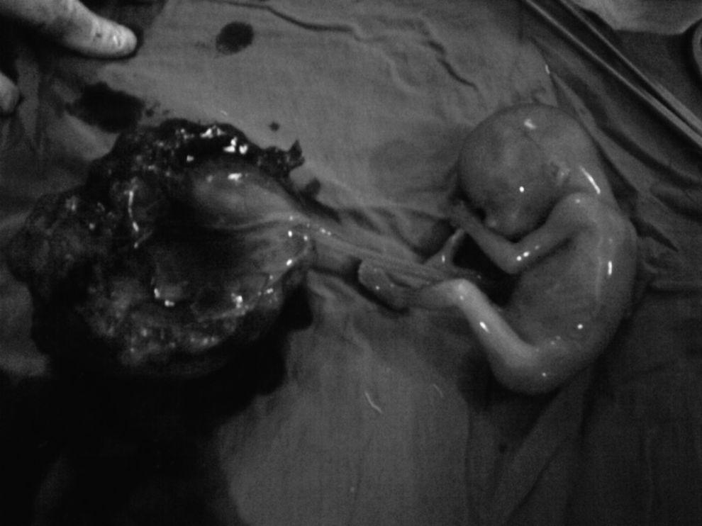
Cut-open section of excised rudimentary horn reveling fetus with attached placenta.
Results
This patient's postoperative period was uneventful, and she was discharged on seventh postoperative day.
Discussion
Pregnancy in a rudimentary horn is one of the rarest and most morbid conditions. A rudimentary horn results from an arrest in the development of one of the Müllerian ducts with inappropriate fusion with the contralateral side. Absence of dysmenorrhea, endometriosis, and hematometra suggests that the rudimentary horn was of a communicating type in the present case. The communication between a rudimentary horn and a unicornuate uterus is usually very thin and not detectable by the naked eye. The endometrium of the rudimentary horn has been described as being thinner than usual and sometimes being nonfunctional. Even pathologic placentation may be seen. 3
The timing of rupture varies from 5 to 35 weeks, depending on the horn musculature and its ability to hypertrophy and dilate. 4 Because of the variable muscular constitution of the wall of the rudimentary horn, pregnancy can be accommodated until late in pregnancy (usually in the second trimester) when rupture occurs, manifesting commonly as acute abdominal pain with a high risk of maternal mortality. In most cases of pregnancy in the rudimentary horn, the pregnancy lasts longer than a tubal pregnancy because of the variable musculature of the horn, with 80%–90% of cases rupturing by midtrimester and 10% going to term with a 2% fetal salvage rate. 5
Massive intraperitoneal hemorrhage because of rupture can be life-threatening to the mother. In the current patient, pregnancy reached 24 weeks without any catastrophe. Delayed diagnosis of this condition is quite common, as there are no definite signs to distinguish it from an intrauterine pregnancy. A careful pelvic examination in the first trimester of pregnancy and a finding of a deviated normal-size uterus with a palpable adnexal mass should arouse the suspicion of a uterine anomaly. This can be confirmed by an ultrasound examination although the sensitivity of this test remains at only 26%. The enlarging horn with thinned myometrium can obscure the anatomical structures and the sensitivity decreases further as the gestation increases. 6
Diagnosis prior to rupture is unusual but could be made with ultrasonography and magnetic resonance imaging (MRI). Tsafrir et al. outlined a set of criteria for diagnosing pregnancy in the rudimentary horn. 7 They are: (1) a pseudo pattern of an asymmetrical bicornuate uterus; (2) absent visual continuity between the tissue surrounding the gestation sac and the cervical canal; and (3) the presence of myometrial tissue surrounding the gestational sac.
In the current patient, the diagnosis was missed and induction of abortion was tried with many medications. Buntungu et al. also reported a rudimentary horn pregnancy in a sixth gravid, with all previous normal delieveries, with a diagnosis of intrauterine fetal demise in which induction with misoprostol failed, leading to suspicion of a rudimentary horn pregnancy. 8 Thus, a thorough ultrasound examination should be performed for which the emphasis should be not only on the details of the pregnancy but also on ruling out any uterine anomaly. Additional MRI can be performed to confirm the diagnosis if doubt exists before an invasive procedure is performed.
Once diagnosed, this condition demands an expeditious laparotomy and excision of that horn in order to avoid maternal mortality caused by a catastrophic rupture. Ipsilateral salpingectomy is recommended to decrease the chances of an ectopic pregnancy in the tube.
Conclusions
It is of paramount importance to diagnose rudimentary horn pregnancy prior to catastrophic rupture so as to have a more favorable maternal outcome and a decrease reproductive morbidity. Thus, a high index of suspicion is required for this rare but a dreadful complication of pregnancy.
Footnotes
Disclosure Statement
No competing financial conflicts exist.