
Abstract
Abstract
Introduction
O
Case
A 24-year-old primigravida, at 35 weeks of gestation, was admitted to the department of obstetrics and gynecology, of the Postgraduate Institute of Medical Sciences, in Rohtak, Haryana, India, for management of her oligohydramnios. She had no prior antenatal care prior to presentation. Approximately 1 week after admission, she went into spontaneous labor. During labor, fetal bradycardia occurred, and meconium-stained liquid was obtained during an amniotomy. Other than that, her labor progressed rapidly and she was able to deliver her neonate vaginally.
Results
The neonate was male and weighed 1.9 kg. However the neonate was limp and cyanosed and died a few hours after his birth. On examination of neonate's external genitalia, it was noted that he had a scrotal sac that was well-developed with normally descended testis. However, the neonate had no penile shaft and nor any urethral opening (Fig. 1). His anus was normal. The neonate also had polydactyly of the right hand. No other congenital anomaly was noted. A detailed evaluation of the mother showed that she had no history of fever, exposure to any teratogenic agents, or radiation during the antenatal period. Both parents refused autopsy of the neonate.
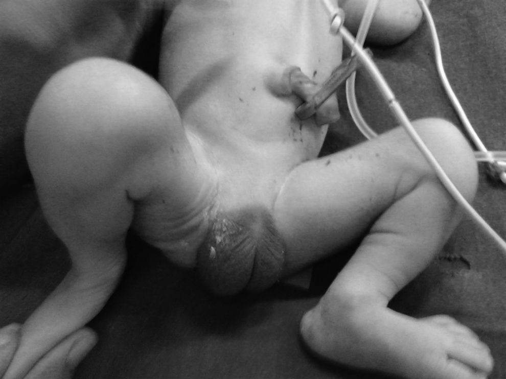
Neonate with penile agenesis.
Discussion
Oligohydramnios refers to amniotic fluid volume that is less than expected for gestational age, defined as, an amniotic fluid index (AFI) of ≤5 cm. 2 AFI is calculated by “dividing” the uterus externally into 4 equal quadrants and using ultrasound to measure the largest vertical pocket in each quadrant in centimeters, which are then added together to calculate the AFI. Oligohydramnios occurs in ∼1%–5% of pregnancies at term. 3
Box 1 lists the conditions associated with oligohydramnios. Approximately 15%–25% of oligohydramnios cases are associated with fetal anomalies, with the most common anomalies being genitourinary defects. 4 Because the amniotic fluid is primarily composed of fetal urine in the latter half of the pregnancy, the absence of fetal urine production or a blockage in the fetus' urinary tract results in oligohydramnios.
Penile agenesis is a rare congenital anomaly, affecting about ∼1 birth in 30 million, in which a male child is born without a penis.5,6 The underlying pathophysiology is thought to result from impaired proliferation of mesenchymal cells from the cloacal eminence forming the genital tubercle, a process that starts during the fourth week of embryologic development. 7 Changes at the early stages of fetal development are likely to affect the cloacal eminence as well as leading to proximal urethral–intestinal communication and other urogenital anamolies. However, changes at the latest stage of fetal development lead to more-distal urethral opening without other proximal anamolies.1,8,9
Cases of penile agenesis may involve an ectopic urethral opening at any point on the perineum in midline, over the pubis, on the anterior aspect of the scrotum, or, most frequently, just anterior to the anus and anterior wall of the rectum. Alternatively, this agenesis may be associated with urethral agenesis, which is often complicated by the Potter sequence. More than 50% of penile agenesis cases have associated anomalies, including developmental defects of the caudal axis, as well as genitourinary- and gastrointestinal-tract anomalies. Cases with urethral agenesis and/or associated renal agenesis/dysgenesis usually present with oligohydramnios in the midtrimester of pregnancy. The lack of amniotic fluid causes compression of the fetal abdomen, which limits fetal diaphragmatic movements. In addition to chest-wall fixation, the lack of amniotic fluid flowing in and out of the fetal lungs leads to pulmonary hypoplasia, which usually has a poor neonatal outcome. 10
• Chromosomal abnormalities • Congenital anomalies • Intrauterine growth restriction • Fetal demise • Post-term pregnancy • Ruptured membranes
• Abruption • Twin–twin transfusion syndrome
• Uteroplacental insufficiency • Hypertension • Preeclampsia • Diabetes
• Prostaglandin synthase inhibitors • Angiotensin-converting enzyme inhibitors
Because of the practical problems of reconstructing a normally functioning penis, therapy for patients with penile agenesis currently includes female gender reassignment, gonadectomy in the neonatal period to avoid a postnatal testosterone surge with possible male gender imprinting, secondary vaginoplasty using scrotal skin or sigmoidal replacement, or anterior urethral transposition, followed by female hormone therapy during puberty.11–14
Conclusions
Any case of oligohydramnios presenting in midtrimester with no history of leakage of liquid and no evidence of any other maternal or placental factors associated with oligohydramnios warrants a detailed sonographic fetal imaging for any associated congenital malformations.
Footnotes
Disclosure Statement
No competing financial conflicts exist.