
Abstract
Abstract
Introduction
N
Case
The case of a 26 year-old morbidly obese, nondiabetic, African American woman, gravida 3, para 1-0-2-1, who presented for planned primary low transverse cesarean delivery with a term breech fetus, is described here. The patient received 2 g of intravenous Ancef prior to skin incision. The cesarean delivery was uncomplicated, and the patient was discharged home on postoperative day (POD) 3. On POD 10 she presented with the complaint of a blister, associated drainage, and pain adjacent to her incision.
On admission, she was afebrile, blood pressure 113/69 mm Hg, heart rate 118 beats per minute. She weighed 191 kg (421 lbs), with a body mass index (BMI) of 72.3. She had a 10 cm area of necrotic, black-purple bullae, with malodorous drainage on the right portion of her pannus above her incision. Her skin incision below the pannus was intact. Her white blood cell count (WBC) was 22.2 with a left shift of 89%, hemoglobin 8.8, creatinine 1.4, sodium 141, and lactic acid 1.2.
There was immediate concern about necrotizing fasciitis; therefore, intravenous vancomycin, clindamycin, and piperacillin/tazobactam were started, and a general surgeon was consulted. The patient was taken to the operating room expeditiously without imaging, and underwent an anterior abdominal wall resection down to the peritoneal cavity. A wound vacuum was placed along the opened abdominal-wall defect. A speculum examination was performed, with no drainage from the cervix.
Postoperatively, the patient remained intubated in the surgical intensive care unit on broad-spectrum antibiotics. On POD 1 from her initial debridement, she became febrile to 101.4°F. Her renal function improved with her creatinine down to 1, but her leukocytosis continued to increase to 25.3. She was taken back to the operating room for a second look, wound debridement, and wound vacuum placement. At the time of her third wound debridement on POD 3, another speculum examination was performed, with no drainage from the cervix. Given this, further exploration of the uterus was not performed.
By POD 6, despite three wound debridements with a stable appearing wound, there was minimal clinical improvement. She continued to be febrile up to 103°F with evidence of persistent sepsis. Wound cultures were polymicrobial (4+ Proteus mirabilis, 3+ Klebsiella pneumoniae, 4+ Pseudomonas aeruginosa, 3+ group C beta hemolytic streptococcus, 4+ diptheroids, 3+ bacteroides [not fragilis], 3+ peptostreptococcus, 3+ lactobacillus). The patient's obesity precluded imaging with a computed tomography (CT) scan, as she exceeded the weight limit of the CT scanner.
Results
Because of continued sepsis, the patient was taken back to the operating room for a fourth debridement on POD 6, with the plan to perform an exploratory laparotomy and possible total abdominal hysterectomy. Intraoperative findings included an enlarged postpartum uterus with necrotic-appearing tissue involving the uterine scar. Given these findings, a hysterectomy was performed. After removal, the uterus was bivalved with necrotic debris noted in the cavity (Fig. 1). Pathology revealed a focus of serosal and myometrial changes, including necrotic debris surround by foamy macrophages, foreign body giant cells, and chronic inflammation. There were no remnants of placental tissue or fetal membranes. The patient began to improve and became afebrile. She was continued on broad-spectrum antibiotics. Her WBC peaked to 29.5 prior to hysterectomy and normalized to 7.9 at discharge. Because of extensive fascial removal, her abdomen was closed with local fasciocutaneous flaps on POD 25.
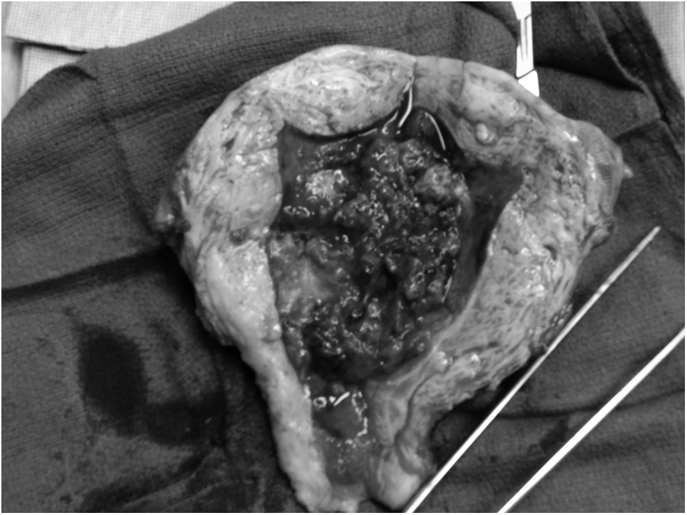
Bivalved uterus at the time of hysterectomy, with necrotic debris seen within the uterine cavity.
Discussion
Necrotizing fasciitis can be difficult to diagnose, resulting in a delay in appropriate management. Clinical findings include crepitus, blistering, discharge, early severe pain disproportionate to cutaneous signs, systemic inflammation, worsening sepsis, septic shock, and rapid progression to local necrosis. 4 Risk factors associated with development include obesity, malnutrition, immunocompromised state, operative trauma, previous radiotherapy, diabetes mellitus, peripheral vascular disease, and hypertension. 3 In a systematic literature review, Goh et al. found that misdiagnosis occurred in 75% of patients. 5 The following clinical features are helpful in early diagnosis: pain out of proportion to the physical findings, failure to improve despite broad-spectrum antibiotics, presence of bullae in the skin, and gas in the soft tissue on plain radiographs (although this occurred in only 24.8% of patients). 5
Based on bacteriology, necrotizing soft tissue infection can be divided into two distinct entities that determine the choice of antibiotics. Type 1 infection involves at least one anaerobic species (Bacteroides, Clostridium, or Peptostreptococcus) isolated in combination with one or more facultative anaerobic Streptococci (other than Group A) and members of the Enterobacteriaceae (i.e., Escherichia coli, Klebsiella, Enterobacter, and Proteus). 6 Type II infection results from Group A streptococcal infection or other beta hemolytic streptococcal infection either alone or in combination with Staphylococcus aureus. 7
Because of the thrombogenic nature of necrotizing fasciitis, timely surgical debridement is paramount, as antibiotics are unable to penetrate necrotic tissue. 7
In this case, without the ability to use ancillary imaging, the uterus was assumed to be the probable persistent source of infection. Imaging modalities may not reliably predict the extent of tissue involvement in women with post-cesarean necrotizing fasciitis. The lack of purulent drainage from the cervix does not exclude involvement of the uterus. Goepfert et al. found that 3 of 9 patients with post-cesarean necrotizing fasciitis had uterine involvement, despite the fact that necrotizing fasciitis usually spares the underlying muscle. 3 Two of these three patients required hysterectomy. 3 Childs et al. reported a case of aggressive debridement of necrotic myometrium, which allowed for uterine preservation. 8
Conclusions
As there may be uterine involvement despite sparing of abdominal muscles or lack of purulent drainage from the cervix, it is necessary to counsel patients that hysterectomy may be needed. A high clinical suspicion is required regarding the uterus and hysterotomy site. Given the significant mortality rate associated with progressive necrotizing fasciitis, inspection of the uterus and hysterotomy site on initial wound debridement is warranted. Hysterectomy may be necessary if the patient's course does not improve.
Footnotes
Disclosure Statement
No competing financial conflicts exist.