




Abstract
Abstract
Introduction
U
Hemoperitoneum caused by leiomyoma is very rare and only 95 cases have been described in the literature since 1861. 2 This article describes the case of a 46-year-old woman who presented to the emergency room with a hemoperitoneum caused by a bleeding subserosal vein overlying an anterior leiomyoma.
Case
A 46-year-old woman, gravida 4, para 4-0-0-4, presented to the emergency room with complaints of abdominal pain and bilateral cold feet. She reported an acute onset of abdominal pain, localized to the periumbilical area, associated with nausea, vomiting, and diarrhea. The patient also reported feeling bilateral cold and painful feet. A review of her body system was otherwise negative. The patient stated this was the first time that she had experienced such pain. Her gynecologic history included menses at age 12, with menstruation every 28 days, of 5 days duration, and consisting of a normal flow. She denied having any history of leiomyomas. The remainder of her medical history was negative for problems, and she had four uncomplicated spontaneous vaginal deliveries.
This patient's vital signs testing revealed tachycardia, but her blood pressure and oxygen saturation were within normal limits, and her hemoglobin and hematocrit were 9.2 and 29.9, respectively. She appeared to be pale and to be experiencing severe abdominal pain. An abdominal examination showed that her abdomen was rigid and diffusely tender, without rebound or guarding. Her bilateral lower extremities were cold and her pedal pulses could not be palpated. After stabilizing the patient, she underwent an emergency computed tomography (CT) angiogram to rule out an aortic dissection or a ruptured abdominal aortic aneurysm.
The CT scan revealed the presence of normal iliofemoral blood flow without occlusion, no evidence of aortic dissection, and a grossly enlarged heterogeneous uterus, possibly representing a degenerating fibroid. There was evidence of hemoperitoneum in her abdomen.
With no clear etiology of the hemoperitoneum seen on the CT scan, this patient was taken to the operating room for an exploratory laparotomy by the hospital's trauma team. After 1500 mL of fresh blood and clots were emptied from the patient's abdomen, the source of her bleeding was visualized. There was a pinhole opening in a superficial vein overlying an anterior fundal leiomyoma that was actively bleeding. An obstetrics and gynecology team was called in at this point. There was no evidence of degeneration of the leiomyoma. Several figure-of-eight stitches were placed on the superficial vein in an attempt to contain the bleeding, but they were unsuccessful because of the friability of the leiomyoma (Fig. 1). A myomectomy was considered; however, because of this patient's rapid blood loss, a total abdominal hysterectomy was performed with a total estimated blood loss of 2100 mL. Five units of packed red blood cells were administered intraoperatively.
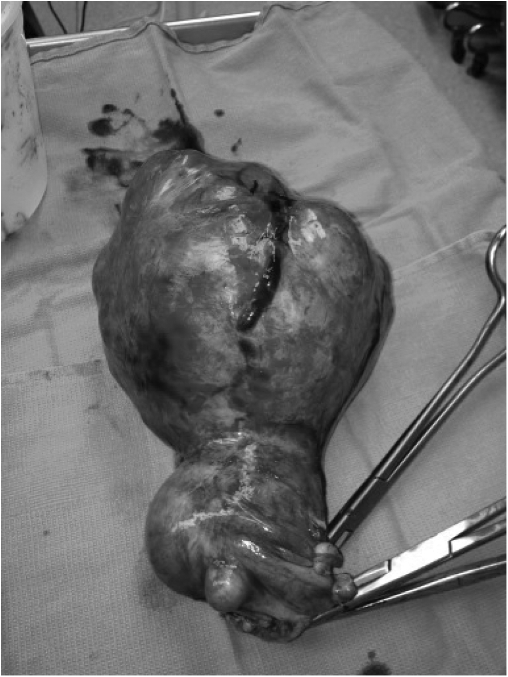
Uterus and cervix. A prominent subserosal vein overlying a large uterine fibroidgrowth, after several attempts at ligation.
Postoperatively, the patient was kept in the surgical intensive care unit. She developed a left iliac-vein thrombus on the second day and was started on heparin for anticoagulation. She also developed a fever on the same day with blood cultures showing a Streptococcus viridans infection, and was treated with intravenous ceftriaxone. Later, on the sixth day, the patient developed ileus and was managed conservatively with a nasogastric tube and she had significant improvement in her condition.
Results
On postoperative day 16, this patient was stable for discharge. She was placed on therapeutic anticoagulation for 6 months for treatment of the left-iliac thrombus.
Pathology testing revealed uterine leiomyomas, with the largest measuring 15 cm, as well as focal adenomyosis, and an unremarkable cervix and Fallopian tubes.
Discussion
Uterine leiomyomas are the most common tumors in women of reproductive age, but these tumors rarely lead to hemoperitonea. According to an evaluation of the literature, only 66 cases of hemoperitoneum caused by rupture of a subserosal vein overlying a uterine myoma have been reported in the literature since 1861. Other reported causes of hemoperitoneum associated with leiomyomas are bleeding from a subserosal artery, 3 an avulsed pedunculated leiomyoma, 4 a lacerated leiomyoma, 5 or a ruptured leiomyoma. 6 While some of the cases were caused by spontaneous rupture of the vein, most were associated with trauma (including coitus), 5 increased blood flow around the uterus, such as in menstruation 3 or pregnancy (such as this case), and actions resulting in increased abdominal pressure, such as defecation. 7 A careful history should be elicited to search for precipitating factors. Management recommended in the literature was equally divided between total or subtotal hysterectomy and myomectomy, and 4 patients died before reaching the operating room. In the overwhelming majority of the reported cases, the diagnosis was not known prior to exploratory laparotomy, despite the previous knowledge of the presence of leiomyomas.
Conclusions
Although rare, a bleeding leiomyomatous vessel should be kept in the differential diagnosis of a hemoperitoneum of unclear origin. The consent for emergent exploratory laparotomy includes the potential loss of any abdominopelvic organ; therefore patients should be made aware of the possibility of infertility following the procedure.
Footnotes
Disclosure Statement
Each author represents and warrants that he or she has no financial affiliation (e.g., employment, direct payments, stock holdings, retainers, consultantships, patent-licensing arrangements, or honoraria) or involvement with any commercial organization with potential financial interest in the subject or materials discussed in this article. There are no other potential conflicts of interest.