
Abstract
Abstract
Introduction
C
Case
A 50-year-old multiparous female presented to the hospital with complaints of swelling in the pubic region for a duration of 1 year. The swelling was insidious in onset and grew slowly. It was painless, and there were no associated symptoms of dysurea, vaginal discharge, or pruritis vulva. No history of trauma or hormonal treatment was elicited. The patient did not have any systemic complaints. Her general physical and abdominal examination was unremarkable.
On local examination, a cystic mass of 15×6 cm was noted over the clitoris on the right side, which was mobile and nontender (Fig. 1). The overlying skin showed no hyperpigmentation or ulceration. The labia minora on the right side were splayed over the cyst. The urethral and vaginal openings were normal. Ultrasound testing revealed a normal urinary tract.
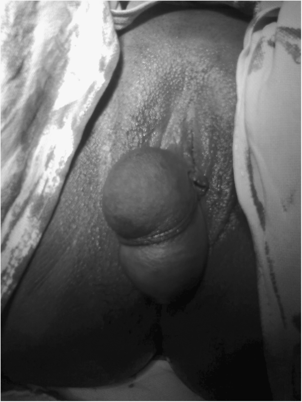
Giant cyst of the clitoris.
The patient was scheduled for for surgical excision of the cyst under anesthesia. Under a subarachnoid block, complete excision of the cyst was performed without damaging the neurovascular bundle of the clitoris (Fig. 2). After achieving complete hemostasis, the redundant portion of the cyst was also excised, dead space was obliterated, and the skin was sutured by interrupted sutures. A normal cosmetic appearance of the external genitalia was restored as much as possible. The patient's histopathology report revealed that the innermost lining of the cyst was lined by columnar epithelium with a demonstrable basement membrane and this histology was consistent with a mesonephric cyst (mucous cyst).
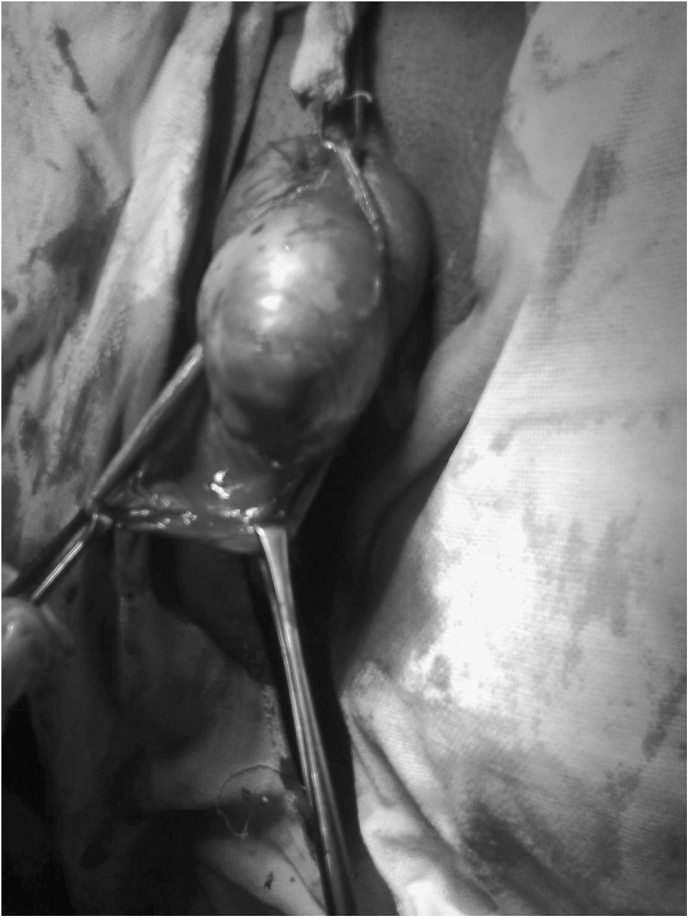
Excision of cyst being performed, while preserving the neurovascular bundle.
Results
The patient's postoperative period was uneventful. No recurrence was noted 17 months postsurgery at a follow-up.
Discussion
According to the present understanding of human genital organogenesis, the vagina is derived from the paramesonephric (Müllerian) ducts and the urogenital sinus. 1 Glands lined by either mucinous or ciliated epithelia are normal constituents of the vulvar vestibule and are derived from the urogenital sinus. The vestibule is formed from the confluence of cells forming the urogenital groove (ectoderm) and those of the urogenital sinus (endoderm). The Müllerian mesoderm does not contribute to this formation. 2
Mucous (dysontogenetic) cysts arise from the minor vestibular gland, or from the mesonephric duct remnants, and may be found at the introitus and the labia minora. Clinically, cysts of embryonic origin are similar. Vaginal cysts of embryonic origin are most commonly of mesonephric and paramesonephric origin. Developmental cysts of the vestibule are most often derived from urogenital sinus epithelia. During embryonic development, these ducts normally become atretic and lose their glandular lining. If parts of the ducts persist and remain functional, secretory activity gives rise to cystic tumors. 3
Embryologically, cysts of the female external genitalia are related to paraovarian cysts of mesonephric origin. A second variety of embryonic cyst—clinically indistinguishable from Gartner's duct cyst, and arising from the vestiges of the paramesonephric ducts—is termed a “paramesonephric cyst,” which can have mucinuous or ciliated linings. A third variety of embryonic cyst, involving the vestibule of the vulva, is the mucous cyst of the urogenital sinus epithelia. 4
Implantation of cysts of the clitoris arise from the invagination of keratinizing squamous epithelium within the dermis, which becomes cystic and filled with laminated keratin. This implantation in the clitoris is most commonly induced by trauma and rarely occurs spontaneously originating from dysontogenetic displacement. 5 Celik et al., described a case of a clitoral epidermoid cyst secondary to blunt trauma in the form of a swing accident in a 9-year-oil girl. 6 In the current case, the formation of cyst was attributed to dysontogenetic displacement, as there was no history of trauma.
Most mesonephric cysts are lined by nonciliated cuboidal epithelia or by low columnar epithelia that may have a demonstrable basement membrane. Occasionally, a partial lining of squamous epithelia is noted. Tanha et al. described a case of large mucous cyst in the clitoris; this cyst was excised and, on microscopy, showed a cyst-wall structure with severe chronic inflammation containing plasma cells—places covered by flat pseudostratified columnar ciliated epithelia. Mucous cysts of urogenital sinus origin are usually lined by a single layer of tall columnar epithelia. 3 Positive nuclear staining for estrogen and/or progesterone receptors also supports the Müllerian histogenetic hypothesis. 7 In the current case; the innermost lining of the cyst was was columnar epithelia with a demonstrable basement membrane.
Conclusions
Mucous (dysontogenetic) cysts are rare causes of clitoral cysts, compared to epidermoid cysts of the clitoris. Tanha et al. presented the first case report of a dysontogenetic clitoral cyst. 3 Given that the gross appearance and symptoms of both of these cysts are not sufficiently characteristic, histopathologic examination is necessary for differentiation and confirmation of diagnosis.
Footnotes
Disclosure Statement
No competing financial conflicts exist.