
Abstract
Abstract
Introduction
L
Abdominal wall leiomyomas are the rarest of the extrauterine leiomyomas with only three cases reported in literature. They may result in diagnostic and surgically perplexing situations if unsuspected. We report a case of de novo origin of a large leiomyoma of the anterior abdominal wall in a vaginally hysterectomized postmenopausal woman.
Case Report
A 60-year-old para five postmenopausal woman reported to the outpatient department of Obstetrics and Gynecology with the feeling of an abdominal lump for 2 months. She had five uneventful vaginal deliveries in the past and had undergone vaginal hysterectomy 10 years ago for uterovaginal prolapse. There was history of passage of loose stools through the vagina since the vaginal surgery. However, there was no vaginal passage of fecal matter when the stools were well formed or hard. Menopause had been achieved 3 years before the vaginal hysterectomy. She had not undergone any other minor or major surgical procedure ever and was not on any drug or hormone replacement therapy (HRT).
The general physical examination was normal. The abdomen was distended with a firm, nontender, oval mass of restricted mobility extending from the symphysis pubis to midway between the xiphisternum and umbilicus. The speculum examination revealed a 4-mm-sized opening 1 cm below the vault on the posterior vaginal wall. The mass could only be tipped on a digital vaginal examination. A rectovaginal examination confirmed the small rectovaginal fistula.
Hematologic and biochemical parameters of the woman were normal. Sonography of the abdomen revealed a large multicystic mass extending from the pelvis to above the umbilicus with both hyperechoic and intervening hypoechoic areas. The ovaries were not visible on sonography. Serum CA-125, alpha-fetoprotein, beta human chorionic gonadotropin, carcinoembryonic antigen, and lactate dehydrogenase levels were within normal limits. The patient was taken up for laparotomy with a provisional diagnosis of ovarian tumor. A self-retaining catheter was inserted in the urinary bladder and the abdomen was opened by a vertical midline incision. However, after separating the recti, the parietal peritoneum was not visible. Instead, a thin-walled bluish area was visible with fluid underneath. Patency of the urinary catheter was reconfirmed. Suspecting ascites now, the thin layer covering the fluid was incised, however, only 80 cc of clear fluid drained and the cavity was found to be blind on all sides. The abdominal incision was extended for a few centimeters above the umbilicus, but attempts to enter the peritoneal cavity through the drained area failed as it was soon realized that only a large cystic area of an otherwise large grayish-white underlying mass had been drained. The mass could be separated digitally from above, from the overlying anterior abdominal wall and recti muscles that had got partially divaricated. Groups of angry looking, tortuous, and dilated blood vessels were interspersed all over the spongy mass and it was separated from the surrounding tissues all around after ligating the feeding vessels that were abundant on the peritoneal side of the mass (Fig. 1) The mass of 25 by 20-cm size lay in the preperitoneal space with no fixity or adherence to any surrounding structure and was enucleated. The parietal peritoneum could now be easily opened to reach the peritoneal cavity and the rectovaginal fistula was repaired by the general surgeons abdominally in the same sitting. Small atrophic ovaries were present in the pelvis. The postoperative period was uneventful. Histology of the mass revealed smooth muscle cells interspersed with cystic areas and stroma (Fig. 2). Immunostaining for smooth muscle actin was positive.
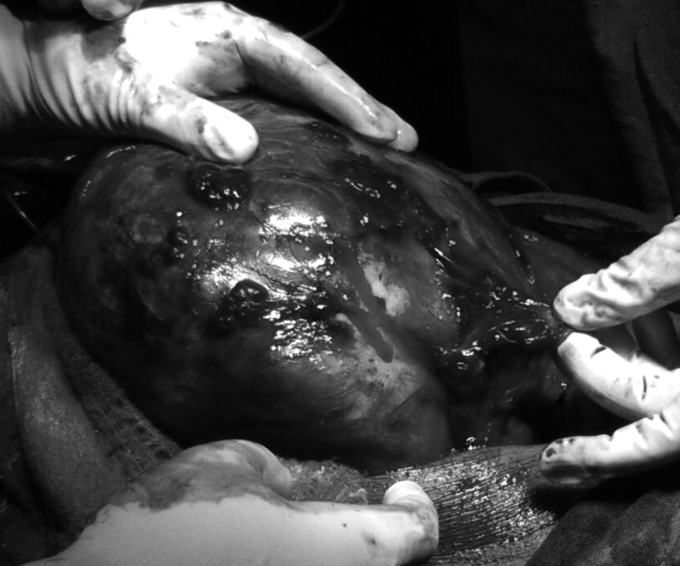
Clinical photograph showing the large vascular tumor in the anterior abdominal wall.
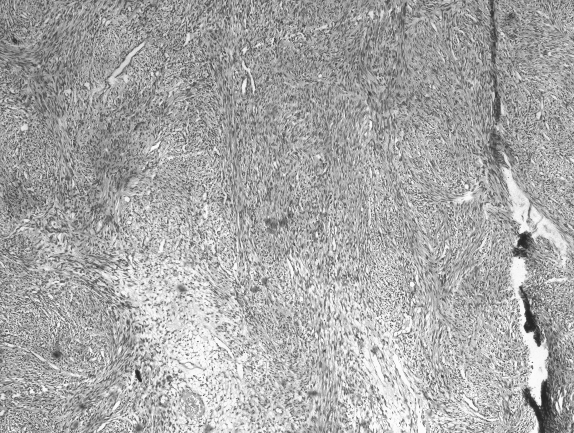
Histologic picture showing the whorled pattern of smooth-muscle bundles with cystic change at places (hematoxylin & eosin, 10×).
Discussion
The uterine leiomyomas are believed to arise from the smooth muscle cells of the uterus while the extrauterine myomas are thought to take origin from the smooth muscles of blood vessel walls or intestine especially in women on HRT. 2 Previous abdominal uterine surgery, exogenous administration of sex steroids, and diabetes mellitus have been regarded as predisposing factors for de novo development of these benign lesions at extrauterine sites. 3 Aberrant implantation and growth of uterine leiomyoma and even disseminated peritoneal leiomyomatosis have been reported after laparoscopic myomectomy, suggesting a role of seeding of smooth muscle cells in the abdominal wall at surgery. 4 They are also known to contain sex steroid receptors and this could explain the promotional effect on their growth of exogenous estrogen in women on HRT.
Only three cases of de novo origin of anterior abdominal wall leiomyomas have been reported in literature and all had one or more of these predisposing factors in them. Al-Wadaani reported the development of anterior abdominal wall leiomyoma in a perimenopausal woman with type 2 diabetes, while Lalor et al. described a preperitoneal lipoleiomyoma in the incisional umbilical scar of a previous nongynecologic abdominal surgery in a postmenopausal woman, both suggesting the etiologic roles of operative seeding of smooth muscle cells and conducive metabolic/hormonal influence.3,5 Schindl et al. reported a preperitoneal lipoleiomyoma in a postmenopausal and abdominally hysterectomized woman who had been on HRT for 5 years, again corroborating the promotive influence of steroidal administration and the seeding hypothesis. 6 Our case is unique in being the first case of de novo development of a leiomyoma in the preperitoneal space in the absence of any prior abdominal surgery, exogenous hormone therapy, any metabolic disorder, or any other known or hypothesized predisposing factor. Moreover, our patient had been hysterectomized long ago and had already achieved menopause by the time of primary surgery, making any significant ovarian contribution to estrogen production unlikely. Besides, the vaginal route of hysterectomy would practically rule out direct seeding of the abdominal wall and that too for a functional (and nontumorous) indication. However, a pertinent question would be: Can any other genital surgery (vaginal hysterectomy in this case) by an extra-abdominal route also cause seeding of smooth muscle cells at a relatively distant and extraperitoneal site like the abdominal wall? Such an occurrence does not find support in literature. However, the hematogenous inoculation and subsequent implantation of the smooth muscle cells at a distant site still remains a theoretical possibility. The likelihood of the tumor in our case being a parasitic leiomyoma that originally arose from the uterus and later got attached to the anterior abdominal wall also does not seem applicable in the absence of the uterus for more than a decade before appearance of the mass. Several other factors with ill defined or as yet unknown etiologic roles resulting in conversion of normal smooth muscle cell(s) into a leiomyoma have been postulated like somatic mutation along with unclear synergistic action of progesterone and insulin-like growth factor, deranged lipid metabolism, adiposite through the tumor necrosis factor TNF-alpha cytokine, and local growth factors.2,3
De novo origin of the tumor from the vascular smooth muscle cells could be a possibility in the present case. The absence of attachment to any surrounding structure and abundance of groups of dilated vascular channels all over the tumor may appear to favor, but not confirm such an origin. Such a suggestion would also make one wonder whether a probable vascular origin of a smooth muscle tumor makes it more vascular. The available literature is silent on this account and given the few cases reported till date of abdominal wall leiomyomas, such a generalization would be premature. The large myomatous mass under discussion also differed from conventional leiomyomas of uterine origin in another gross characteristic-variegated consistency with cystic areas interspersed with loose solid areas giving it a cheese-like appearance. Given the paucity of literature on these extremely rare extraperitoneal tumors, no definite reason is known for this appearance, although ample availability of space in the preperitoneal area and smooth muscle to stroma ratio within the tumor may have some role to play in it. We could not find any specific histologic features reported in literature about this variety of leiomyomas, but would suggest a specific look at the amount of smooth muscle content in comparison to the stromal, cystic, and vascular components in such cases.
In nonhysterectomized individuals, uterine leiomyomas would form the commonest differential diagnosis of these tumors followed by ovarian neoplasms in practically all age groups. Superficiality, mobility of these masses, and fixity to surrounding structures should specifically be looked for and may help differentiate abdominal from extra-abdominal tumors. Leiomyomas originating from the extraperitoneal part of round ligament may also present as similar masses, although they commonly present with an inguinal swelling if originating from the more distal part of the ligament. A desmoid tumor arising from the rectus abdominis muscle is an important, but a rare possibility that accounts for 0.3% of all abdominal wall neoplasms. 7 It affects young women more commonly, especially after childbirth, and may involve any skeletal muscle even though the abdominal wall continues to be the most favored site. Despite being histologically benign, desmoid tumors may become adherent, infiltrate locally, and may recur after an incomplete excision. Immunostaining with vimentin, alpha smooth muscle actin, muscle actin, and desmin may help differentiate them from other tumors. A lipoleiomyoma arising from the peritoneum could be another rare possibility. 8
Lack of knowledge and suspicion about the extraperitoneal location of leiomyomas can cause surgical problems like difficulty in locating and opening the peritoneal cavity, entry into the cystic areas or substance of the tumor and seeding of surrounding areas, injury to underlying urinary bladder that forms the bed of the tumor in the lower part, and hemorrhage. The presence of a fluid-filled swelling beneath the rectus muscles may be confused with a full urinary bladder or an encysted peritoneal collection. Opening of such a cyst after ensuring the emptiness of bladder will not result in opening of the peritoneal cavity, as happened in the present case, which can further perplex the surgeon if not suspected.
Footnotes
Disclosure Statement
No competing financial interests exist.