
Abstract
Abstract
Introduction
A
Case
A 51-year-old (para 2, live births 2) woman presented with a complaint of pain in her abdomen, that started one day prior. This pain was continuous in nature, and radiated to her back with no aggravating or relieving factors. This patient had no history of fever, vomiting, or urinary or bowel complaints. She had had regular menstrual cycles in the past. She did not have any other comorbidity.
On physical examination, the patient's vital signs were noted to be stable. A tender 20-week gestational-size mass was felt in her abdomen. There was second-degree uterine prolapse with supravaginal elongation of the cervix noted during a speculum examination. A 10-cm mass was felt in the right forniceal region vaginally.
A transabdominal scan revealed a normal-sized uterus with an endometrial thickness of 16 mm. There was a heterogeneous lesion of 9.6 × 6 × 5.2 cm in the midline suprapubic region; this lesion was suggestive of either a complex ovarian cyst or a degenerated exophytic uterine fibroid. A computed tomography (CT) scan was performed, which revealed the presence of a large hypodense lesion, with a small foci of calcification in the right lower abdomen, and adnexa of 9.5 × 6 cm, suggestive of a right ovarian cystic neoplasm (Fig. 1).

Computed tomography scan image of large hypodense lesion, with a small foci of calcification in the right lower abdomen, and adnexa of 9.5 × 6 cm suggestive of a right ovarian cystic neoplasm.
Her CA-125 level was 49.65 U/mL. Other laboratory test results were within normal ranges. A Papanicolaou smear and endometrial biopsy that had been performed at a different hospital were negative for atypia or malignancy. A total laparoscopic hysterectomy with a bilateral salpingo-oophorectomy were planned, because of this patient's complex ovarian mass, uterovaginal prolapse, and increased endometrial thickness.
During a diagnostic laparoscopy, (Fig. 2) the patient's uterus was noted to be normal in size. The right ovary had a simple cyst of 2 × 1 cm (Fig. 3). The left ovary and bilateral Fallopian tubes were normal. There was a 9 × 5 cm gangrenous appendicular mass, which was not adherent to surrounding structures (Fig. 4). There was no ascites, nor evidence of malignancy or metastasis. A laparoscopic appendectomy was performed by a general surgeon. A specimen was retrieved in an Endobag,

Port placement.

Intraoperative image showing right simple ovarian cyst.
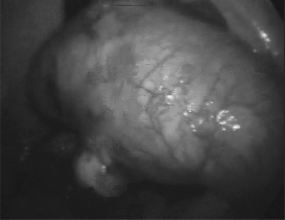
Intraoperative image showing appendicular mass.
Results
The histopathology report results showed that this patient had a simple mucocele of her appendix with chronic appendicitis. There was no evidence of atypia or malignancy on shown on histopathology testing. The endometrial sample was unremarkable. This patient was discharged on postoperative day 4, and her postoperative period was uneventful.
Discussion
A mucocele of the appendix is a rare occurrence, with a 0.224% incidence. The benign variety of this lesion is most common. 1 Malignancy is encountered in 1 in 4300 surgically removed appendices.2,3 A mucocele causes nonspecific symptoms, such as abdominal or pelvic pain, nausea, fever, or as a mass in the abdomen.4,5 However, in 23%–50% of cases, this mass can be asymptomatic, making a preoperative clinical diagnosis difficult. Mucoceles are more common in females, with a female-to-male ratio of ∼4:1. Age >50 years is also a risk factor.4,5 The lesion can be diagnosed via barium enema, ultrasound, CT scanning, or colonoscopy. A mucocele is the most common lesion to be misdiagnosed as an ovarian cyst on ultrasound scans.
On a single-contrast barium enema, a mucocele shows as a smooth, broad-based filling defect in the cecum near the ileocecal valve. On an air-barium double-contrast enema, the mass appears as a smooth filling defect in the submucosal area, which indents and deviates the cecum. 6
On ultrasound, variable findings are seen. A cystic mass with echogenic layers (i.e., an “onion-skin” sign) is diagnostic of a mucocele of the appendix. 6 However, this sign can be seen in cases of other mucinous cysts (e.g., a mucinous cyst of the ovary). 5
CT scanning of the abdomen usually shows a hypodense, cystic well-encapsulated mass with an occasional finding of wall calcification in the right lower quadrant. 6
Magnetic resonance imaging (MRI) can show areas of local inflammation and can be used to exclude the possibility of ovarian cysts. 5
Colonoscopy shows the characteristic “volcano sign.” The appendiceal opening is seen in the center of a bulbous, smooth submucosal lesion protruding into the cecum. Movement of the mass with respiration is diagnostic. 6
Fine-needle aspiration cytology to determine the nature of the mass has been abandoned because of the associated risk of rupture.
Tumor markers such as carcinoembryonic antigen, CA-125, and CA-19-9, are elevated when patients have malignant lesions. These markers can serve as prognostic markers and for diagnosis of recurrences following surgical intervention. 7 However, these levels cannot be used to make a diagnosis of appendiceal tumors, as these markers are also raised in cases of cystadenoma (false–positive results). 5
Despite all the imaging options that are available, a mucocele is often diagnosed during surgery. In such cases, a frozen-section sample should be sent to rule out malignancy. A simple appendectomy is sufficient for benign cases, and extensive surgeries are reserved for malignancy cases.4,5 Laparoscopy offers the advantage of inspection of the entire abdominal cavity. In addition, intraoperative blood loss is minimal and postoperative recovery is rapid. 5 However, it is very important to remove the appendix intact, as spillage of its contents may lead to pseudomyxoma peritonei. If malignancy is suspected, it is advisable to convert the procedure to a laparotomy, as laparoscopy carries a risk of spillage of the contents and inadequate resectioning.
In the current case, a mucocele of appendix was diagnosed intraoperatively, and the option of a frozen section was not available. As there were no signs of malignancy, a laparoscopic simple appendectomy was performed.
Conclusions
A mucocele of appendix, although rare, should be considered when a patient presents with a right adnexal mass. For a gynecologist, this finding represents a challenge in surgical approach and sometimes requires the support of a surgeon and medical oncology specialists. Simple appendectomy is the surgery of choice for a benign mucocele of the appendix. Laparoscopic appendectomy can be considered as an option, but care should be taken to prevent appendiceal rupture. However, conversion is advised in cases of rupture, suspicion of malignancy, or anticipated spillage of contents.
Footnotes
Acknowledgments
Dr. Nilopher Surti was responsible for literature search and compilation of this article. Dr. Makum Ramesh was the primary surgeon, and Dr. Saraswathi Ramesh assisted him during surgery.
Disclosure Statement
No competing financial conflicts exist.