
Abstract
Abstract
Introduction
H
Risk factors for uterine inversion include fundal attachment of a tumor, thickness of the pedicle, tumor size, thinning of the uterine wall, and dilation of the cervical canal. 5 Nonpuerperal uterine inversion is often chronic. Researchers reported that 8.6% of nonpuerperal uterine inversions occurred suddenly. 6 This current case was a nonpuerperal acute inversion caused by a submucosal leiomyoma in a young nulliparous woman. This case is being reported because of the rare occurrence of nonpuerperal inversion.
Case
A 28-year-old nulliparous female was admitted to the Bowen University Teaching Hospital, in Ogbomosho, Oyo State, Nigeria, with a history of vaginal bleeding, lower abdominal pain, and a mass protruding from her vagina of 8 hours duration. She had a prior history of menorrhagia and dysmenorrhea over the past 5 years. Two weeks prior, she went to an adult emergency room with severe menstrual pain and underwent ultrasound scanning. The ultrasound revealed that she had a bulky uterus with a 65 mm × 50 mm × 40–mm, fundal, submucosal fibroid tumor. She was treated with analgesics and counseled on the need for a subsequent myomectomy. She was given an appointment to attend the gynecology clinic but did not come in for this appointment.
A general examination of this patient revealed tachycardia with a pulse rate of 120 beats per minute, blood pressure of 90/60 mm of Hg, and pallor. An abdominal examination did not show any abnormalities. During a vaginal examination, a 12 × 8–cm, firm mass was noted; it protruded out of her vagina and had a bossellated surface with patchy white flakes. Touching the mass caused it to bleed.
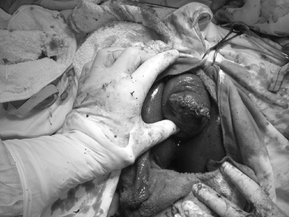
The patient's cervix was visible (Fig. 1). A cup-shaped depression was felt instead of the uterine fundus during a rectal examination. Hence, the clinical diagnosis was acute uterine inversion.

Protrusion of submucosal fibroid tumor. The surgeon's finger points to the uterine fundus.
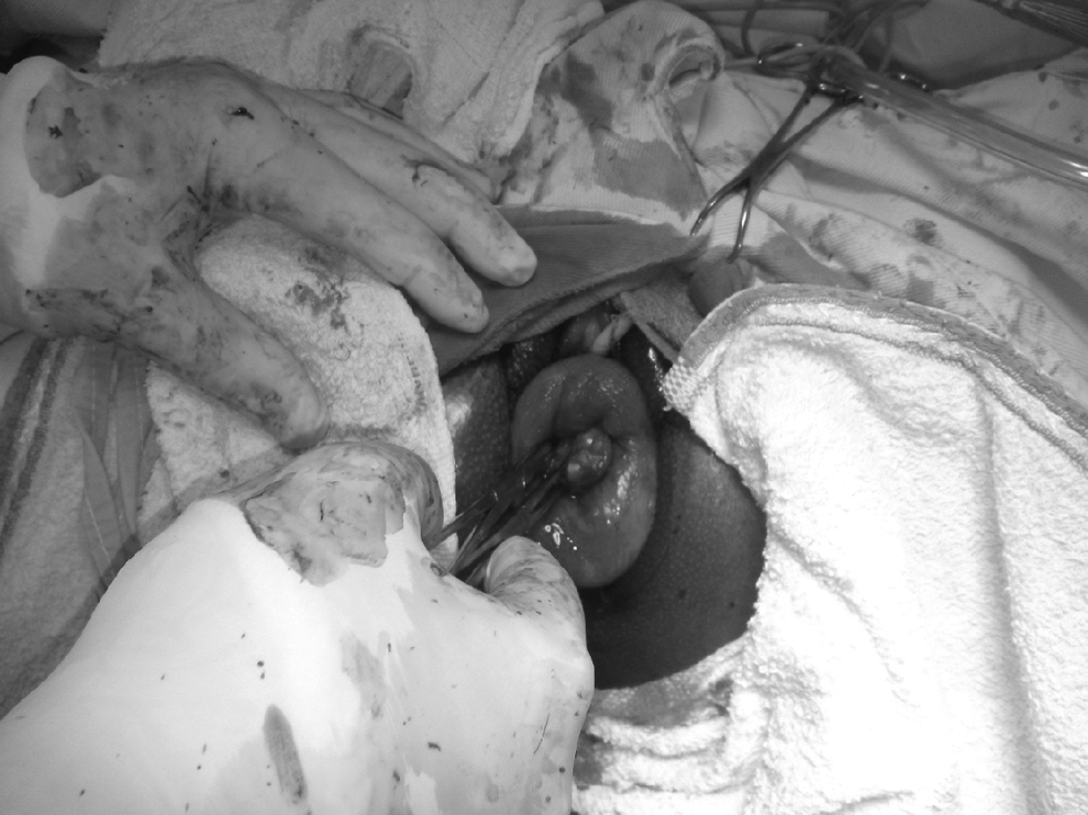
The patient was in shock; hence, she was resuscitated, and then appropriate surgery was performed under general anesthesia, with endotracheal intubation using an inhaled agent (i.e, isofluorane) for adequate uterine-muscle relaxation. The first procedure was a vaginal myomectomy (Fig. 2). The fundus of the uterus was then repositioned manually through a vaginal route with the aid of a ring forceps (Fig. 3). Anesthesia was then reversed, with the forceps kept in the uterus during the anesthesia reversal to reduce spontaneous inversion. A pelvic examination was then conducted, and the success of the manual repositioning was thus confirmed (Fig. 4).
Fundus of the uterus after vaginal myomectomy. The surgeon's thumb points to the rim of the dilated cervix.
Manual repositioning of the inverted uterus vaginally with the aid of a ring forceps.
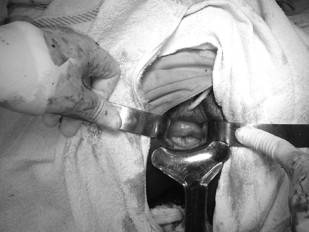
Dilated cervical os following successful manual repositioning of the uterus.
Results
The patient's postoperative period was uneventful. Histopathology testing of the tumor revealed that it was a leiomyoma, weighing ∼150 g (Fig. 5). She recovered fully and resumed her normal activity after 2 weeks.
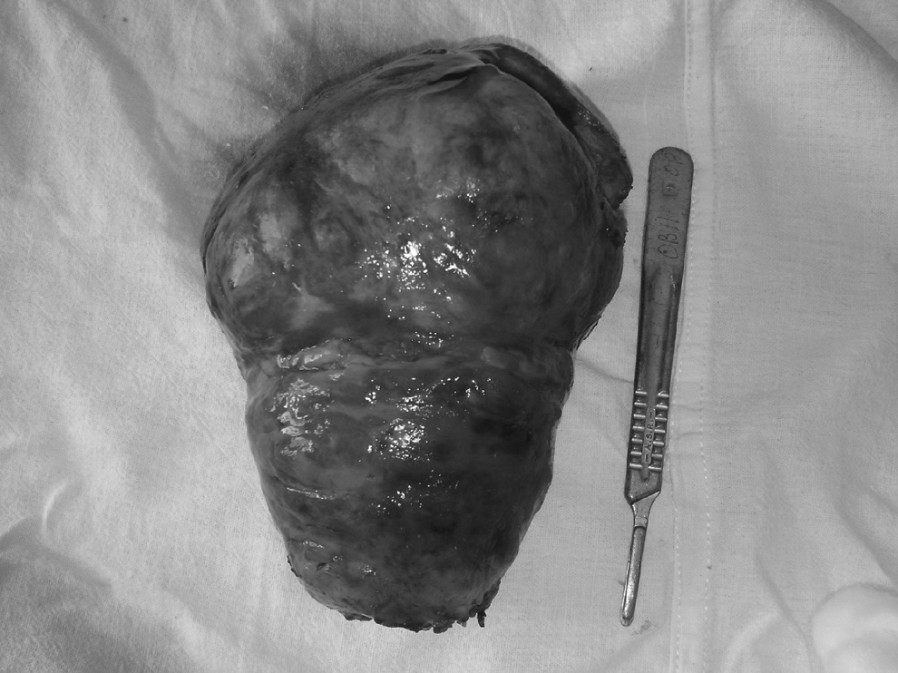
Submucosal 150-g fibroid tumor after vaginal myomectomy.
Discussion
Uterine inversion is an unusual entity, with few cases reported in the literature. The condition may be classified as puerperal or obstetric and non-puerperal or gynaecologic inversion. 5 The incidence of puerperal inversion varies considerably and can range from 1 case in 2000 to 1 case in 50,000 births. 1 The occurrence of nonpuerperal inversion of the uterus is extremely rare, with <200 cases reported in the literature since 1887. These cases represent 5% of all cases of uterine inversion. 3 Nonpuerperal inversion can be classified as chronic or acute, based on the onset and evolution of the condition.
Generally, uterine inversion can be graded as follows: stage 1, inverted fundus remaining in the uterine cavity; stage 2, complete inversion of the uterus through the cervix; stage 3, inverted fundus protruding through the vulva; and stage 4, inversion of the uterus and vagina through the vulva. 7 The current case was a stage 3, acute nonpuerperal inversion.
Nongravid uterine inversion is usually associated with uterine pathology. Prolapse and extrusion of fibroid tumors, especially submucosal myoma of the fundus tends, to be the most common factor causing the inversion. Other less-common causes are endometrial polyps1–3 and inversion associated with uterine neoplasms. Leiomyoma, leiomyosarcoma, rhabdomyosarcoma, endometrial carcinoma, all have been known to be preceding factors.4,5 Mwinyoglee et al. reported that 97.4% of uterine inversion were associated with tumors, 20% of which were malignant. 7 This emphasizes the need to perform biopsies prior definitive treatment.
Risk factors for uterine inversion include fundal attachment of tumors, thickness of tumor pedicle, tumor size, thin uterine wall, and dilation of the cervix, according to Lascarides and Cohen. 5
The current case had similar risk factors. Most cases of nonpuerperal uterine inversion are chronic, and only 8.6% present with sudden onset. 6 The current case occurred acutely. This kind of rare case is often difficult to diagnose. 6 Symptoms associated with nonpuerperal uterine inversion include vaginal bleeding, vaginal tumor, lower abdominal pain, and urinary disturbance. The current patient presented with shock, which is sometimes associated with puerperal uterine inversion.
Magnetic resonance imaging (MRI), computed tomography, and ultrasonograhy are useful diagnostic tools. MRI can be used to examine the characteristics of a uterine inversion. Lewin and Bryan reported that, in T2-weighted MRI scans, a “U” shaped uterine cavity, a thickened and inverted uterine fundus on a sagittal image, and a “bullseye” configuration on an axial image are indicative signs of a uterine inversion. 8
The morbidity and mortality associated with uterine inversion correlate with the degree of hemorrhage, the rapidity of diagnosis, and the effectiveness of treatment. Manual repositioning through the vaginal route was described by Kochenour 9 and is possible with acute inversion. Saline hydrostatic pressure reduction was also described by O'Sullivan (cited by Ward) 1 and modified by Ogueh and Ayida. 10 Delay in treatment of acute uterine inversion causes dense constriction ring formation, progressive edema, hemorrhage, and tissue necrosis; thus the uterus cannot be repositioned by vaginal manipulation. In the current case, manual repositioning was possible after vaginal myomectomy.
In chronic inversions and, occasionally, in acute inversions, manual repositioning through a vaginal route might not be successful. Therefore, such conditions are treated surgically. Fertility-sparing surgical treatment is ideal for patients with this condition. In young patients, conservative operations are performed either via an abdominal or a vaginal approach or via a combined abdominovaginal approach. Huntington and Haultain procedures are the commonly used abdominal approaches and Kustner and Spinelli procedures are the commonly used vaginal approaches.8–10
Huntington's approach
The round ligaments are pulled after laparotomy. Allis forceps are placed on the round ligament ∼2 cm below the insertion on both sides. Gentle traction is exerted, clamps are advanced 2 cm below the previous clamps, and the process is repeated until reduction is complete.
Haultain's operation principle
After opening the abdomen, the constriction ring is divided posteriorly, the inversion is corrected, and incision is closed in two layers.
Spinell's operation for chronic inversion of the uterus
The bladder is dissected away from the inverted uterus. A midline split is made in the cervix and it is separated carefully from the bladder. The anterior wall of the everted uterus is split. Using pressure of the surgeon's index fingers and thumbs, the uterus is turned “outside in.” The myometrium is reapproximated with two layers of running suture, and the serosal surface is reapproximated with a single layer. The vaginal skin is reapproximated with interrupted sutures, as is the full thickness of the cervix.
Kustner's operation for chronic inversion of the uterus
After opening the posterior cul-de-sac, the cervix and posterior wall of the uterus are incised. Then, thumb pressure along the sides of the uterus produce reversion, Interrupted sutures are used to close the incisions and the uterus replaced in the pelvic cavity. This is followed by closure of the colpotomy.
Conclusions
Nonpuerperal uterine inversion is a very uncommon clinical condition; hence, it is a diagnostic challenge. It can be fatal if not managed properly. With a tumor protruding from a patient's vagina or through her vulva, uterine inversion must be considered unless proven otherwise.
Nonpuerperal uterine inversion may occur in malignant cases; thus, preoperative diagnosis is essential. A high index of suspicion is always required for proper diagnosis and a successful outcome.
Footnotes
Acknowledgments
Written informed consent was obtained from the patient for publication of this case presentation and accompanying images.
The current authors acknowledge useful contributions from Adesegun Fatusi MBChB, FWACP, Funmito Fehintola, MBBS, FWACP, Olufemi Ogundele, MBChB, FWACP, of the community health department of the Obafemi Awolowo University Teaching Hospitals Complex, in Ile Ife, Osun State, Nigeria for their input regarding this article.
Author Disclosure Statement
The authors declare that they have no competing interests.