
Abstract
Abstract
Introduction
E
LPD was first described by Willson and Peale in 1952. 1 It is a rare disease of unknown etiology seen mainly in women of reproductive age. LPD is characterized by multiple smooth-muscle nodules of various sizes, either subperitoneal or peritoneal, and located on the omentum or the rest of the peritoneal surface. It has been suggested that the origin could be from submesothelial multipotential cells, although it is not clear if the stimulus for smooth-muscle differentiation is genetic or hormonal or both.1,2
BML was first named by Steiner in 1931. 3 He referred to this entity as a tumor for which the primary growth and the metastasis are composed histologically of benign-appearing smooth-muscle cells and dense connective tissue. BML may present as lesions in the lymph nodes, deep soft tissues, mesentery, bones, central nervous system, and heart. Most commonly, BML presents with lesions in the lungs and can be confused with leiomyosarcoma. 4
Case
A 42-year-old multiparous woman presented for a routine gynecologic evaluation. She had an ultrasound result of a fibroid uterus. On general examination, it was noted that she had no pallor or edema. An abdominal examination revealed that her uterus was enlarged to ∼16 weeks' size. A vaginal examination showed the uterus enlarged to 14–16 weeks with decreased mobility.
Transabdominal and transvaginal scans showed a normal-sized uterus with few fibroids. The largest of these tumors was 3.1 × 3.6 cm, and also, posterior to her uterus, there was a cyst, which was multiloculated with a “ground glass” appearance. No color flow was seen in the cyst and both ovaries were not visualized. Multiple hypoechoic and heterogenous masses were visualized in the left adnexa extending to the left iliac region, with the largest mass measuring 7.6 × 5.2 cm. No vascularity was seen. There was minimal pelvic ascites with few septations. Minimal free fluid was seen in the pelvis. Her CA-125 was 92.25. She was advised to undergo surgery, with the probable diagnosis of an endometriotic cyst and a fibroid uterus.
She was scheduled for an abdominal hysterectomy and bilateral salpingo-oophorectomy for the fibroid uterus and endometriotic cyst, and underwent the surgery 10 months later, as she was lost to follow-up in the interim period.
Intraoperatively, there was minimal free fluid in the abdomen. The uterus had multiple fibroids of 3 × 3 cm and 2 × 2 cm. A chocolate cyst was seen arising from the left ovary 8 × 10 cm; it was adhered to the bowel posteriorly. There was a large fibroid of 6 × 10 cm, which was adherent to the bowel and ureter medially, and the sigmoid mesocolon inferiorly; this tumor was also burrowing into the posterior peritoneum (Fig. 1). There was another fibroid of 4 × 4 cm in the broad ligament. The rectosigmoid was pulled up and had multiple fibroids of 4 × 4 cm and 3 × 2 cm infiltrating the wall and a 5 × 5–cm fibroid in the sigmoid mesocolon. The omentum had multiple fibroids and the peritoneal surface had a few fibroids. There were small 1 × 1.5–cm fibroids on the liver surface.
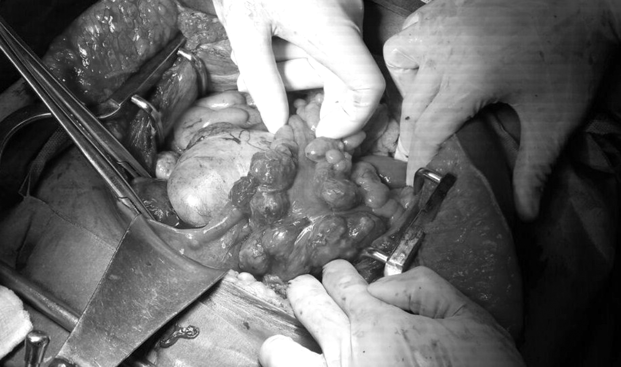
Multiple fibroids growths on the uterus, broad ligament, and sigmoid mesocolon.
The omentum was sent for frozen section and was reported as a benign leiomyoma, probably metastasizing leiomyomatosis. The surgeon proceeded with a hysterectomy after identifying the ureters on both sides. As the left ovarian cyst and broad-ligament fibroid were adherent to the bowel and surrounding structures, it was decided to resect the bowel with the tumor and ovarian cyst en masse. The sigmoid colon was resected and anastomosed. An appendectomy was performed, as the appendix had a tumor on it. Blood loss was ∼1 L, and she was transfused. Otherwise, this patient's postoperative period was uneventful.
Histopathology was reported as multiple leiomyomata involving the uterus, fallopian tube, and left ovary. Leiomyomata of the mesocolon, omental nodules, and peritoneal were noted on biopsy. The appendix had leiomyoma in the serosal aspect. Figure 2 shows Hematoxylin and Eosin staining of the omental nodules. It shows adipose tissue with bundles and fascicles of smooth muscle. High-power magnification revealed that there was no presence of nuclear atypia, necrosis, or mitotic figures (Fig. 3). Immunohistochemistry showed that these tumor cells had diffuse and strong nuclear positivity for estrogen receptor (ER) (Fig. 4).
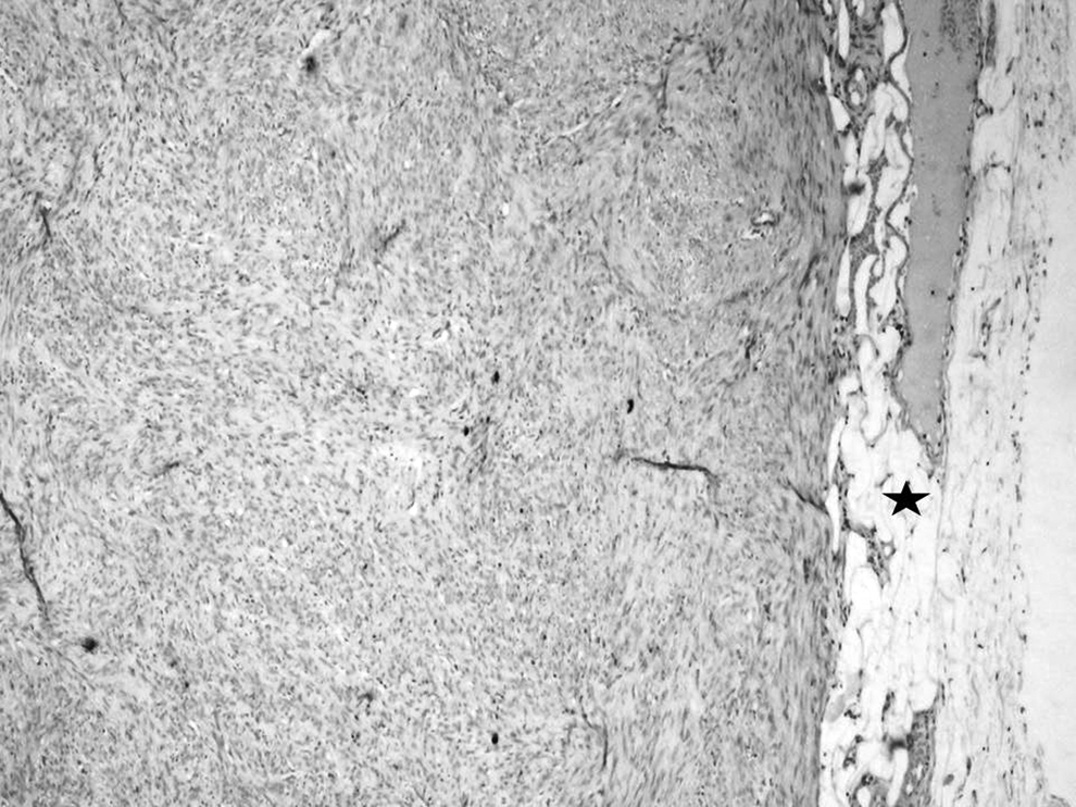
Section from omentum showing bundles and fascicles of benign smooth-muscle cells. Hematoxylin and Eosin stain (4 × magnification). Black star shows adipose tissue.

Nuclear atypia, mitotic figures or necrosis are not seen in high-power view (40 × magnification).

Tumor cells show diffuse and strong nuclear positivity for estrogen receptor (10 × magnification).
Postoperatively she was advised to undergo computed tomography (CT) of her thorax to look for lesions in the lung. The CT scan showed a few lung nodules of 3 mm, 4 mm, and 5 mm in the superior segment of the left lower lobe and right middle lobe. There was no pleural effusion, no cardiomegaly, no pericardial effusion, no mediastinal, axillary or supraclavicular adenopathy, no aggressive-appearing focal bone lesions, and no abdominal or pelvic lymphadenopathy. She was prescribed 2.5 mg of letrozole once a day.
Results
Follow-up at 15 months showed no evidence of recurrence. She had a repeat CT scan of the thorax, which showed no increase in the size of the lesions.
Discussion
Uterine leiomyomas, the most common gynecologic neoplasm in women of reproductive age, can be found in ∼50% of women above age 30, and result from clonal proliferation of uterine smooth muscles. 5 BMLs are uterine leiomyomas which histologically appear to be benign with a low mitotic rate but are associated with the development of similar tumors in distant sites. There is still controversy about where to draw the line between benign tumors and their malignant counterparts. It is recognized that the mitotic index, the degree of cytologic atypia, and the presence or absence of coagulative necrosis are the most important features of tumor behavior.6,7
The pathogenesis of BML has been controversial. Vascular dissemination has been the most-accepted theory. There are some cases in which the uterine tumor has been found even after the metastasis. 8 In some explanations, a multifocal origin has been put forward.9,10 Smooth-muscle neoplasms can actually develop in virtually any region from vascular smooth muscle. However, extrauterine leiomyomas are uniformly ER-negative, while most BMLs are ER-positive.11,12
In most reported cases, pulmonary involvement is present. 13 The extrapulmonary sites where lesions have been documented are the lymph nodes, 14 deep soft tissues,8,10,15,16 omentum, and mesentery,9,10 bone, skull base and spine,17–19 and heart. 20
There have been many reports on the hormone dependency of BML.9,13 At the same time, there has been regression of lesions once estrogen levels fell.13,21 In view of this, various hormone therapies have been tried on patients with BML. Bilateral oophorectomy has been reported to be effective for treating growth of tumors. 22 Medical methods have also been sought to resolve this predicament. The use of long-acting gonadotropin-releasing hormone (GnRH) analogues have been described as having good results.17,19,23,24 GnRH analogues suppress endogenous gonadotropin secretion, which is needed for gonadal steroid production. Progesterone treatment has also been shown to be effective as a prophylaxis against recurrences and for inducing regression of leiomyomatous tumors.13,25 Anastrazole, an aromatase inhibitor, and raloxifene, a selective estrogen-receptor modulator, have also been used successfully to treat BML. 4 Compared to the other agents used, these drugs block steroid synthesis and action directly at the target-tissue level.
Conclusions
Aromatase inhibitors have been shown to decrease uterine fibroid volume in studies.26,27 They are being used more in ER-positive breast cancers. There are not many reports of the use of aromatase inhibitors for BML.28,29 This report aimed to find a suitable role for aromatase inhibitors in the treatment of BML. Aromatase inhibitors decrease production of estrogen from extragonadal sources. Given the rarity of the condition, a randomized controlled trial will not be possible. More case studies and reports will be needed to find the definitive place of aromatase inhibitors for treating BML.
Footnotes
Acknowledgment
The authors acknowledge the contribution of the department of Obstetrics and Gynecology, Christian Medical College, Vellore, Tamilnadu, India.
Author Disclosure Statement
The authors declare no conflict of interest.