
Abstract
Abstract
Background:
Primary vaginal cancer is rare, comprising 1%–2% of all gynecological malignancies. It is commonly seen in women aged 60–80. A primary vaginal carcinoma presenting in conjunction with pelvic organ prolapse is even more rare. This carcinoma usually involves the upper one-third of the anterior or posterior wall of the vagina. Primary vaginal carcinoma involving the lower one-third of the posterior vaginal wall and associated with uterovaginal prolapse is extremely rare and only 1 case has been reported in literature until now. This article reports a very rare case of a primary vaginal carcinoma in a long-standing isolated rectocele involving the posterior lower third of vaginal wall.
Case:
A 62-year old postmenopausal female presented with complaints of a mass protruding through her vagina for 22 years and bloodstained, foul-smelling vaginal discharge for 2 months. She had a history of chronic constipation for 12 years. Her vulva was healthy. A 4 × 4-cm exophytic growth, 1 cm from the fourchette and well away from the cervix, which bled on touch, was present over a grade 2 rectocele.
Results:
Histopathology of the vaginal biopsy demonstrated poorly differentiated squamous cell carcinoma. A diagnosis of International Federation of Gynecology and Obstetrics stage 1 primary carcinoma of the vagina with a rectocele was made. She was referred to the radiotherapy department for further treatment.
Conclusions:
In all patients with long-standing uterovaginal prolapse and postmenopausal bleeding, although the first differential diagnosis is a decubitus ulcer, the possibility of vaginal cancer should also be kept in mind. (J GYNECOL SURG 33:268)
Introduction
P
Case
A 62-year-old woman—para 3, postmenopausal for 10 years, and sexually inactive—presented with complaints of a mass protruding through the vagina for 22 years and a bloodstained, foul-smelling vaginal discharge for 2 months. She had a history of chronic constipation for 12 years. All of her infants were home deliveries, with her last childbirth occurring 27 years ago. Her medical, surgical, and oncologic histories were insignificant. She was a nonsmoker but had a history of heavy weight lifting.
On inspection of her external genitalia, it was noted that her vulva was healthy. A 4 × 4-cm exophytic growth 1 cm from the fourchette and well away from the cervix, which bled on touch, was present over a grade 2 rectocele (Fig. 1). The growth was hard in consistency with well-defined borders limited to the posterior lower third of the vaginal wall. Speculum examination in the office revealed a healthy looking cervix, and no obvious discharge was visible. On bimanual examination, her uterus was atrophic and firm, the fornices were free, and no adnexal mass was palpable. A rectal examination revealed normal mucosa without infiltration.
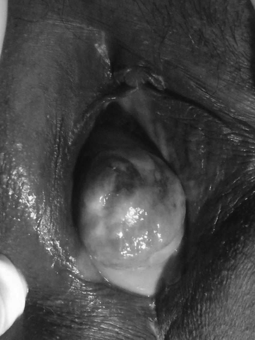
A 4 × 4–cm exophytic growth present over a grade 2 rectocele in the lower third of the posterior vaginal wall.
Results
Histopathology of a vaginal biopsy revealed poorly differentiated squamous-cell carcinoma. Colposcopic evaluation of the cervix was normal. A detailed work-up of this patient included a chest X-ray and ultrasounds of the abdomen and pelvis, which did not reveal any metastasis.
A diagnosis of International Federation of Gynecology and Obstetrics stage 1 primary carcinoma of the vagina with a rectocele was made. She was referred to the radiotherapy department for further treatment, where she was lost to follow-up.
Discussion
Eighty-four percent of vaginal cancers are secondary to the cancer arising from the adjacent organs, cervix (32%), endometrium (18%), colon and rectum (9%), ovary (6%), or vulva (6%). 1 Due to the rarity of primary vaginal carcinoma, it is a diagnosis of exclusion of other coexistent gynecologic cancers. In general, uterine prolapse combined with vaginal cancer is a very uncommon condition. After Howat et al. 4 reported a case of a patient with an enterovaginal fistula, several other reports were published subsequently (Table 1).3–17 However, there has been no report of a primary vaginal carcinoma arising from an isolated rectocele only.
FIGO, International Federation of Gynecology and Obstetrics.
Risk factors are bacterial infection, trauma (especially after pessary or prolapse), and human papilloma virus (HPV) exposure. Diethylstilbestrol (DES) exposure is associated with a clear-cell subtype of vaginal cancer. Daling et al. reported that the invasive vaginal cancer has a strong correlation to HPV infection and HPV DNA was detected in 60% of such patients in a population-based study. 18
Vaginal carcinoma is a disease of older women, with ∼70% of patients diagnosed after the age of 70 years. Often, the diagnosis is delayed because of the subtlety of the symptoms, especially in sexually inactive women. 8 The most-frequent clinical symptom is vaginal bleeding, but dysuria and pelvic pain are also common. 19 Less frequently, lesions involving the anterior vaginal wall may lead to dysuria, hematuria, or urgency. Alternatively, constipation may result from posterior vaginal-wall cancers. The current patient presented with a bloodstained vaginal discharge of 2 months duration. In a similar case of a stage 1 primary vaginal cancer of the posterior third of the vaginal wall associated with third-degree uterovaginal prolapse reported by Kumar and Bulusu, 3 no history of bloodstained discharge was present, but a vaginal mass and constipation were present for 2 years and 6 months, respectively. As in the current case, a vaginal mass and constipation were present for 22 and 12 years, respectively. Moreover in the current case, only an isolated rectocele with an exophytic growth was present, compared to Kumar and Bulusu case, which had a third-degree uterovaginal prolapse with a second-degree cystocele and rectocele. 3
In vaginal carcinoma associated with uterovaginal prolapse, procidentia has been present for 10 years or more in 60% of cases. 20 In an Indian case series comprising 11 cases of primary carcinoma of the vagina during a 10-year period, 6 cases were associated with third-degree uterine prolapse. All patients came to the hospital at a late stage despite having third-degree uterine prolapse. Bloodstained discharge and ulceration on the prolapsed part, and irreducible prolapse with urinary retention and marked edema of local and surrounding tissues were the presenting symptoms. The lesions on the patients' vaginas varied in size from 5 cm to 15 cm. 5 It is believed that continued irritation and chronic inflammation of the exposed vagina contribute to the occurrence of vaginal cancer. In most of the cases, the prolapse is the reason that a patient seeks a gynecologist's advice; the diagnosis of vaginal carcinoma is usually made as a coincidence. 8 In the current case, the patient was not troubled by her long-standing rectocele of 22 years or her chronic constipation but sought a gynecologist's help for the troublesome bloodstained discharge.
Primary vaginal cancer has two major histopathology types: squamous-cell carcinoma (80%) and adenocarcinoma (15%). Melanoma, lymphoma, and sarcoma are highly unusual, comprising the remaining 5% of these cases. 21 In the current case, it was a poorly differentiated squamous-cell cancer. Of note, only 1 of 6 patients described in Rao et al.'s 1984 case series had poorly differentiated squamous-cell cancer and the remaining 5 had squamous-cell carcinoma. 5
Squamous-cell carcinoma is more common in postmenopausal females, whereas adenocarcinoma commonly affects younger patients (median age: 19) and is more likely to metastasize to the lungs and lymph nodes. 22 One subtype, clear-cell adenocarcinoma, is classically associated with in utero exposure to DES and is found in 2% of exposed females. 23 However, Sachan et al. reported a case of clear-cell adenocarcinoma of the vagina in a 27-year- old woman associated with a third-degree uterovaginal prolapse without DES exposure in utero. 13
The choice of treatments for vaginal cancer is determined by lesion size, location, and degree of infiltration; age; stage; and histology. Lesions located in the lower region of the vagina wall at clinical stage I are treated with a combination of external beam and intracavitary radiation therapy, whereas lesions located in the upper region are treated with a similar dose of radiation and therapeutic range as would be applied to cervical carcinoma. 14 Furthermore, brachytherapy may not be technically feasible without prior palliative surgery such as vaginal hysterectomy or colpocleisis to reduce the prolapse.
There are no recommendations in the literature to guide this particular treatment decision. 24 Surgical therapy is effective for treatment of stage I cancer and primarily consists of radical hysterectomy and dissection of the pelvic lymph nodes. Lesions in the lower vagina are treated by resection of the inguinofemoral lymph nodes, including the external genitalia. 14 In the current case, as only an isolated rectocele was present with a primary vaginal carcinoma, primary radiotherapy was given without risk of radiation injury to adjacent organs. Surgery was avoided in order to avoid injury to the rectum, although Tjalma et al. reported that patients who have stage I and II squamous vaginal cancer have good outcomes in terms of survival and local tumor control if they are managed by initial surgery followed by selective radiotherapy. 25
Conclusion
In all patients with long-standing uterovaginal prolapse and postmenopausal bleeding, although the first differential diagnosis is that of a decubitus ulcer, the possibility of vaginal cancer should also be kept in mind.
Footnotes
Author Disclosure Statement
No financial conflicts of interest exist.