
Abstract
Abstract
Background:
Lichen simplex chronicus (LSC) is an eczematous disease caused by cell-mediated autoimmune inflammation that presents as an erythematous and distressingly pruritic area that develops into a thickened, indurated, and atrophic area. U.S. prevalence is ∼0.5% and most commonly involves the labia minora. Often, an itch–scratch–itch cycle perpetuates this condition. Scratching causes intense pleasure, which could lead to excoriation, lichenification, depigmentation, and chronic dermatitis. Itching may be intensified at night. Common triggers are sweating, stress, walking, irritation from clothing and panty-liner–based infections.
Case:
A 22-year-old nulligravida presented in a clinic for emergency room follow-up with a diagnosis of an ovarian cyst and pyelonephritis that were treated with proper antibiotics. A wet mount was diagnostic for bacterial vaginosis, so the patient was treated with the indicated antibiotic and was scheduled for follow-up at 1 month. After multiple months of treatment with no improvement, she was referred to a vulvovaginal-specialized clinic.
Results:
At the specialized clinic, an abnormal lesion involving the anal and intergluteal areas, with a small portion of the vulva were seen. These lesions were described as soft, whitish, and hypopigmented. A punch biopsy was performed, and a pathology test confirmed LSC. She was prescribed a high-potency topical steroid, and 2 weeks later her symptoms were almost completely resolved with great improvement in the vulvar and perianal areas.
Conclusions:
LSC is an eczematous disease that usually presents in middle-aged women with the chief complaint of pruritus that is relieved by scratching. (J GYNECOL SURG 33:273)
Introduction
P
Evaluation of anogenital pruritus should include description of the pruritus, patient age, food and/or drug allergies or side-effects, grooming, bathing, skin care habits, and relevant interventions.1–3 Additionally, infectious causes that should be ruled out include condyloma acuminate, herpes simplex lesion, herpes zoster, gonorrhea, chlamydia, and trichomonas. Moreover, careful inspection of the skin and vaginal rectal examination to look for evidence of erythema, swelling, lichenification, and hyper- or hypopigmentation are crucial in narrowing the diagnosis. Wet mount preparation should be performed to rule out vaginitis.2,4 Secondary irritant contact dermatitis and autoimmune chronic dermatitis should be ruled out. 1 If no apparent causes are determined, biopsy of the skin area involved should be performed.3,4
Differential diagnosis includes hepatic disease, leukemia, diabetes mellitus, and anal cancer. Other disorders, such as anal fissures, fistulae, and internal or external hemorrhoids, should also be considered. A large portion of anogenital pruritus is caused by dermatologic infection, such as Coryneacterium minutissimum, streptococcus, sexually transmitted infection, syphilis, dermatophytosis, and pinworms.1,3
LSC is an eczematous disease caused by cell-mediated autoimmune inflammation that presents as an erythematous and distressingly pruritic area that develops into a thickened, indurated, and atrophic area. 2 Prevalence in the United States and Western Europe is ∼0.5%. This condition most commonly involves the labia minora and presents at 30–50 years of age. LSC can be primary (arising from normal appearing skin) or secondary. 3 There is often an itch–scratch–itch cycle that perpetuates the condition and that can lead to chronic dermatitis. Severe scratching can lead to excoriation, lichenification, and depigmentation. Secondary LSC can be triggered by vulvar candidiasis or may be idiopathic in origin. Scratching causes intense pleasure, which may lead to the itch–scratch–itch cycle, eventually causing skin alteration. Itching may be intensified at night and the patient may not be aware of scratching. Common triggers may include sweating, stress, walking, irritation from clothing, and panty-liner–based infections. There is an additional rare risk of malignant transformation.2,3
According to some reports, there seems to be a psychologic component in the causation and perpetuation of this condition. In addition, it has also been correlated with anxiety, depression, and obsessive–compulsive disorders.5–7
Histopathologic diagnosis often shows prominent fibroblasts, zones of pale epithelium, and thickening of the epidermis. More importantly, this disorder shows hyperplastic vulvar epithelium in contrast to specific atrophic changes typical of lichen sclerosus.8,9 On physical examination, epidermal thickening of lichenified plaques may be seen as well as excoriations. 4
Treatment includes a short dose of high-potency steroids (e.g., clobetasol) followed by short-term use of medium- or mild-potency steroids. Prolonged topical steroids should be avoided. Additional therapies that should be considered include topical calcineurin inhibitors (pimecrolimus cream), or local anesthetics. Moreover, subdermal Decadron® is an alternative treatment, which, when used appropriately, does not result in skin atrophy.3,10–12
Additionally, to break the itch–scratch cycle, antihistamines, such as Benadryl® or hydroxyzine, may be given to prevent night scratching. Night-time sedation with amitriptyline and doxepin can be used to diminish rapid eye movement sleep and night-time scratching. Other adjunctive treatments include symptomatic antipruritic treatment with topical anesthetic, avoidance of irritants, and treatment and identification of other symptoms.3,9 It is also recommended that patients address the psychologic component frequently associated with this condition.5,7,11
Additionally, silk and cotton underwear (nonirritating); water only for cleaning; white petroleum as an emollient; and avoidance of scented soaps, lubricants, or perfumes are also recommended.9,12 The patient should be followed-up monthly.
A Medline® review of the literature, using the terms
Case
A 22-year-old nulligravida presented to the Department of Obstetrics and Gynecology, of Texas Tech University Health Sciences Center at the Permian Basin, Odessa, TX, for emergency-room (ER) follow up with a diagnosis of an ovarian cyst and pyelonephritis that had been treated with proper antibiotics. She had been intensely anxious because of her ovarian cyst diagnosis and was referred to the psychiatry department, where she had been started on a selective serotonin reuptake inhibitor and an anxiolytic. This patient complained of having an irritating vaginal discharge with vaginal and perirectal pruritus. A wet mount was diagnostic for bacterial vaginosis, so she was treated with the indicated antibiotic and was scheduled for follow-up after 1 month. At this follow-up, her complaints persisted but she had not taken her medications as indicated and was then prescribed a topical antifungal.
Two months later her symptoms were still persistent and were accompanied by rectal bleeding and dyschezia. Her perineal examination revealed edematous erythematous fourchette with perirectal involvement and excoriations. She also had a positive hemoccult test but no other rectal findings. Additionally, due to the presence of fungi in the vagina, an oral antifungal and a topical steroid were prescribed.
One month later, given that her symptoms were not relieved, she was referred to a vulvovaginal-specialized clinic. At the specialized clinic, an abnormal lesion involving the anal and intergluteal areas with a small portion of the vulva were seen (Figs. 1 and 2). These lesions were described as soft, whitish, and hypopigmented. A punch biopsy was performed and the pathology diagnosis confirmed LSC. She was prescribed a high-potency topical steroid and, 2 weeks later her symptoms were almost completely resolved with great improvements in the vulvar and perianal areas.

Erythematous thickened and indurated area involving the labia majora.
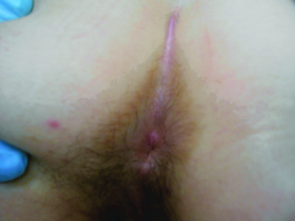
Perianal and interfluteal excoriations, lichenification, and depigmentation typical of lichen simplex chronicus.
Results
There were several unusual aspects regarding this patient's presentation. First, her recent history of antibiotic use, positive wet mount, and absence of symptoms prior to her ER visit pointed toward an infectious etiology of the pruritus. Additionally, her age and the location of her lesion did not fit the classic description of LSC. Her pruritus and dermatoses were mainly in the perianal region and only involved a small portion of the perineum. 3
The uncommon diagnosis of perianal LSC can be difficult to make because, as in this case, it could masquerade as vulvovaginal conditions including infections and/or candidiasis. Therefore, if symptoms of a skin condition persist or progress after ruling out poor compliance with the prescribed therapy, a punch biopsy must be strongly considered to clarify the diagnosis. 4 Although anogenital pruritus is a common complaint seen in the young adult population, chronic atopic dermatitis is not often high in the potential differential diagnoses.1,8 This case also highlights the usual tortuous road to the correct diagnosis of LSC and its associated psychologic component.5,6,14 A timely diagnosis is crucial to avoid discomfort for the patient and reactive skin changes in the vulva, which can, in the long term, lead to architectural distortions of the vulva. Once properly diagnosed, the correct treatment can lead to prompt relief of symptoms and lesions, as described in this case report.
Conclusions
LSC is an eczematous disease that usually presents in middle-age women with the chief complaint of pruritus that is relieved by scratching. This disorder is often hard to diagnose, especially when it presents in the perianal area, but there is usually a history of an itch–scratch–cycle. Prompt diagnosis, using clinical examination findings and tissue pathology, can lead to appropriate treatment with high-potency topical steroids, and resolution of the condition.
Footnotes
Author Disclosure Statement
None of the authors has any conflicts of interest to report.