
Abstract
Abstract
Background:
Uterine rupture is a catastrophic obstetric emergency with potentially life-threatening maternal and fetal outcomes. Malpresentation can add more risk factors for occurrence of this catastrophe.
Case:
Spontaneous uterine rupture occurred at 38 weeks of gestation in a 26-year-old woman with no risk factors except for malpresentation. Repair of fundal transverse tear was performed with an emergency laparotomy.
Results:
The patient's postoperative period was uneventful. Her postoperative hemoglobin level was 9.5 g/dL. She was discharged on day 4 after the operation with no complaints.
Conclusions:
Malpresentation alone can be a risk factor for spontaneous, third-trimester uterine rupture in an unscarred uterus. This occurrence is associated with poor fetal and maternal outcomes. Surgeons should explain to their patients the hazards of probable risk of recurrent uterine ruptures in future pregnancies. (J GYNECOL SURG 34:89)
Introduction
U
There are several conditions that cause increased risks of uterine rupture during pregnancy. Some of these risk factors include uterine anomalies5,6 use of misoprostol, 7 uterine curettage, multiparity with short interdelivery interval, 8 uterine scar from cesarean section, and laparoscopic and hysteroscopic uterine surgeries.9–12 The most common risk factors for uterine rupture is previous cesarean section in a scarred uterus and cephalopelvic disproportion in an unscarred uterus. 13
Rupture of an unscarred uterus is a rare event. The prognosis of uterine rupture if developed in an unscarred uterus is even worse. 14 Additionally, this rupture is more hazardous with a higher rate of mortality to fetuses, compared to their mothers. 15
This article reports a case of a primigravida, who was pregnant at 38 weeks, with a breech fetus, who presented to the Women's Health Hospital in Assiut, Egypt. This patient had a spontaneous prelabor rupture of her unscarred uterus.
Case
In May of 2017, a 26-year-old woman, primigravida, presented to the emergency unit of the Women's Health Hospital, at 38 weeks of gestation (based on her last menstrual period). She was experiencing acute onset of severe lower abdominal pain and absent fetal movements for 2 hours. This patient had no history of previous cesarean section, any other uterine surgery, or intrauterine device insertion. During her pregnancy, she made several prenatal visits, and sonographic examinations showed that she had frank breech presentation up till 1 week prior to this presentation. The patient had not been offered a trial of external cephalic version, as all of her prenatal visits were in a rural primary health care unit with no experienced obstetricians in such maneuvers.
On general examination, the patient noted to be conscious and pale, with a pulse of 120 beats per minute, a blood pressure of 100/60 mm Hg, and a temperature of 37°C. An abdominal examination revealed generalized tenderness, rebounding, easily felt fetal parts, shifting dullness, and inaudible fetal heart sounds. A pelvic examination showed that the cervix was closed and not effaced; there was no vaginal bleeding.
An emergency ultrasound (US) revealed an empty uterus with a single nonviable fetus present outside the uterine cavity and marked intraperitoneal fluid collection. The patient's preoperative hemoglobin level was 7 g/dL, and her coagulation profile was normal. The patient was counseled concerning the possibility of uterine rupture, and informed consent for abdominal exploration was obtained.
An exploratory laparotomy though a Pfannensteil incision was performed under general anesthesia. The peritoneal cavity was filled with ∼2000 cc of blood and the fetus was found to be completely extruded from the uterine cavity through a large transverse fundal defect of ∼8 cm, with irregular actively bleeding edges (Fig. 1). A dead male fetus weighing 2800 g was delivered.
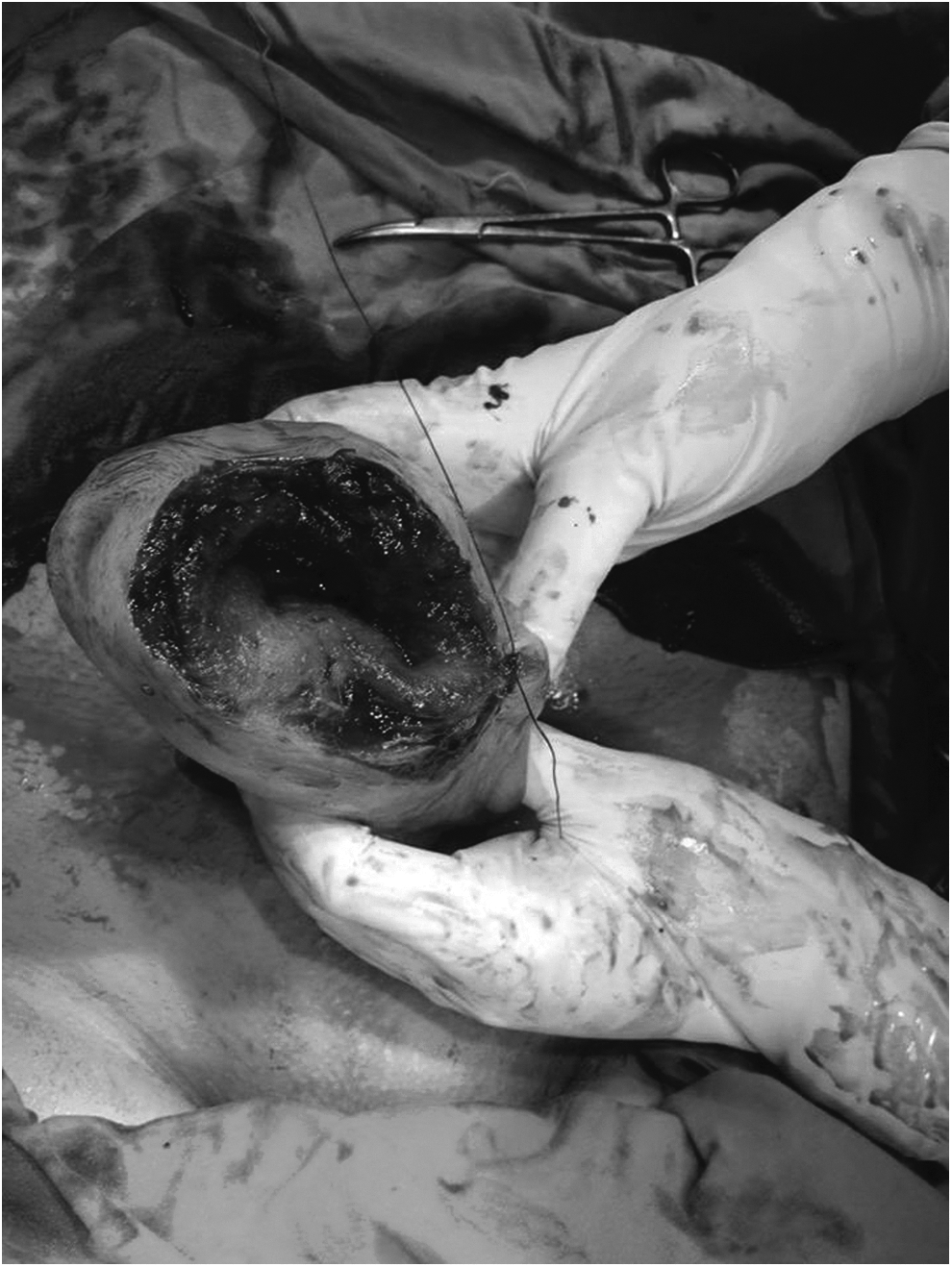
Ruptured uterus at the fundus.
As the patient had no living children, the uterine tear was repaired without tubal ligation. The uterine tear was repaired with a double-layer closure using synthetic continuous Vicryl® nonabsorbable sutures (Fig. 2). The abdomen was closed in layers after normal saline lavage with insertion of an intraperitoneal drain. She received 4 units of packed red blood cells during the surgery.

The uterus after repair of the tear in two layers.
Results
This patient's postoperative hemoglobin level was 9.5 g/dL. She was discharged on day 4 after the operation with no complaints.
Discussion
Uterine rupture is a frequent complication of pregnancy in developing countries. However, it is extremely rare in developed countries. In the United States, the incidence varies between 1:8000 and 1:15,000 births. 16 Risk factors for uterine rupture are lower-segment cesarean section or upper–uterine segment surgeries, including hysterotomy, classical cesarean section, and myomectomy. 17
Rupture of an unscarred uterus is relatively rare; however, a rupture can occur especially in multiparous women, and when there is abnormal placentation, malpresentation, instrumental delivery, and labor induction by misoprostol in the third trimester. 18 Chronic use of corticosteroids can lead to impaired collagen synthesis causing uterine rupture. 19
In the current case, the patient was primigravida with no defined risk factors for uterine rupture. She had no previous intrauterine instrumentation or surgeries on the uterus, no medical history of steroid use, and, during her prenatal visits her US showed a breech fetus with a normally situated placenta.
The lower uterine segment, which is the weakest part, is a common site of unscarred third-trimester uterine rupture, 20 while fundal rupture is rare and is associated with late diagnosis due to free blood collection in the intraperitoneal space. 21 In the current case, the uterine rupture was fundal.
Despite the relative rarity of uterine rupture in primigravidae, this obstetric emergency is life-threatening to mothers and fetuses with late grave consequences on the reproductive outcome. Therefore, investigating the rupture's etiology and pathogenesis is of major importance. Unscarred uterine rupture in a primigravida with no apparent causes or risk factors is extremely rare. Over a period of 60 years, Walsh and Baxi identified only 36 cases of uterine rupture among primigravidae in a study, 22 and only 2 of the cases had no apparent risk factors for uterine rupture.
Spontaneous uterine rupture during pregnancy can be diagnosed on a scarred uterus by performing a US scan. When a spontaneous uterine rupture occurs, there can be a projection of membranes at the site of the scar. If possible, external electronic fetal monitoring should be carried out in each case when there is abdominal pain occurring during a pregnancy, because, in cases of uterine rupture, there will be fetal heart rate abnormalities. 23
When there is late presentation as in the current case, there is abdominal pain and signs of shock, at which time the US scan will reveal an intraperitoneal pregnancy. In these cases, the fetus is already dead and efforts are made to save the mother.
Abdominal exploration is the only key for successful management of uterine rupture. It should be managed in a tertiary-level hospital. Full thickness repair with proper suture materials is mandatory. An effective method of contraception must be used for at least 1 year after uterine repair 24 with close monitoring of the patient's future pregnancies.
Conclusions
This case highlights that malpresentation alone can be a risk factor for a spontaneous third-trimester uterine rupture in an unscarred uterus. This occurrence is associated with poor fetal and maternal outcomes due to difficult predictions by obstetricians in the absence of risk factors. Obstetricians must keep this potential in mind when a pregnant woman presents with an acute abdomen in late pregnancy even with the absence of any risk factors.
Footnotes
Author Disclosure Statement
No financial conflicts of interest exist.