




Abstract
Abstract
Background:
There are many types of leiomyomata; however, this case report is about an anterior, isolated, large, rare vaginal myoma that is not classified in the International Federation of Gynecology and Obstetrics (FIGO) leiomyomata subclassification system. In this case, the patient was treated with a simple vaginal myomectomy without any abdominal skin incisions.
Case:
A 41-year-old female came to the hospital because she had an increasing amount of a watery, yellowish, odorless vaginal discharge for ∼1 month. An isolated vaginal solid mass, measuring ∼5.9 × 5.3 cm, with a normal uterus, was found on transvaginal ultrasonography. Magnetic resonance imaging showed a well-defined, homogenous, isolated vaginal mass, measuring 5.9 × 5.3 × 5.0 cm. The mass was located over the superior segment of the vagina, was removed through vaginal excision, and was sent for pathology testing.
Results:
Pathologic testing revealed that the mass was a vaginal leiomyoma.
Conclusions:
An isolated vaginal myoma—a rare type of myoma—can be managed with a simple vaginal myomectomy.
Introduction
T
Magos et al., who were the first to report about vaginal myomectomy in 1994, showed a vaginal route for approaching a uterine myoma surgically by making a vaginal incision through which the myoma was excised and uterine suturing was performed.3,4 Vaginal myomectomy is an alternative procedure for the treatment of vaginal myomas. This current report is about a vaginal myomectomy approach to an anterior, vaginal isolated myoma, of ∼5.9 cm, without making any abdominal skin incisions.
Case
A 41-year-old female (gravida 2, para 2, with both normal deliveries), with a regular menstrual cycle, came to an outpatient clinic due to having an increasing amount of watery, yellowish, odorless vaginal discharge for ∼1 month. Transvaginal ultrasonography, as shown in Figure 1, revealed an anterior, isolated, rare vaginal myoma, measuring 5.9 × 5.3 cm; it compressed the urinary bladder, urethra, and cervix, and had an endometrial thickness of 1.58 cm. As shown in Figure 2, magnetic resonance imaging (MRI) revealed a well-defined, homogenous, isolated vaginal mass, measuring 5.9 × 5.3 × 5.0 cm, arising from the anterior superior segment of the vagina. The MRI imaging features suggested that the mass was most likely to be a leiomyosarcoma of the vagina, with a differential diagnosis of a leiomyoma with myxoid degeneration and no evidence of nodal or distant lesions.

Transvaginal ultrasonography showed an anterior, isolated, rare vaginal myoma that was 5.9 × 5.3 cm; it compressed the urinary bladder, urethra, and cervix.
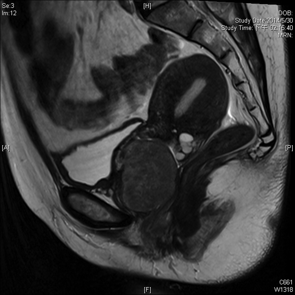
Magnetic resonance imaging showed a large, isolated, rare, anterior vaginal myoma that was 5.9 × 5.3 × 5.0 cm; it compressed the urinary bladder, urethra, and cervix.
In the operative theater, upon inspection of the vagina, a nodular myoma of ∼6 cm was found anteriorly in the vaginal wall and very close to the bladder base. The mass was separated from the urethra. Normal sodium saline solution (3 cubic cm) was injected into the mass. A 2-cm transverse vaginal incision was made below the vaginal rugae and was deepened down to the plane of the vaginal myoma. The edges of the mass were excised by using bipolar scissors and electrocauterization. The homogeneous vaginal mass weighed 52 g. The edges of the vagina were sutured with Vicryl 2-0. The vaginal myomectomy operation was performed completely. The mass was sent for pathologic diagnosis.
Results
Pathologic testing revealed that the mass was a leiomyoma of the vagina.
Discussion
In 1994, there was a report about a vaginal procedure as an alternative approach to abdominal or laparoscopic surgery for myomectomy; this report stated that this approach produced superior cosmetic results. 3 Conventional, rather than miniature, instruments can be used for a vaginal procedure. Moreover, these instruments are more efficient for handling tissues with gross pathology. Stronger grasping forceps can be used, and suturing the uterus is tremendously easier, which should make the suture repair stronger and less likely to dehisce during pregnancy in the future. Furthermore, surgeons who perform vaginal myomectomies surely have all the tactile advantages of conventional surgery, which are absent from endoscopic procedures. 5 There have been some reports of uterine ruptures in future pregnancies after patients underwent laparoscopic myomectomy; these ruptures were due to the uterine repairs being performed with low-strength laparoscopic suturing.6,7
In the current case, the MRI of the vaginal myoma—with the differential diagnosis per that MRI of the leiomyosarcoma of the vagina—showed that the mass totally filled the vaginal cavity. Thus, other surgeons might have thought that it was not safe to do a vaginal procedure. Other surgeons might have used an abdominal procedure to perform a hysterectomy with removal of the vaginal myoma via an abdominal route.
The current case report was about an isolated, anterior vaginal myoma, measuring ∼5.9 cm, that was excised with a vaginal myomectomy. This study shows that isolated anterior vaginal myomectomy can be a safe vaginal route for selected patients, with no morbidity, great success rates, and no uterine ruptures in subsequent pregnancies.
Conclusions
A rare, large, isolated vaginal myoma—which is not included in the FIGO classification system—can be managed by vaginal myomectomy. This is a safe procedure that can be performed with no abdominal skin incisions, minimal blood loss during the procedure, and fast recovery after the operation.
Footnotes
Acknowledgments
The authors thank the Asia-Pacific Association for Gynecologic Endoscopy and Minimally Invasive Therapy (APAGE) for providing the International Fellowship Endoscopy Training Program at Chang Gung Memorial Hospital at Linkou, Tao-Yuan, Kweishan, Taiwan, for Dr. Chatchotikawong.
Author Disclosure Statement
No financial conflicts of interest exist.