
Abstract
Background:
Struma ovarii, an ovarian tumor, is diagnosed when >50% of the teratoma is thyroid tissue. Struma ovarii's incidence is <2% of mature teratoma.
Case:
We present a rare case report of struma ovarii of a 40-year-old female who presented with lower abdomen pain having right ovarian cyst on imaging study. Her cancer antigen (CA)-125 level was 18.70 units/mL and histologically proved to be struma ovarii.
Results:
Multicystic 7 × 6 cm ovarian mass was diagnosed as struma ovarii.
Conclusion:
Struma ovarii though a rare entity should always be kept in mind during ovarian neoplasm excision and should be cautiously searched for any malignant transformation.
Introduction
Struma ovarii, an ovarian tumor, is diagnosed when >50% of the teratoma is thyroid tissue. Struma ovarii's incidence is <2% of mature teratoma. 1 Malignant transformation is <5% with the presence of papillary thyroid carcinoma (PTC) and follicular thyroid carcinoma as the most common type. 2 Most patients are euthyroid and asymptomatic and patients present with symptoms related to the mass. However, thyrotoxicosis has been reported in 5%–15% of struma ovarii cases. 3 Most malignant struma ovarii have poor iodine uptake with less secreting thyroid hormones.4,5
Case Report
This case report is written in view of rarity of presentation. A 40-year old female patient presented with chief complaints of lower abdominal pain for 1 month that was gradually progressive in nature and radiating to the back. There was no history of any bleeding per vagina. On examination, the abdomen was soft and suprapubic tenderness was present. Per vaginal examination showed healthy cervix and retroverted normal sized uterus with a cystic mass palpable in the right fornix. Her cancer antigen (CA)-125 level was 18.70 units/mL.
The patient underwent right oophorectomy and her intraoperative finding was with a 7 × 6 cm multiloculated cyst in the right ovary. No solid mass was seen in the uterus and left ovary was normal. The right oophorectomy specimen was sent for histopathology evaluation. Right oophorectomy specimen received was a cut opened cyst weighing 200 grams and measuring 7 × 6 × 3 cm in size. Outer surface was smooth and cut section showed a multiloculated cyst filled with brownish inspissated material (Fig. 1).

Right oophorectomy specimen shows gray white cyst measuring 7 × 6 cm with a smooth outer surface. Cut surface shows a multiloculated cyst filled with brownish inspissated material.
Histopathologic examination of multiple sections showed the multicystic tumor predominantly composed of thyroid follicles filled with colloid comprising ∼90% of the area. Small foci of respiratory epithelium with areas of calcification and fibrosis were also noted. There was no cellular atypia, nuclear pleomorphism, mitotic activity, and vascular and/or capsular invasion, and a final diagnosis of struma ovarii was made (Fig. 2).
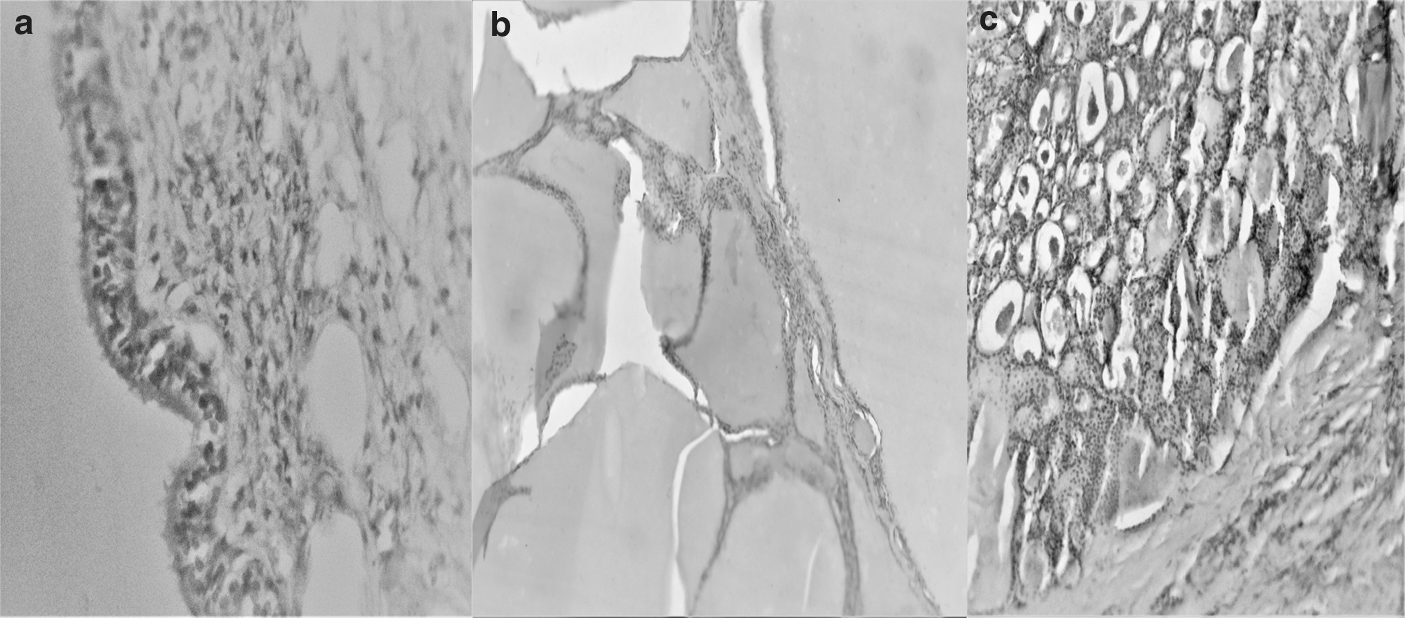
Microphotograph showing cyst wall lined by pseudostratified ciliated columnar epithelium
Discussion
Mature teratoma in the ovary originates from a single germ cell after the first meiotic division. Thyroid tissue can be found in 5%–20% cases of mature teratoma.6–8 Struma ovarii is the most common type of monodermal teratoma, accounting for ∼3% of all ovarian teratomas. 3 It typically consists of normal-appearing thyroid tissue composed of thyroid follicles of various sizes.
PTC is the most common thyroid malignancy to occur in struma ovarii. 9 The diagnostic criteria for PTC in thyroid gland include demonstration of diagnostic nuclear cytology, such as nuclear elongation, overlapping with chromatin clearing, irregular nuclear membrane, intranuclear grooves, and pseudoinclusions. 10 The diagnosis of follicular carcinoma requires vascular and/or capsular invasion. Whether these criteria used for the thyroid gland are applicable to the cases of struma ovarii is still controversial. 11 Devaney et al. reported 13 cases of malignant struma using thyroid gland criteria, but only 1 of them actually proved to be of biologic significance that was spreading beyond the ovary. 2
Robboy et al. reported 27 biologically malignant struma ovarii: 12 arose from histologically malignant tumors and 15 tumors arose from histologically benign lesions, that is adenomas (13 cases) or unremarkable thyroid tissue (2 cases). 11 In contrast, Schmidt et al. reported B-raf protooncogene (BRAF) mutations were present in three of five PTCs from struma ovarii and none of the nine benign struma ovarii, in which they applied the PTC diagnostic criteria of thyroid gland. 12 The findings suggested a common pathogenesis for PTCs from thyroid gland and struma ovarii.
It is not uncommon to find thyroid tissue embedded in either ovarian or teratomatous stroma, which may mimic infiltrative growth of a follicular carcinoma. The only reliable diagnostic criterion for follicular carcinoma should be vascular invasion in struma ovarii. Adequate sampling is particularly important in this setting; this may explain why some adenomas had malignant behavior in Robboy's study. 11
The term proliferative struma denotes a lesion that comprises areas of densely packed follicles or papillary formations lacking nuclear features of PTC. The histologic spectrum of proliferative struma ovarii includes multinodular adenomatous proliferation, resembling follicular adenoma, including so-called embryonal adenoma of the thyroid. 2
In our case, struma ovarii had normal thyroid follicles in the stroma comprising ∼90% of the tumor with small foci of the respiratory epithelium, calcification, and fibrosis. After thorough sampling, the diagnostic criteria of malignant struma ovarii such as cellular atypia, nuclear pleomorphism, mitotic activity, and vascular and/or capsular invasion were not seen. The patient is asymptomatic without any recurrence after 1 year of follow-up.
Conclusion
Struma ovarii though a rare entity should always be kept in mind during ovarian neoplasm excision. Although the diagnostic criterion of malignant struma ovarii is not yet uniformly standardized, thorough sampling and histologic criteria mentioned in this report should be followed in all the cases. Patients without malignant transformation should be followed up.
Footnotes
Authors' Contributions
P.P. collected the data, collected the reference, and typed the article. C.S.B. provided gross and histopathological opinion and collected and compiled the data. H.N. examined and operated the patient. R.V.B. edited the article.
Acknowledgments
We thank the Faculty of Department of Pathology and Gynaecology of Indira Gandhi Medical College, Puducherry, for their support.
Author Disclosure Statement
No competing financial interests exist.
Funding Information
No funding was received for this article.