




Abstract
Background:
One of the most common surgical procedures, cesarean section, can cause adhesion bands in the abdomen. Intestinal injury is a complication of repeat cesarean sections, particularly in the presence of adhesion bands. Occurrence of extensive cervical, subcutaneous, and mediastinal emphysema following colon perforation is a rare condition. This report presents a very rare case of cervical and mediastinal emphysema as the primary sign following colon perforation caused by a cesarean section.
Case:
A 27-year-old woman undergoing cesarean section was referred to an emergency room due to crepitus palpation. A physical examination revealed that she had “snowball” crepitation in her neck and upper chest. A chest-X ray revealed that she had mediastinal emphysema. In an abdominal exploration, her sigmoid colon was found sticking to her abdominal wall in the left corner of a Pfannenstiel incision while that colon had a small laceration and fistula to the abdominal wall. The sigmoid colon was released from the fascia and repaired with some Gambee stitches. Then, the pneumomediastinum space was drained from her neck by a collar incision.
Results:
The patient's granulation tissue gradually appeared at the incision line over the intestine and lateral wound. The skin in the upper and lower parts of the midline incision were approximated, and the lateral wound was left open for spontaneous closure. A few days after the surgery, the subcutaneous and mediastinal emphysema disappeared. She was discharged with instructions on taking a daily bath and dressing the lateral surgical wound. After 4 weeks, the lateral surgical wound had closed.
Conclusions:
Adhesion bands occur in repeat cesarean sections due to intestinal injuries, even though an optimal technique is used. A sufficient pressure gradient between the intestinal duct and the subcutaneous space following perforation and the presence of dead spaces in fascia resulting in surgical wounds are important factors in development of subcutaneous emphysema due to intestinal perforation.
Introduction
Cesarean section is a major surgical procedure that can be associated with short- and long-term complications. 1 Understanding the long-term and short-term effects of cesarean section for women is subject to constraints. These limitations can include study design, the inadequacy of a study, and inappropriate selection of the groups to be compared.2–4 Bleeding, pelvic adhesions, and surgical injuries to the bowels and pelvic viscous are among the most-common complications of cesarean sections.3,5
Mediastinal and subcutaneous emphysema is the presence of inappropriate and outward air in mediastina, subcutaneous, or interstitial tissue. 6 Subcutaneous emphysema is a pathologic condition that can be secondary to many events. In general, iatrogenic, traumatic, infectious, or spontaneous injuries can play roles in the formation of cervical, subcutaneous, and mediastinal emphysema. 7 Intubation, mechanical ventilation, vaginal delivery, dental surgeries, surgical procedures, and colon perforation are also reported causes of mediastinal emphysema.7–10
The occurrence of extensive cervical and mediastinal emphysema as the presenting sign following colon perforation is very rare. 11 This report presents a rare case of cervical and mediastinal emphysema as the first clinical manifestation following colon perforation caused by a cesarean section.
Case
A 27-year-old woman underwent cesarean-section surgery. She was discharged 48 hours after the surgery and,12 hours later, she came back to the emergency room due to crepitus palpation. On physical examination, her vital signs were as follows: pulse rate, 110 beats per minute; temperature, 37.5°C; blood pressure, 110/60 mm Hg; and respiratory rate (RR), 22.
A physical examination revealed that she had “snowball” crepitation in her neck and upper chest that suggested subcutaneous emphysema in these areas. Other examinations including the abdomen and incision line yielded normal results. An electrocardiogram only showed sinus tachycardia, but a chest-X ray revealed mediastinal emphysema.
This patient was referred to the thoracic surgery department for an investigation to see if she had mediastinal emphysema and to rule out esophageal perforation. Three hours after admission to the ward, she was febrile (temperature: 39°C), with a prothrombin ratio of 120/minute and an RR of 25/minute. She was conscious but seemed to be ill and complained of having difficulty breathing and swallowing.
Her surgical history, included the fact that she had had a previous cesarean section. Yet, she did not have any complaints regarding her abdomen. Her incision line was normal without any discharge or edema, and her bowel sounds were also normal.
There were two questions: (1) Was the main pathology in the chest due to any esophageal or tracheal perforations? Or (2) was it in the abdomen? To rule out esophageal perforation, an esophagogram was performed with gastrographin and barium. Computed tomography (CT) and the chest and abdomen was also performed. The esophagogram yielded normal results, but the CT scan showed that she had mediastinal emphysema (Fig. 1). There were chest-wall emphysema and air in the abdominal-wall muscles without any gases in the intra-abdominal space (Figs. 2 and 3). During these investigations, erythema, edema and other signs of cellulitis appeared on the patient's the left flank. Accordingly, the diagnostic dilemma was resolved with the appearance of this left-flank cellulitis and with the CT results.
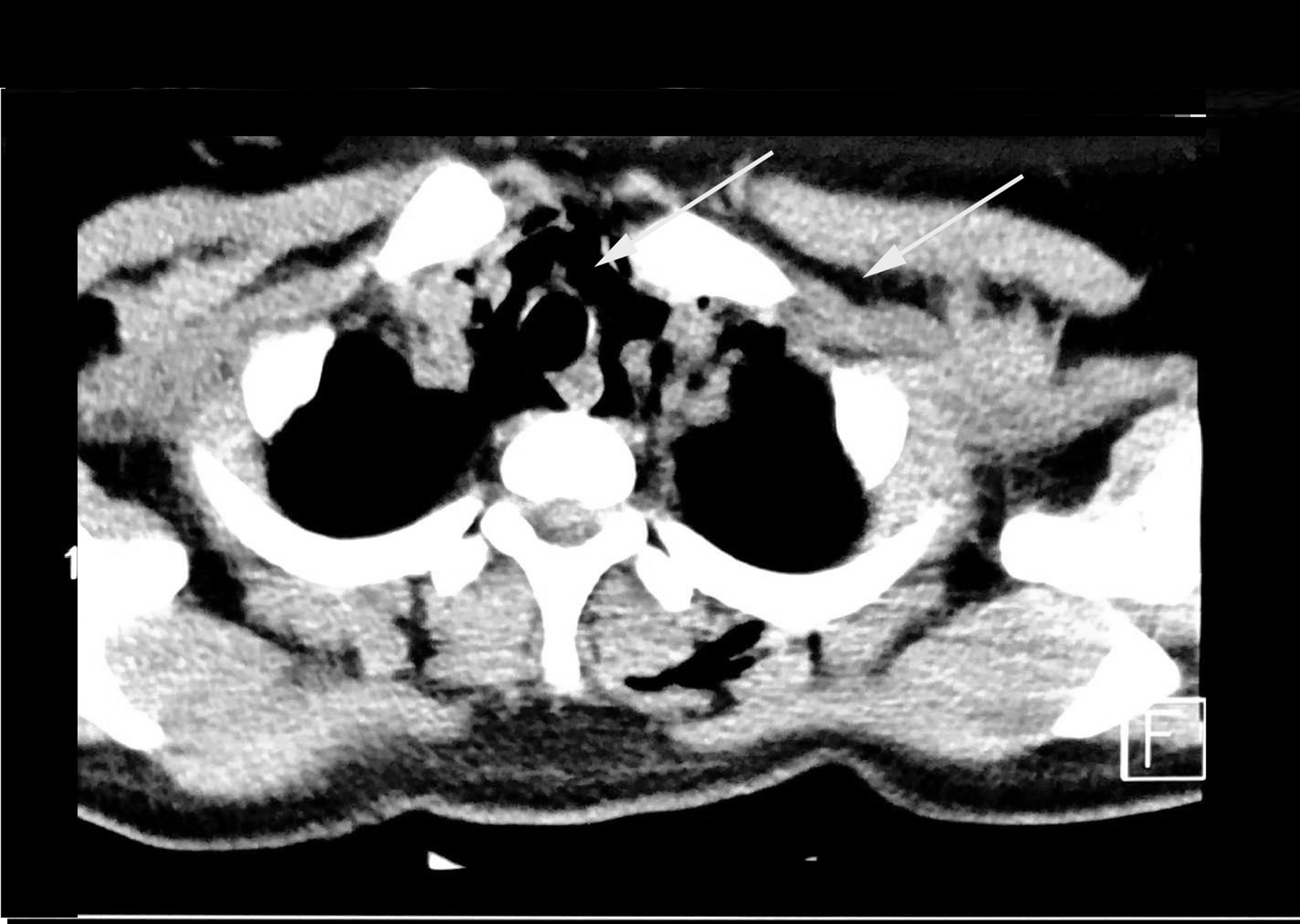
Chest computed tomography in the axial plane (at the level of the apex of the lungs) showing mediastinal and cervical emphysema (arrows).

Abdominal computed tomography scan in the axial plane showing abdominal-wall subcutaneous emphysema in the left flank (arrow).

Chest computed tomography scan in the axial plane showing chest-wall subcutaneous emphysema (arrows).
Findings
A diagnostic laparotomy was performed. After making a midline incision and abdominal exploration, there were no intra-abdominal findings, but her sigmoid colon was adhering to her abdominal wall in the left corner of a previous Pfannenstiel incision. Therefore, the cesarean section incision line was opened and, when the surgeons reached the fascia, malodorous secretions (resembling dishwater) were noted. On a corner of the Pfannenstiel incision, where the sigmoid colon was attached to the fascia, there was a small laceration in that colon; this made colonic content drain into the muscular and subcutaneous layers, as would occur in a colostomy retraction.
The sigmoid colon was released from the fascia. In a reevaluation, it was noted that the colonic laceration was ∼10 mm in size. Given that there was no contamination in the abdomen and the laceration was minimal, the colon was repaired with several Gambee stitches. Then, another incision was made in the lateral abdomen of the skin and subcutaneous tissue. After elevating the soft tissue and debridement of necrotic muscles, vigorous irrigations were performed, the omentum was placed over the intestines, and the abdomen was left open for managing as an open abdomen.
Due to the presence of emphysema in the mediastinum, after changing the surgical instrument set, a collar incision was made to drain the mediastinum from the neck. There were no pus and signs of infection or inflammation in the mediastinum. Then, the wound caused by the surgical incision at the neck was closed. Abdominal-wound irrigation, debridement, and dressing were performed twice per day in the operating room.
Results
Granulation tissue gradually appeared at the incision line over the intestine and lateral wound. At this point, the skin in the upper and lower parts of the midline incision were approximated, and a lateral wound was left open for spontaneous closure. A few days after the surgery, the subcutaneous and mediastinal emphysema disappeared.
The patient was discharged with instructions to take a daily bath and was also given instructions for dressing the lateral surgical wound. After 4 weeks, the lateral surgical wound had closed.
Discussion
Neck emphysema and pneumomediastinum emphysema are complications that occur for various reasons, including head-and-neck surgery and trauma. 8 In particular, subcutaneous and mediastinal emphysema can be a very rare symptom of colonic perforation. In this condition, despite the presence of colonic perforation, diagnosis and treatment can be delayed due to the absence of peritoneal inflammation and typical complaints.12,13
Cesarean sections are among the most-common surgical procedures increasingly performed around the world. Occurrences of adhesion bands in repeat cesarean sections and intestinal injuries have been seen, although an optimal technique for cesarean sections still exists. 3
Intestinal perforation, a sufficient pressure gradient between the intestinal duct and the subcutaneous space, and the anatomical location of the perforation are among the most important factors leading to subcutaneous emphysema with diffusion from the gastrointestinal (GI) tract.7,14 In the current case, the patient's sigmoid colon was attached to the incision site by an adhesion band and, due to reincision, a laceration was created at the point where the colon was attached to the old incision site, and the sigmoid colon was also still connected to the abdominal wall. After closure of the Pfannenstiel incision, formation of a dead space in Camper's fascia along with tissue movement in the wound area leads to an increased pressure gradient and development of emphysema.
Yet, it seems that the association between the endothoracic fascia and Camper's fascia in the abdomen can be considered as a transmitter of emphysema from the subcutaneous to mediastinum spaces. More importantly, the presence of free communication of the neck with the retropharyngeal, carotid, and visceral spaces—as well as with the mediastinal space can be considered as a causative agent of pneumomediastinum. 15
In a case report and review study by Oetting et al., 14 although the exact cause of perforation was different, intestinal perforation in different parts of the intestine was considered as a causative agent of emphysema. The most-common causes of emphysema with GI origin among the cases reviewed by Oetting et al. 14 and Muronoi et al. 12 were gastric ulcer and diverticulitis, respectively. Other reported factors included intestinal traumatic perforations, appendicitis, carcinoma, ulcerative colitis, stercoroma, and gastric and rectal cancer. Similar to the case described here, in that review study, the most commonly reported perforation site was the sigmoid colon.
Several case reports were reviewed by Muronoi et al. 12 In these cases, retroperitoneal emphysema was also reported, while, in the current case, unlike previous reported cases, no gas was seen in the retro- or intraperitoneal spaces. It seems that because the fistula created between the intestine and the abdominal wall had no leakage to the abdominal space, no retro- or intraperitoneal emphysema occurred.
None of the cases reviewed by Oetting et al. 14 and Muronoi et al. 12 showed signs of infection and generalized peritonitis before the onset of the occurrence of emphysema. Similar to previous case reports, in the present case, there was no evidence of infection and generalized peritonitis during preoperative examinations.
Similar to a case reported by Lee et al., 15 in the present case, respiratory distress along with difficulty in swallowing, was noted, which could be related to the cervical and mediastinal emphysema.
For managing mediastinal emphysema, in numerous cases reviewed by Muronoi et al., 12 only a chest drain was placed. In the current case, the pneumomediastinum emphysema was managed by drainage through a collar incision.
Conclusions
Adhesion bands are among the complications of various surgical procedures, including cesarean section. Considering the high probability of delivery through cesarean section in patients who have had previous cesarean sections, this can increase the possibility of damage to the intestines in the presence of adhesion bands—especially in cases when cesarean section is of an emergency nature—that might be missed and involve severe consequences to the patient. In this situation, if subcutaneous emphysema occurs, given that it might not be associated with peritonitis and other symptoms, the damage may be misdiagnosed. Therefore, in these cases, in the event of cervical subcutaneous and mediastinal emphysema following various surgical procedures, the possibility of perforation and emphysema with an intestinal origin should be considered.
Footnotes
Author Disclosure Statement
No financial conflicts of interest exist.
Funding Information
No funding was received for this article.