




Abstract
Background:
Pure osteosarcoma arising from the uterus is extremely rare. It is difficult to diagnose and predict disease progression with blood markers, because of the rarity. In this study, we describe a case of pure osteosarcoma arising from the uterus, which showed that alkaline phosphatase (ALP) was more sensitive than lactate dehydrogenase (LDH) for the diagnosis and observation of disease development.
Case:
A 78-year-old woman was referred to our hospital due to calcification of the uterine mass. Pelvic magnetic resonance imaging revealed a mass lesion (10 cm in diameter) showing a low-intensity area on both T1- and T2-weighted images. Positron emission tomography/computed tomography revealed 18-fluoro-2-deoxy-
Results:
Two weeks postsurgery, some metastatic regions arose. Along with metastatic progression, ALP levels were continually elevated, whereas LDH did not show a constant tendency.
Conclusion:
The novel finding of this report is that ALP might be more sensitive and useful than LDH in diagnosing osteosarcoma arising from the uterus and aid in evaluating the recurrence and disease progression. (J GYNECOL SURG 36:205)
Introduction
Pure osteosarcoma arising from the uterus is extremely rare with only about 20 previously reported cases. Computed tomography (CT) and magnetic resonance imaging (MRI) with contrast enhancement are commonly used for the diagnosis of this kind of uterine tumor. 1 Osteosarcoma arising from the uterus was described as a solid uterine tumor with calcification and it is often difficult to differentiate from uterine myoma with calcification. In blood samples, lactate dehydrogenase (LDH) is used in the diagnosis of uterine sarcoma. 2 In the field of orthopedic surgery, there is no unanimous agreement on the usefulness of both LDH and alkaline phosphatase (ALP) for the diagnosis and prognosis of osteosarcoma.3–5 However, ALP is considered to be more sensitive and accurate for the diagnosis of osteosarcoma arising from extremities compared with other tumor markers such as LDH and cancer antigen 125 (CA125). 6 Therefore, ALP tends to be used primarily as a prognostic marker for the recurrence of the disease. However, it is still controversial whether LDH and/or ALP could be used as accurate blood markers to reflect the disease development and recurrence of uterine pure osteosarcoma, as it rarely arises from the uterus.
In this article, we describe a case of pure osteosarcoma arising from the uterine body, which showed that ALP was more accurate and sensitive than LDH for the diagnosis and determining the disease development.
Case Report
A 78-year-old woman, gravida 2, para 2, presented with lower abdominal pain that started a month before visiting our hospital. She had previously undergone transthoracic esophagectomy with radical lymphadenectomy due to esophageal cancer when she was 77 years of age. Pelvic MRI revealed a mass lesion (10 cm in diameter) showing a low-intensity area on both T1- and T2-weighted images. The T2-weighted images showed a high-intensity area inside the mass lesion, suggesting a necrotic change. In addition, the mass lesion showed the contrast effect broadly in the tumor (Fig. 1a, b). CT imaging showed a mass lesion (10 cm in diameter) with calcification in the pelvis and some disseminated lesions with calcification in the abdominal cavity (Fig. 1c, d). Positron emission tomography/CT (PET/CT) revealed 18-fluoro-2-deoxy-
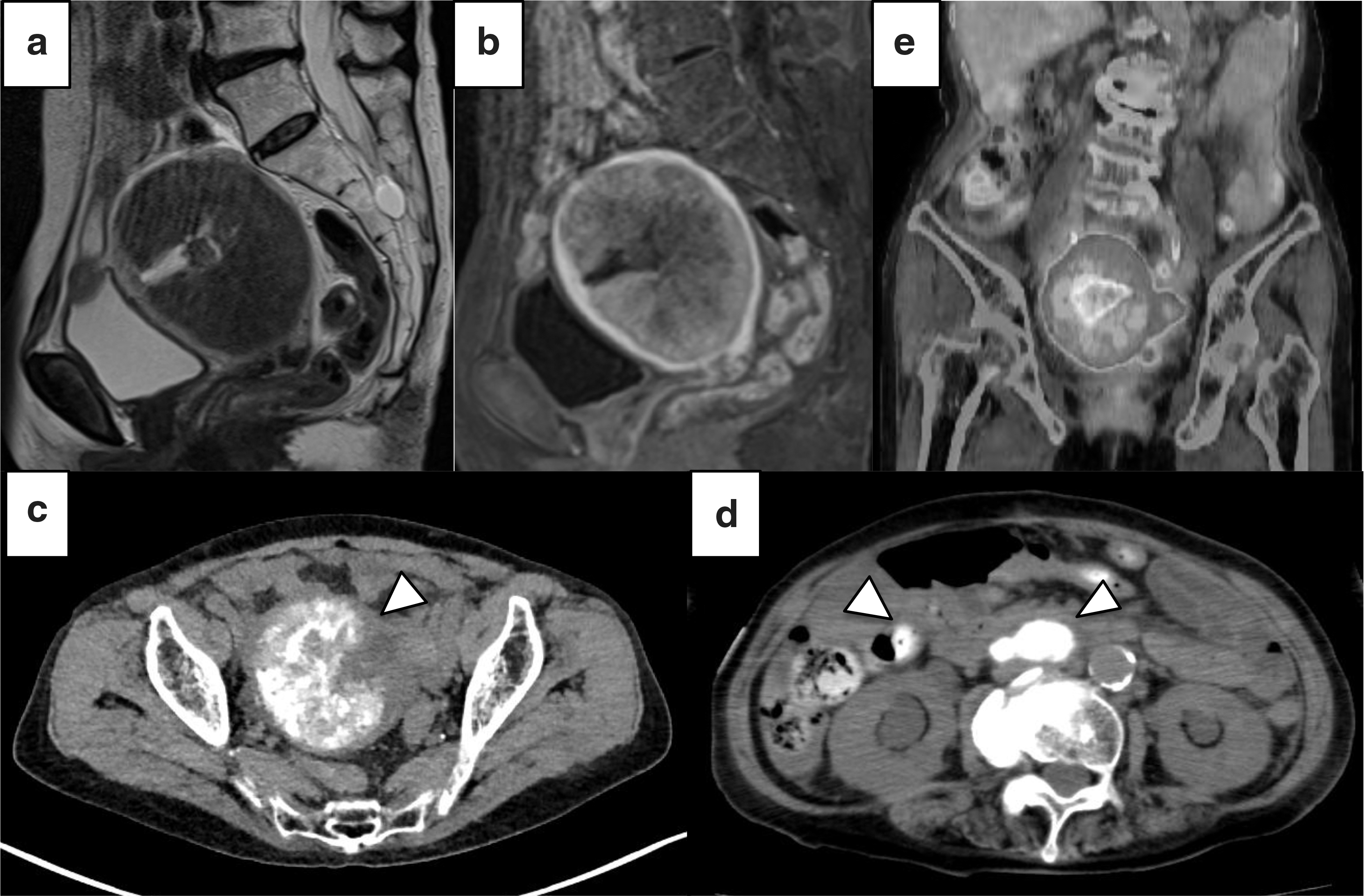
Imaging findings.
Macroscopically, the uterus (10 × 11 × 7.5 cm in size) was markedly swollen and the endometrial cavity was narrowed by the uterine corpus tumor protruding into the lumen. The tumor was a hard mass (9 × 9 × 6 cm in size) and most of the uterine corpus was replaced by the tumor (Fig. 2a). Histologically, the dense proliferation of atypical spindle cells with irregular size and admixture of osteoclast-type giant cells and with neoplastic bone formation were noted in all tumor tissues (Fig. 2b, c); however, epithelial structure was not noted. Immunohistochemical staining revealed neoplastic spindle cells positive for vimentin (Fig. 2d) and cyclinD1 (Fig. 2e) but negative for cytokeratin (Fig. 2f). The ascites cytology was positive. The final diagnosis was pure osteosarcoma arising from uterine corpus.
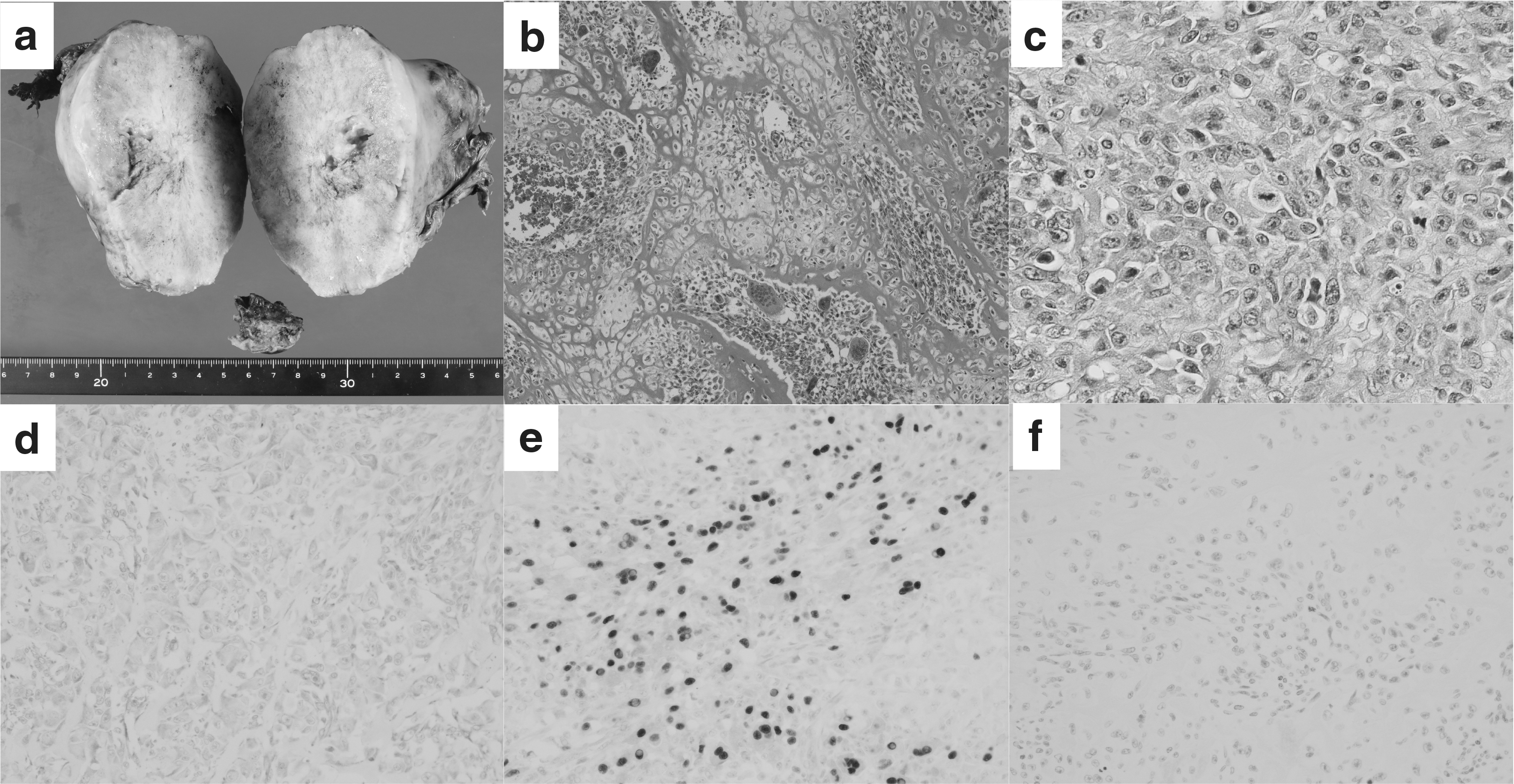
Pathological findings.
She was designated stage IV (pT1bNxM1) after surgery. Two weeks after the debulking surgery, we found many recurrent lesions with calcification in the abdominal cavity and lung (Fig. 3a, b). Laboratory data showed that ALP was elevated constantly along with the spread of metastatic osteosarcoma, whereas LDH did not show constant elevation (Fig. 3c). The patient and family had no desire for adjuvant chemotherapy and the patient died 40 days after surgery due to progression of the disease.

Time course of the imaging findings and laboratory data.
Discussion
Generally, osteosarcoma arising from the uterus is extremely rare and its aggressive nature leads to a poor prognosis (mean survival, 8.1 months; range, 20 days–37 months) 7 compared with osteosarcomas arising from other primary organs such as extremities. In the gynecologic department, measuring levels of LDH is commonly used for the diagnosis and evaluation of the recurrence and therapeutic effect in uterine sarcoma. 2 However, in this case, the LDH level did not show constant elevation in relation to the metastatic spread. This finding showed that LDH was not an accurate serum marker to evaluate the disease aggression in osteosarcoma of the uterus. In the field of orthopedic surgery, ALP is considered to be more sensitive and accurate for the diagnosis of osteosarcoma arising from extremities than LDH. 6 Kim et al. reported that the sensitivity and specificity of ALP for diagnosis were 53.2% and 90.1%, respectively. 6 In addition, the specificity of ALP for metastasis was 78.2% at 15 months postoperatively and 90.0% at 3 years postoperative. This finding is thought to be based on the organ specificity of ALP, whereas LDH elevation is caused by the systemic abnormality. The specificity of the ALP for the osteosarcoma derived from the ALP3 elevation, which was one of the isozyme reflected the bone component. In this patient, the ALP3 ratio was elevated in 78% (normal range: 25%–59%). In this case, the ALP level changed constantly according to the disease aggression from the preoperation period to the period after recurrence. To the best of our knowledge, this is the first article showing the benefit of measuring ALP in the diagnosis and prediction of osteosarcoma arising from the uterus and its aggressiveness. This ALP trend suggests that ALP might be a more sensitive and accurate serum marker in diagnosing pure osteosarcoma arising from the uterus, as well as osteosarcoma arising from other organs such as in extremity bones.
In conclusion, serum ALP may be more sensitive and useful than LDH for the diagnosis of osteosarcoma arising from the uterus, and for evaluating the recurrence and disease aggression.
Footnotes
Acknowledgment
We thank Dr. Masaharu Fukunaga, Department of Pathology, Shinyurigaoka General Hospital, for providing the advice of the pathological findings.
Author Disclosure Statement
No competing financial interests exist.
Funding Information
No funding was received for this article.