
Abstract
Background:
Pyometra is an unusual condition commonly occurring in elderly postmenopausal females. Spontaneous perforation of pyometra is rare and challenging situation to diagnose for clinicians. A high index of suspicion is needed to make an appropriate diagnosis, as the presentation is generally vague and most patients are treated for acute abdomen, considering it as a surgical emergency. This article reports on 3 cases of spontaneous perforation of pyometra at the Post Graduate Institute of Medical Sciences, Rohtak, Haryana, India..
Cases:
Retrospective analysis of the medical records of 3 cases of pyometra perforation are presented. These cases of spontaneous perforation of pyometra are described in this article.
Results:
All 3 patients underwent exploratory laparotomy followed by hysterectomy, bilateral salpingo-oophorectomy, and peritoneal lavage. Postoperative extensive care in intensive care unit (ICU) and broad spectrum antibiotics helped the patients' recovery.
Conclusions:
Spontaneous perforation of pyometra should be considered as a differential diagnosis when a postmenopausal woman presents with an acute abdomen. Timely detection and early intervention can prevent associated morbidity and mortality that can occur with perforated pyometra.
Introduction
Pyometra is an unusual condition commonly occurring in elderly postmenopausal females. Spontaneous perforation of pyometra is a rare and challenging situation to diagnose for clinicians. 1 A high index of suspicion is needed to make an appropriate diagnosis, as the presentation is generally vague and mostly patients are treated for acute abdomen, considering this condition as a surgical emergency. This article presents a series of 3 cases of spontaneous perforation of pyometra at the Post Graduate Institute of Medical Sciences (PGIMS), in Rohtak, Haryana, India.. All 3 patients underwent exploratory laparotomy, followed by hysterectomy, bilateral salpingo-oophorectomy (BSO), and peritoneal lavage. Postoperative extensive care in the intensive care unit and broad-spectrum antibiotics helped these 3 patients to recover.
A PubMed literature search was conducted and articles were reviewed including the current authors' cases for clinical presentation, management, associated etiology, and outcomes, which had been described.
Cases
This retrospective analysis was conducted to evaluate the clinical presentation, management, and outcomes of patients presenting with acute abdomen after spontaneous perforation of pyometra at a tertiary-care center in India. The medical records of all the patients who underwent surgery for pyometra perforation in our institution (PGIMS) during 2010–2019 (9 years) were reviewed. There were 3 patients. Their clinical records were studied to analyze and compile data on their cases.
Case 1
A 60-year-old, parous postmenopausal female presented in the emergency department of PGIMS with a history of abdominal pain and vomiting for 5 days. Her pain was intermittent, nonradiating, and noncolicky. The vomiting was nonprojectile, frequent, and constituted of food particles. She gave a history of a vaginal discharge that was yellow and foul-smelling. There was no history suggestive of any bowel trouble nor was there any urinary problems. She had no history suggestive of intrauterine copper device insertion, dilation and curettage, postmenopausal bleeding, or any sexually transmitted diseases. She was a chronic smoker.
On examination, she was conscious and her vitals were stable. Bilateral basal crepitations were present on chest auscultation. Her cardiovascular system was normal. On abdominal examination, there was distension with diffuse tenderness, guarding, and rigidity. Bowel sounds were absent. Speculum examination revealed a cervix flush with the vault, with slight bleeding from her vagina. Bimanual examination revealed that her uterus was soft with slight tenderness in the fornices.
Blood testing revealed mild anemia with leukocytosis. Her blood urea was 113 mg/dL, serum creatinine was 2.3 mg/dL, and serum electrolytes were normal.
Sonography showed that her abdomen was flatulent with free fluid and her uterus had minimal fluid in the endometrial cavity. Bilateral adnexa were normal. On a computed tomography (CT) scan, the uterus had hypodense areas. She underwent an emergency laparotomy as perforation peritonitis was suspected.
Operative findings revealed a uterine perforation present at the fundus with foul-smelling pus coming out of the perforation. Approximately 100 cc of pus was present in the peritoneal cavity and pus flakes were present on all guts. On exploration, however, her gut was normal. Subtotal hysterectomy with BSO was performed along with peritoneal lavage. The patient's abdomen was closed with an abdominal drain in situ.
She subsequently developed wound sepsis, which was managed by injectable antibiotics and serial dressings. Resuturing was done on the 30th postoperative day. She also developed shortness of breath on the 4th postoperative day and was found to have restrictive lung disease on high-resonance CT; the condition was managed by a pulmonologist.
On histopathology, the uterine corpus showed suppurative inflammation with gangrenous changes and gangrenous slough was replacing the endometrial cavity. Bilateral tubes and ovaries showed chronic nonspecific salpingo-oophoritis.
She was subsequently discharged in a healthy state.
Case 2
A 58-year-old postmenopausal multiparous woman came to the emergency department of PGIMS with complaints of fever and pain in her abdomen that was nonradiating and noncolicky for 7 days. Her fever was insidious in onset, of a moderate grade associated with chills and rigor. She had a history of a yellow discharge from her vagina for the last 10 days. Her gynecologic history was unremarkable.
On examination, she was febrile with temperature of 102°F, a pulse rate of 120 beats per minute (bpm), and a blood pressure (BP) of 130/86 mm Hg. Her chest was normal. Abdominal examination revealed abdominal distention with tenderness, guarding, and rigidity. Bowel sounds were absent. On speculum examination, the cervix and vagina was normal. On vaginal examination, the uterus was ∼12 weeks' size with tenderness in all fornices.
She had mild anemia. Her total leukocyte count was 14,000/cm of blood. Liver and renal function tests yielded normal results. Sonography of her abdomen showed that her uterus was enlarged with thick liquid and air content in the endometrial cavity. A contrast-enhanced CT of her abdomen revealed a ruptured uterus with pyometra, with a few foci of gas in the pelvis (Fig. 1).
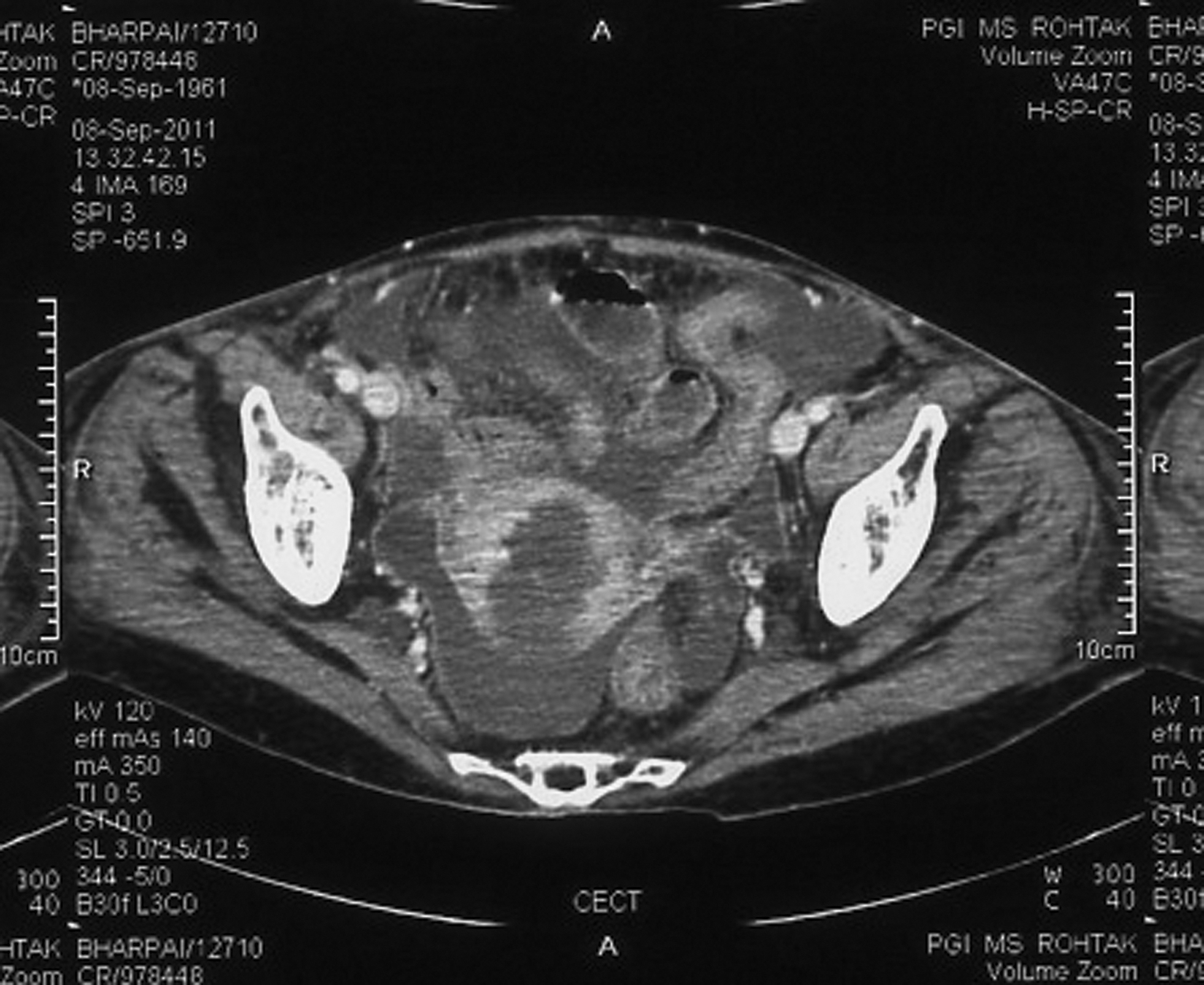
Computed tomography image showing perforation of a uterus.
She underwent an emergency laparotomy because pyometra perforation peritonitis was suspected. On laparotomy, her uterus was enlarged to a 10-week size and purulent fluid oozed from a uterine perforation on the posterior surface of her uterus ∼1 cm below the fundus (Figs. 2 and 3). Pus was present in the peritoneal cavity and all viscera was covered with pus flakes. Her gut was healthy. Subtotal hysterectomy with peritoneal lavage was done and her abdomen was closed with an abdominal drain in situ.

Operative finding of perforation of a uterus on its posterior surface.

Cut section of a subtotal hysterectomy specimen.
On postoperative day 5, this patient developed a fever and respiratory distress with bilateral basal crepitation. She was shifted to the intensive care unit (ICU) and given broad-spectrum antibiotics. She developed wound dehiscence on day postoperative day 10.
Histopathologic examination of the excised specimen revealed that the endometrium was infiltrated with acute inflammatory cells and granulation tissue with no evidence of malignancy
Case 3
A 60-year-old, postmenopausal parous woman presented to the emergency room of PGIMS with a fever and abdominal pain for last 10 days. Her pain was intermittent, nonradiating, and noncolicky. The fever was associated with rigor and chills. There was a history of mild abdominal distension for 3 days.
On examination, she was dehydrated with a pulse rate of 110 bpm, a BP of 90/60 mm Hg, and a respiratory rate of 20 inspirations per minute. Complete blood count testing revealed mild anemia with leukocytosis. The chest and cardiovascular system appeared normal. On abdominal examination, there was slight distension with diffuse tenderness, guarding, and rigidity. Bowel sounds were absent. On speculum examination, her ectocervix appeared normal. Vaginal examination revealed a soft uterus of normal size with slight tenderness in all fornices. Ultrasound revealed that her abdomen was flatulent with free fluid, and her uterus had a minimal amount of fluid in the endometrial cavity. An X-ray of the abdomen show that it was normal. Because of the absence of flatulence under her diaphragm, perforation of her uterus was ruled out.
Within 6 hours of admission, the general condition of this patient deteriorated, and she started gasping for breath. She was shifted to the ICU. However, her condition continued to deteriorate. CT scanning of her abdomen and pelvis were done. There was a perforation in her uterus with multiple pelvic and intra-abdominal collections of flakes. Laparotomy was planned. Operative findings revealed a perforation measuring 1.1 cm on the posterior side of the uterine fundus sealed by pus flakes with multiple pelvic and abdominal adhesions. Total abdominal hysterectomy (TAH) with a BSO were performed, along with peritoneal lavage
Cut-section specimens of her uterus and cervix did not reveal any associated pathology.
Histopathologic examination of the surgical specimens had features of a necrotic endometrium with no evidence of malignancy.
Results
All 3women were postmenopausal and each presented as a case of acute abdomen in the PGIMS emergency department. After preoperative assessment, laparotomy was done in all 3 patients, considering them as pyometra perforation peritonitis and, only in 1 case, the preoperative diagnosis of pyometra perforation was delayed. There was no associated malignancy found in any case.
Discussion
Pyometra is an accumulation of pus in the uterine cavity, mainly occurring in elderly postmenopausal women. It is an uncommon condition with an incidence of 0.01%–0.5% in gynecological patients. Pathophysiology of development of pyometra is mainly due to interference of natural drainage. 1
Causes of pyometra include:
Classical triad of symptoms associated with pyometra is postmenopausal bleeding, vaginal discharge and lower abdominal pain but none of these symptoms is specific for pyometra and some patients remain asymptomatic. 2
A PubMed literature search was performed on pyometra perforation in humans and data were collected. Table 1 summarizes 64 cases of perforation of pyometra to date, including the three cases in this current article.2–48 All cases were elderly females, postmenopausal, mostly above 60 years of age. The most-common presentation seen in almost all of these cases was abdominal pain (60 cases, 93%). Fever (21 cases, 32%) and vomiting (20 cases, 31%) are other common complaints. Four cases also had vaginal discharge at the time of presentation. Five cases presented as shock (3.2%). Genital bleeding occurred in 2 cases. The current 3 patients also presented with abdominal pain with either fever or vomiting.
Spontaneous Perforation of Pyometra from Literature Review and Current Cases
AP, abdominal pain; VD, vaginal discharge; DP, diffuse peritonitis; TAH, total abdominal hysterectomy; BSO, bilateral salpingo-oophorectomy; PGIT, perforation of gastrointestinal tract; cx, cervix; FIGO, International Federation of Gynaecology and Obstetrics; F, fever; V, vomiting; PP, pyometra perforation; VB, vaginal bleeding; SCC, squamous cell carcinoma; CC, cancer cervix; N, nausea; PMB, postmenopausal bleeding; AAP, acute abdominal pain; STH, subtotal hysterectomy; ND, not defined; SVH, supravaginal hysterectomy; RA, rheumatoid arthritis; DM, diabetes mellitus; HT, hypertension; CVA, cardiovascular accident; ICU, intensive-care unit.
The correct diagnosis of pyometra perforation has been rarely made preoperatively and the most common preoperative diagnoses were generalized peritonitis and perforation of gastrointestinal (GI) tract. Given that the patients presented with features of acute abdomen, a large number of these patients were misdiagnosed preoperatively as having gastrointestinal perforation or peritonitis. GI perforation was suspected preoperatively in 28 cases (43.7%), diffuse peritonitis in 18 cases (28.1%), appendicitis in 1 case (1.5%), pneumoperitoneum in 2 cases (3.1%), and mesenteric artery ischemia in 1 case (1.5%).
In 1 of the current cases because of the absence of air under her diaphragm, a diagnosis of perforation of the uterus was delayed after basic investigations and was suspected on seeing a CT scan. Vyas et al. also reported that multidetector CT with sagittal and coronal reformatted images may aid preoperative diagnosis of a ruptured pyometra. 21 Thus, it is suggested that a high index of suspicion and imaging studies by CT or magnetic resonance imaging is required for diagnosing a case of pyometra perforation. 11
There was documentation of associated malignancy in 22 cases of the 64 cases analyzed, with the most being carcinoma of the cervix (19 cases, 86%), and others were endometrial adenocarcinoma (1 case, 4.5%), rectal carcinoma (1 case, 4.5%), and sigmoid colon cancer (1 case, 4.5%). Other comorbidities associated with pyometra perforation were diabetes mellitus (6 cases), acute endometritis (1 case), fibroid uterus (1 case), and renal failure (1 case). Yildizhan et al. had also reported that the most-common etiology of pyometra is malignant diseases of the genital tract and the consequences of their treatment (radiotherapy). 3
The site of perforation was mentioned in 49 cases of 64 cases. The most-common sites of perforation were the fundus (39 cases, 80%), anterior surface of the uterus (12%), and posterior surface of the uterus (8%).
Preferred immediate treatment of suspicious pyometra perforation is urgent laparotomy and TAH with BSO with thorough drainage and irrigation of the abdominal cavity. Broad-spectrum antibiotics and intensive care postoperatively, followed by definite management according to etiology helps in fast recovery. Cervical cancer is managed according to stage of disease in consultation with a gynecologic-oncologist. 4 Of 64 cases, 58 patients (90%) underwent hysterectomy either complete or subtotal and 4 cases (6%) underwent drainage of pus and conservative management.
Prognosis was documented in 56 cases. Forty-two patients (75%) were treated and survived after extensive management of their perforation of pyometra. Fourteen patients died subsequently even after treatment, with the most-common cause being sepsis. Ikeda et al. also analyzed prognoses in 44 patients and found that 25% of patients died either because of pyometra perforation and its complications postoperatively or comorbidities associated with this condition. 13
Perforation of pyometra is a life-threatening condition and a high index of suspicion is needed to diagnose it, as its presentation mimics other surgical causes of acute abdomen. Aggressive treatment with laparotomy and removal of septic foci along with adequate coverage with broad spectrum antibiotics are required for speedy recovery.
Conclusions
Perforation of pyometra spontaneously is a very rare condition and it should be as considered as a differential diagnosis when a postmenopausal woman presents with an acute abdomen. Timely detection and early intervention can prevent the associated morbidity and mortality.
Footnotes
Funding Information
No funding was received for this article.