




Abstract
Objective:
This research compared the effectiveness of Papanicolaou (Pap) smears with biopsies for diagnosing atypical squamous cells of undetermined significance (ASCUS) accurately in postmenopausal women. Another goal was to determine the diagnostic value of Kiel(Ki)–67 expression to exclude negatives in atypical Pap smears of postmenopausal women.
Materials and Methods:
A prospective study was conducted in the departments of gynecology and pathology of the Indira Gandhi Medical College and Research Institute, in Puducherry, India. Eighty postmenopausal women were examined with liquid-based cytology (LBC), and this was compared with conventional cytology for detection of histologically confirmed cases. Ki-67 immunocytochemistry (ICC) was performed on atypical smears of postmenopausal women to determine its diagnostic value for detecting abnormal smears.
Results:
There was a reduction in the frequency of ASCUS in LBC, compared to conventional Pap smears in postmenopausal women. The specificity of LBC (82.7%) was higher than conventional Pap smears (51.7%) for diagnosing ASCUS accurately. Ki-67 ICC staining in atypical smears showed 100% sensitivity and specificity as diagnostic biopsy shows.
Conclusions:
In comparison to a conventional Pap smear, a liquid-based Pap smear has high specificity, and Ki-67 expression with LBC can exclude false positivity, which can substantially reduce diagnostic biopsies and subsequent Pap smears in postmenopausal women with atypical Pap smears.
Introduction
Cervical cancer is mainly implicated in developed countries and is the second most-frequent cancer among women in low-income and lower-middle-income countries. 1 In developing countries, such as India, cervical cancer is one of the most-common cancers, and nearly 74,000 deaths occur due to this disease. Yet, it is a preventable cancer that can be diagnosed early before patients present with invasive carcinomas.2,3 George C. Papanicolaou, MD, developed the Papanicolaou (Pap) test, which is the cytologic examination used to identify precancerous lesions of the cervix. Even today, it is the most simple and cost-effective screening tool. However, the conventional Pap test has limitations due to the presence of mucus, blood cells, and overlapping epithelial cells, which can reduce the test's specificity.
To overcome these limitations, an alternative method—liquid-based cytology (LBC)—was developed.4,5 In postmenopausal women, tissue atrophy occurs due to decreased estrogen levels in the body. This often yields inadequate cervical smears and might also mimic atypical squamous cells of undetermined significance (ASCUS) or low- and high-grade cervical intraepithelial neoplasias that also produce high false-positive results in precancerous conditions.6,7 LBC is an advanced technique that enables cells to be suspended in a liquid medium that can be spread in a monolayer after centrifugation. This has the advantage of improving specimen adequacy and detection of precursor lesions, compared to conventional Pap smears.
The residual cell suspension can also be used for ancillary techniques, such as immunocytochemistry (ICC).8,9 Commonly seen cytologic abnormalities in postmenopausal women are ASCUS on Pap smears. The possibility of false-positive results are high in postmenopausal women. This has a negative impact on their lives and will burden these patients unnecessarily during follow-up procedures. 10 In abnormal conditions, such as dysplasia and carcinoma, Kiel(Ki)–67 protein is overexpressed in the upper third of the basal epithelium; thus the number of positive cells increases. 11 Ki-67 proteins present on proliferating cells, and the percentage of cells with positive nuclei is a reliable tool for rapid evaluation of the growth fraction. ICC investigation of Ki-67 expression in a prepared Pap smear is a simple noninvasive, safe, and cost-effective method that can reduce diagnostic biopsy and follow-up Pap smears in postmenopausal women with atypical Pap smears.11,12
Many studies have been conducted to compare the two Pap methods, but very few studies have been done with postmenopausal women. This article highlights the advantage of LBC smears and the significance of Ki-67 application for detecting precancerous and cancerous lesions in postmenopausal women.
Materials and Methods
This prospective study was approved by the Ethics Committee on Human Research at the Indira Gandhi Medical College and Research Institute, in Puducherry, India, and was conducted in the departments of gynecology and pathology at this institute for 2 years.
Eighty postmenopausal women attending the outpatient department with various complaints were included in the study. After obtaining written consents, cytologic and biopsy specimens were collected from the patients. Most of them had undergone hysterectomy for various other reasons and, in some patients, colposcopic biopsies had been performed.
Cytologic specimens were collected before surgery. Each patient was placed in a modified lithotomy position. The cervix was exposed with a bivalve vaginal speculum. An endocervical brush (U Prep LBC) was inserted into the endocervical canal, rotated 360°, and the sample obtained was placed on a slide to prepare a conventional Pap smear. Then the brush was detached and placed into a vial containing fixative and the vial was closed and shaken to produce homogeneous mixing. Two mL of the separator solution given by the manufacturer and 5 mL of the sample were centrifuged at 2100 rpm/minute for 10 minutes. The cell button was taken and smeared on another slide. The slides were fixed further by dipping them in 95% alcohol for 15 minutes and staining them with a Pap stain. All slides were examined under a light microscope. Classification of cytology was performed according to the Bethesda system of 2014. The final cytologic diagnosis was confirmed by consensus of 2 independent cytopathologists.
ICC analysis was performed directly on slides with manual LBC smears previously stained with the Pap technique. Only the slides with a sufficient amount of cells were subjected to ICC analysis. The ICC procedures were performed after removing coverslips from the slides in xylene and then rehydrating the smears in decreasing concentrations of ethyl alcohol and distilled water. Antigen retrieval was performed using a 10-mM concentration of citrate buffer (pH 6.0) in a pressure cooker for 4 minutes. The slides were allowed to cool until they reached room temperature and then were subjected to immunostaining. ICC analysis of samples was conducted using an anti-Ki-67 antibody incubated for 30 minutes. The secondary antibody was added for 25 minutes. A streptavidin-biotin complex reagent was added for 25 minutes and the formed solution was placed under a diaminobenzidine–hydrogen peroxide solution. Samples were washed using phosphate saline.
After light counterstaining in Harris's hematoxylin, the slides were mounted and analyzed using a light microscope. The number of Ki-67 immunoreactive cells per 1000 cells was counted and their percentage was calculated. The cells were considered as positive, when their nuclei became stained in spotted form or homogeneously. Later, the diagnosis was compared with the histopathologic reports of all the patients with abnormal atypical Pap smears.
The data were analyzed using Statistical Package for the Social Sciences software, trial version 20. Quantitative data were presented as mean ± standard deviation. The validity (accuracy) of each test was assessed for sensitivity and specificity. The diagnostic power of the test was assessed for predictive accuracy.
Results
In the present study, 80 postmenopausal women were randomly included. Of 80 smears, 20 smears were inadequate and lost during follow-up. Sixty smears were included as the final study population. Of the 60 smears, 30 with normal cervical cytology were taken as quality controls for Ki-67 ICC. The mean age of presentation in the current study was 58.7 years, and the ages ranged from 45 to 88. The predominant symptom noted was uterine prolapse followed by postmenopausal bleeding. The percentage of various symptoms presented by patients is shown in Figure 1.
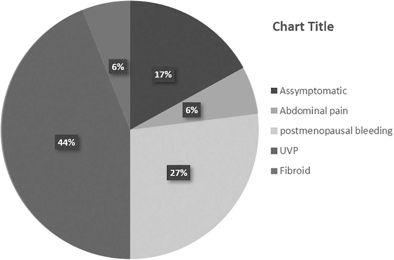
Distribution of cases in relation to various presenting complaints. UVP, uterovaginal prolapse.
Among the 60 postmenopausal women of the study group, conventional Pap smears and liquid-based cytology smears were compared (Fig. 2). The 30 patients with the diagnosis of ASCUS were evaluated further by examining LBC smears. Only 12 of these showed abnormalities. Of these 12, 11 had ASCUS and 1 had a high-grade squamous intraepithelial lesion (HSIL). All of these patients underwent biopsy—the gold standard for diagnosis. The sensitivity of both conventional Pap and LBC was 100%. The specificity of conventional Pap smears and LBCs were 51.7% and 82.7%, respectively. The positive predictive value (PPV) of conventional Pap smears was 6.67%; for LBC, PPV was 16.6%. The diagnostic effectiveness of conventional Pap smears was 53%; for LBC, it was 83%.
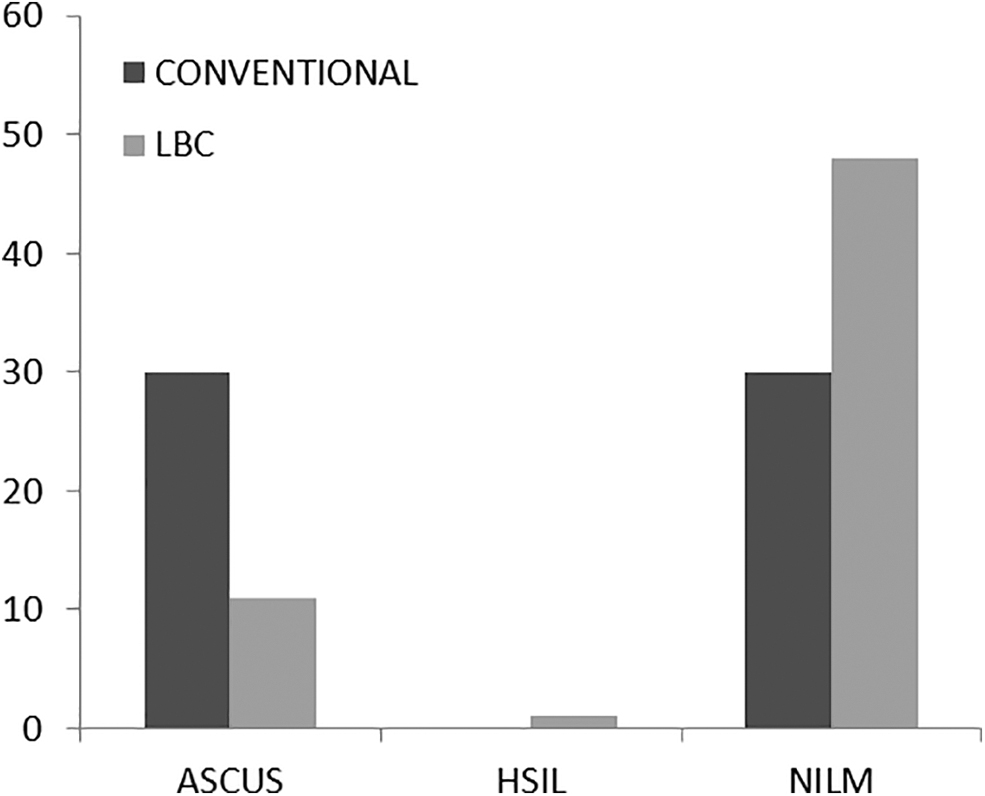
Comparison of microscopic findings in conventional Papanicolaou smear and liquid-based cytology.
Among 60 postmenopausal women, 30 smears were normal and were considered as the control group for ICC. The 30 smears with the ASCUS diagnosis on conventional Pap smears, for which LBC smears were performed, were considered as the test group for ICC. In both the test and control groups, the Ki-67 ICC was performed. Of the 30 subjects in the test group, only 2 smears were positive for Ki-67; all the control smears were negative for Ki-67.
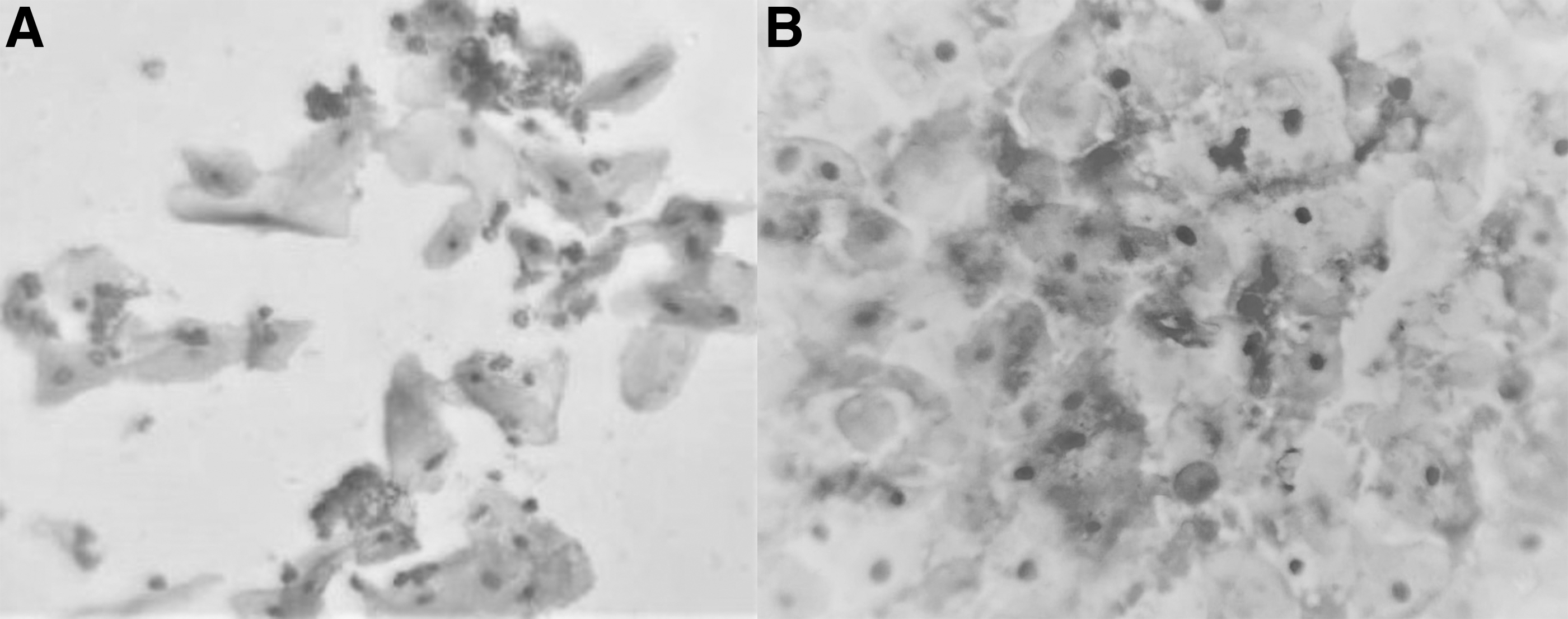
The smear from a 58-year-old woman who was diagnosed with ASCUS on a conventional Pap smear and as HSIL on LBC had 5% of cells showing positivity for Ki-67, which were later also diagnosed as HSIL on biopsy (Fig. 3). Another smear from a 62-year-old woman diagnosed as ASCUS on both a conventional Pap smear and LBC showed 20% positivity for Ki-67, which later turned out to be squamous-cell carcinoma on biopsy (Figs. 4 and 5). The sensitivity and specificity of Ki-67 ICC was 100%, in comparison with diagnostic biopsy, which is considered as the gold standard investigation (Fig. 6).
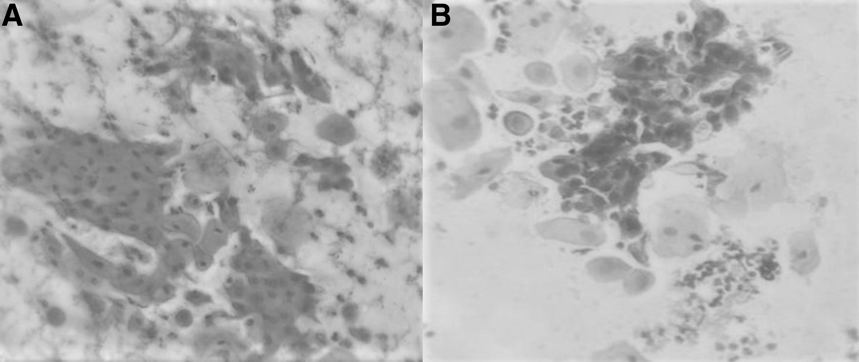
Microphotograph (Papanicolaou [Pap] stain; 10 × ) of conventional Pap smear
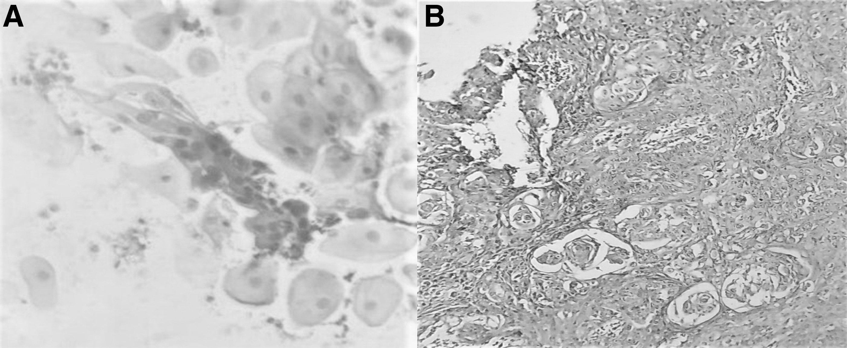
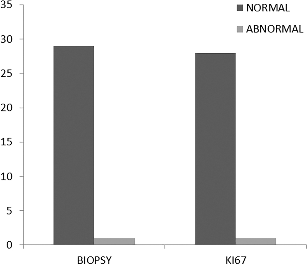
Comparison of Ki-67 immunohistochemistry and biopsy.
Discussion
The Pap smear examination is one of the best screening methods widely used for identifying early precancerous lesions. In postmenopausal women, because of atrophy, the yield of cells for Pap smears will usually be less, compared to other populations of women. The predominant population of cells in atrophic smears will be parabasal cells. Most of these smears might mimic those of neoplastic lesions of the cervix; thus it is a diagnostic challenge to diagnose normal and cancerous lesions accurately in Pap smears of postmenopausal women.12,13 Ki-67 protein is highly expressed in patients with dysplasias and carcinomas. ICC is a simple method that can be performed by testing for immunocytochemical expression of Ki-67 protein in abnormal smears to identify neoplastic lesions in postmenopausal women. This avoids unnecessary biopsies further in patients who indeed have false-positive Pap smears. 11
The mean age of presentation in the current study population was 58.7 ± 9 years. In a study by Akshatha et al., the mean age of the postmenopausal women was 53.2 ± 4.2 years. 14 The most-common complaint in the current study population was uterine prolapse, which accounted for ∼44% of the patients, followed by postmenopausal bleeding. In a study by Velu et al., predominant cases were asymptomatic followed by uterine prolapse. 15 A study conducted by Pooja et al. showed that a common complaint was a white discharge from the vagina, followed by postmenopausal bleeding. 16
The incidence of ASCUS is between 4% and 5%, and the incidence of squamous intraepithelial lesions is 17.2% in postmenopausal women. The incidence can be high in postmenopausal women who have not been screened by Pap tests during these women's pre- and perimenopausal ages.17,18 It is unlikely for a postmenopausal woman to acquire new infections due to atrophy of the cervical and vaginal cells and retraction of the transformation zone. 19
In the current study, many smears were reported as ASCUS when examined via a conventional Pap method due to the paucity of cells and unclear backgrounds. However, in LBC smears, because of the monolayer distribution of cells with clear backgrounds, many smears were deemed as normal. This shows that LBC detected more true-negative cases and eliminated false-positive cases shown on conventional Pap smears. A study by Shobana and Saranya also showed similar findings, with more ASCUS seen on conventional Pap smears, compared to LBC. 20 The conventional Pap smear with LBC showed 100% sensitivity; the specificity was 65% and 90%, respectively, in that study. Bolick and Hellman reported the sensitivity of conventional Pap smears and LBC to be 85% and 95.2%, respectively, and specificity was 36% and 58%, respectively. 21 Another study by Sherwani et al. showed sensitivity and specificity of LBC as 97.6% and 50%, respectively, and the conventional Pap smear as 53.7% sensitivity and 50% specificity. 22 Beerman et al. also showed increased sensitivity and specificity of LBC over conventional Pap smears. 23
LBC, in comparison to conventional Pap smears, can be useful for detecting more nonmalignant cells in patients treated for carcinoma of the cervix during their follow-up screenings. Singh et al. reported a PPV of 83.33% and a negative predictive value of 100% in cervical carcinoma during follow-up screening. 24 Yet, the rates of detection of epithelial abnormalities in both conventional and LBC methods was similar in a study by Pankaj et al. 25
The oncogenes of human papillomavirus cause premalignant and malignant lesions by affecting the host genes, such as p16 and Ki-67, involved in cell-cycle regulation and cellular proliferation. 26 Ki-67, named after its city of origin Kiel, is a nuclear nonhistone protein that helps RNA transcription of ribosomes during nuclein formation and regulates cell-cycle progression at multiple sites. Ki-67 expression can be seen in the normal basal layer, in HSIL, and in malignant lesions of the cervix.
However, benign endocervical cells, tuboendometrial metaplasias, or squamous metaplasias do not express Ki-67. The level of Ki-67 expression predicts the onset of squamous intraepithelial lesions (SILs) and malignancies.27–29 Several studies in the literature suggest higher specificity for Ki-67 application in differentiating HSILs and carcinomas from ASCUS and low-grade SILs in Pap smears of younger patients.30–32 In the current study, the sensitivity and specificity of Ki-67 ICC were 100% and the PPV was also 100%. A study by Fakhrjou et al. showed a specificity and a PPV of 99.3% and 100%, respectively, which were almost the same as those found in the current study. 11 Ejersbo et al. concluded that use of Ki-67 helped detect nonneoplastic lesions in atrophic samples and decreased false-positive results up to 86%. 33
Conclusions
In comparison to a conventional Pap smear, LBC has high specificity. However, differentiating atrophic epithelial cells and cells undergoing neoplastic changes based on cytomorphologic criteria might be very difficult in postmenopausal women and can lead to diagnostic as well as therapeutic errors. ICC for Ki-67 expression with LBC can exclude false positivity; this approach to testing can substantially reduce diagnostic biopsies and subsequent Pap smears in postmenopausal women with atypical Pap smears.
Footnotes
Acknowledgments
The authors acknowledge the faculty and paramedical staff of the departments of the pathology and gynecology of the Indira Gandhi Medical College and Regenix for their support.
BCS designed the study, collected the data, performed the literature search, provided microscopic pictures, and edited the article. SP examined the patients and collected smears from the patients. DM helped with statistical analysis and writing the article, RVB edited the article.
Author Disclosure Statement
No competing financial interests exist.
Funding Information
No funding was received for this research and article.