




Abstract
Background:
A lipoleiomyoma is a rare benign tumor that is comprised of both smooth-muscle and adipose tissue.
Case:
A 26-year-old nulligravida presented with pelvic pain, bloating, constipation, urinary urgency, and weight gain. Imaging revealed a 16-cm pelvic mass with differentials including lipoma, lipoleiomyoma, liposarcoma, and teratoma. Hoping to avoid a midline laparotomy, this patient was referred to a tertiary-care center where diagnostic laparoscopy identified this mass as ovarian in nature. She underwent a laparoscopic salpingo-oophorectomy, with specimen removal through an U.S. Food and Drug Administration–approved tissue-extraction bag. The lesion's pathology was consistent with a lipoleiomyoma.
Results:
This patient was able to be discharged to go home the day of the procedure and recovered without any issues. She was able to maintain fertility.
Conclusions:
Despite a differential including malignancy, minimally invasive surgical techniques can be utilized safely, especially in woman requesting fertility preservation.
Introduction
Pelvic masses are not uncommon in women of reproductive age. The prevalence of uterine fibroids has been documented to be as high as 70%–80% by age 50. 1 However, it is important to have broad differentials when evaluating patients with pelvic masses to ensure proper management. A lipoleiomyoma is a rare, benign tumor that is comprised of both smooth muscle and adipose tissue. 2 This article reports on a case of a young, reproductive-age woman with an ovarian lipoleiomyoma.
Case
A 26 year-old nulligravida presented to her gynecologist with several weeks of “bulk” symptoms. She reported having pelvic pain, bloating, constipation, urinary urgency, and feeling as though she had gained weight. One year prior, she had a levonorgestrel intrauterine device (IUD) placed and had been amenorrheic since that time. Examination revealed that she had a soft, nondistended abdomen with a 15-cm mobile mass in the midline extending from the pelvis to the umbilicus.
Transvaginal ultrasound showed an enlarged heterogeneous uterus measuring 17 cm × 4.7 cm with distorted anatomy. Follow up magnetic resonance imaging showed a 11.0 × 10.6 × 16.3–cm heterogeneous mass occupying the majority of the pelvis (Fig. 1). There was clear mass effect on the bladder, uterus, ovaries, and rectosigmoid colon with possible pedunculated origin from the left anterior uterine body, although the organ of origin could not be identified definitively. The IUD was confirmed to be in situ. Differential diagnoses included leiomyoma, lipoma, liposarcoma, lipoleiomyoma arising from the uterus, and mature/immature teratoma arising from the ovary.
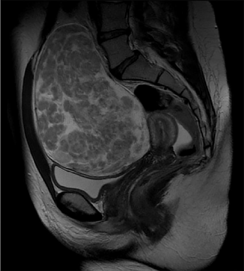
Sagittal magnetic resonance imaging showing a 11.0 × 10.6 × 16.3–cm heterogeneous mass occupying the majority of the pelvis.
This patient was initially scheduled for a laparotomy with her diagnosing gynecologist; due to her strong desire to avoid a midline laparotomy if possible, she was referred to a tertiary-care center for a second opinion. Her case was discussed with a gynecologic oncologist who supported the plan for an attempt at a minimally invasive procedure, given her young age and the noninvasive appearance of the mass. She was then scheduled for diagnostic laparoscopy and removal of the pelvic mass.
The abdomen was entered via an umbilical incision, using the open Hasson technique. Upon entry of the laparoscope, the patient was found to have a 16-cm right ovarian mass, which was solid, consisting of adipose-type tissue. The uterus, fallopian tubes, and left adnexa were normal in appearance. Two additional ports were placed, one in each lower quadrant of the patient's abdomen. There was no differentiable ovarian tissue, thus, a right salpingo-oophorectomy was necessary. The mass was placed in a 17-cm Applied Medical (Rancho Santa Margarita, CA) tissue-extraction bag and hand morcellated through a 4-cm Pfannensteil incision. The patient tolerated the procedure well and was discharged to go home that same day.
The specimen that was sent to pathology was a 1224-g multinodular tan–yellow mass with myxoid cut surfaces adherent to a grossly normal ovary. On microscopic examination, the lesion had a spindle-cell appearance consistent with a smooth-muscle neoplasm with admixed adipose tissue, supporting a final diagnosis of a lipoleiomyoma (Fig. 2).

Lipoleiomyoma, hematoxylin and eosin, 100 × .
Results
This patient underwent a minimally invasive, fertility-sparing procedure and was able to be discharged the day of her procedure. She recovered well without any issues.
Discussion
A lipoleiomyoma is an extremely rare benign tumor composed of both smooth-muscle and adipose tissue. This kind of tumor has been found in the uterine corpus, cervix, ovary, broad ligament, and retroperitoneum. 3 It is unclear if the current patient's mass was an ovarian primary or if it developed a parasitic blood supply from the right ovary after undergoing stalk necrosis from a uterine origin.
Most cases of lipoleiomyomas occur in postmenopausal women, and 90% of reported cases occur in women older than age 40. 4 There are two main theories regarding the pathogenesis of these tumors; it has been suggested that they arise either through (1) direct metaplasia of smooth muscle or through (2) metaplasia of pluripotent mesenchymal cells. 5 Because the vast majority of these cases occur in peri- or postmenopausal women, it is thought that estrogen deficiency might play a role in the abnormal intracellular lipid storage involved with these tumors.5,6 This current case is believed to be the youngest woman reported to have a lipoleiomyoma. Her youth exemplifies the need to consider other means of pathogenesis and to include these tumors in the differential diagnoses of pelvic masses in reproductive-age women.
Given their large size, increased prevalence in postmenopausal women, and adipose contents, lipoleiomyomas can mimic liposarcomas. The current case was discussed with the gynecologic oncologists at the current authors' institution when planning surgical management. Due to the patient's age and the fact that the mass was well-circumscribed and appeared to be noninvasive, suspicion for malignancy remained very low, and a minimally invasive approach was supported.
Previously reported cases of lipoleiomyomas, including those believed to have ovarian origin, have been managed with hysterectomy.2,4,7,8 Because the suspicion for malignancy was low, this case was managed with a salpingo-oophorectomy via laparoscopy and a minilaparotomy. The patient was discharged to go home on the day of surgery and recovered quickly. She was also able to maintain her fertility, which was important to her and which is very critical to many young women. The mass was removed via a small Pfannensteil incision, as opposed to an extension of the umbilical-port site, for cosmetic reasons.
It is reasonable to manage large lipoleiomyomas with exploratory laparotomy via midline incisions, given the uncertainty of the origin of these lesions. The minimally invasive surgical threshold varies widely based on surgeon experience/training. In this particular case, the current authors were prepared to perform a unilateral salpingo-oophorectomy, if the mass was significantly adherent to the ovary; or ligation of a stalk, if a pedunculated myoma was identified. The referral to a tertiary-care center afforded the patient the opportunity to have a minimally invasive procedure with the ability to convert to a more-extensive operation if necessary.
Conclusions
This case demonstrates the need to keep an open mind when evaluating a young woman with a pelvic mass. It is important to start with wide differentials and to consider minimally invasive and fertility-sparing management options if the suspicion for malignancy is low.
Footnotes
Author Disclosure Statement
No financial conflicts of interest exist.
Funding Information
No funding was received for this case report.