
Abstract
Background:
Elephantiasis is a rare disorder involving lymphatic channels of the affected part of the body. It is characterized by huge enlargement of the particular body part and likely to affect limbs, scrotum, and trunk. Genital elephantiasis, also known as esthiomene, is a rare dramatic end result of lymphatic obstruction. Although mainly associated with filariasis and sexually transmitted diseases such as lymphogranuloma venereum and donovanosis, it could also be an uncommon complication of tubercular lymphadenitis.
Case:
A 40-year-old multiparous woman presented with progressively increasing vulval swelling over a period of 3–4 years. The swelling was soft and huge. She was managed successfully by surgical excision with vulvoplasty. Other possible differential diagnoses were excluded, and ancillary tests were performed to reach a conclusive diagnosis of vulval elephantiasis on histopathology.
Conclusion:
Vulval elephantiasis due to filariasis is rare and diagnosis on histopathology is more often by exclusion.
Introduction
Elephantiasis is a chronic inflammatory condition due to obstruction of lymphatic channels characterized by gross enlargement of the particular body part commonly affecting limbs, breast, and trunk. External genitalia can also be involved and genital elephantiasis, also known as esthiomene, is a rare event. Although mainly associated with filariasis and sexually transmitted diseases such as lymphogranuloma venereum and donovanosis, it could also be due to tubercular lymphadenitis.1,2 Radical hysterectomy, pelvic lymphadenectomy, irradiation, tumor invasion, and direct trauma are less common causes. Females have lower incidence of filarial infection. Vulval elephantiasis due to filariasis is still rarer and a rough estimate of its incidence is ∼1%–2% of total cases of filarial elephantiasis. 3 It is difficult to make histopathologic diagnosis alone, as the parasite is usually not identified in tissue sections. Identification of microfilariae in night samples of peripheral blood or seropositivity for filarial antigen is requisite for the correct diagnosis. So here we are reporting a case of vulval elephantiasis due to filariasis.
Case
A 40-year-old multiparous woman presented with progressively increasing vulval swelling over a period of 3–4 years with difficulty in walking. There was no history of chronic cough and chyluria. On general physical examination, the patient was afebrile and there was no lymphadenopathy. Local examination revealed a huge well-defined growth measuring 20 × 15 and 10 × 10 cm arising from the right and left labia majora, respectively (Fig. 1). The growth was firm, and the overlying skin showed rugosities. She was not a known case of tuberculosis or sexual transmitted disease. Her routine investigations were normal, except that the peripheral smear showed microcytic hypochromic picture with eosinophillia. Her veneral disease research laboratory test and tuberculosis polymerase chain reaction tests were negative. A clinical diagnosis of elephantiasis of vulva was made.

Vulval elephantiasis.
She was managed successfully by surgical excision of the mass with vulvoplasty (Figs. 2 and 3). The cut section was gray–white to gray–pink and firm in consistency. The postoperative period was uneventful. Histopathology showed diffuse and focal lymphocytic infiltrate with perivascular infiltration and dilated lymphatic channels to reach a probable diagnosis of vulval elephantiasis. Other possible differential diagnoses were excluded, and ancillary tests such as filarial antigen test were performed that was positive.

Postexcision of the mass.
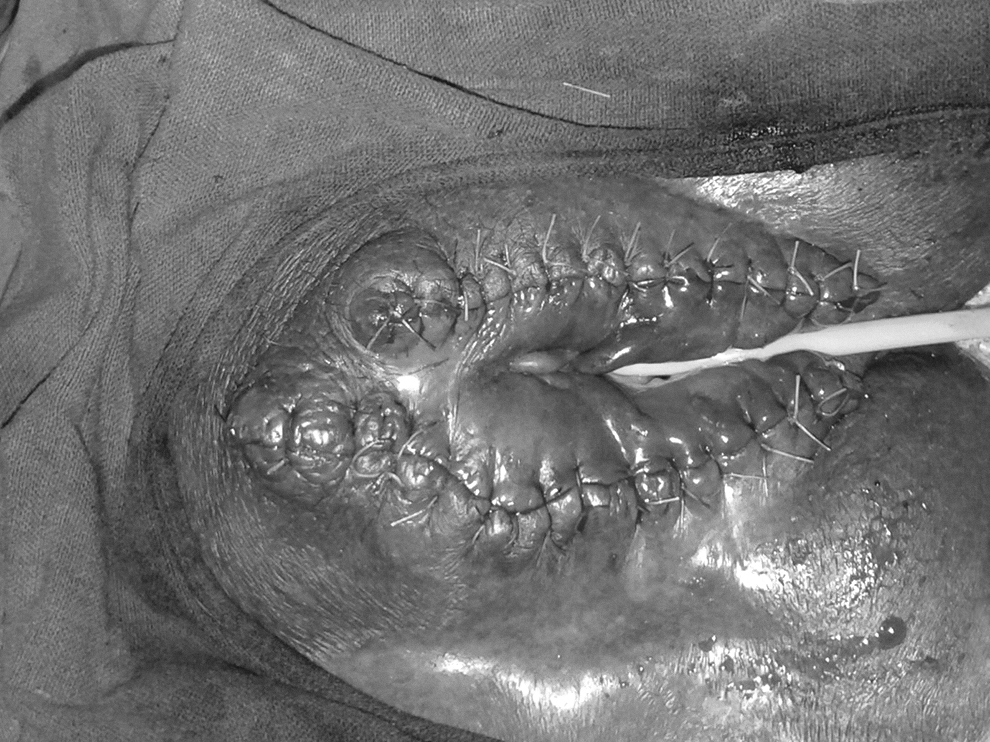
Vulvoplasty.
Discussion
Celsius first coined the term “elephantiasis” to describe an elephant-like appearance of the legs. Later it was used to describe similar enlargement of arm, chest, breast, penis, scrotum, and vulva. 4 Filariasis mostly results from infection with Wuchereria bancrofti (98%) and the remaining due to Brugia malayi (2%). The death of adult worms provokes acute inflammation and lymphatic dysfunction, leading to permanent obstruction of lymphatic channels, lymph stasis, and stimulation of fibroblasts, resulting in lymphedema and elephantiasis. Filariasis can present either as asymptomatic/subclinical microfilaremia or acute disease characterized by lymphadenitis or lymphangitis, or chronic manifestation as lymphedema or elephantiasis. The order of involvement of lymphatic filariasis is lower limb, trunk, breast, upper limb, and genitelia. Vulval elephantiasis has been reported to be caused by a variety of etiologies such as lymphatic filariasis, lymphogranuloma venereum, donovanosis, and tuberculous lymphadenitis. It can also occur after radical hysterectomy, pelvic lymphadenectomy, and radiation therapy.
Palanisamy et al. presented a 45-year-old lady who had elephantiasis of leg and vulva, and she was treated for filariasis in the past. But in our patient there was no history of filariasis and diagnosis was done only on the basis of positive filarial antigen test. 5
Mohan et al. presented a similar case of a 18-year-old unmarried woman with a unilateral vulval growth of 5 × 6 cm who was from the Uttar Pradesh state in India, which is a highly endemic area of filariasis (14.6%). 6
Sharma et al. presented huge vulval elephantiasis in a 28-year-old primigravida woman who was diagnosed as elephantiasis of the vulva of an unclear etiology, as serologic tests to agents that usually cause vulval infections with elephantiasis were negative. 7 Lu et al. in a case–control study of elephantiasis concluded that a combination of lymph stasis promoting factors (trauma, obesity, infection, and inflammatory disorders) produces localized elephantiasis. 8 So pregnancy may also lead to development of elephantiasis due to increased intra-abdominal pressure. So there is a varied etiology for vulval elephantiasis, it is a rare occurrence due to filariasis.
Conclusion
Neither the micro filarial was seen in peripheral smear nor the filarial worm was demonstrated in the tissue section in our case. It was diagnosed only on the basis of positive filarial antigen assay. Although genital elephantiasis is rare in developed countries, it is still a challenge for the tropics and subtropics. It significantly affects physical, social, and mental status of the patient. Excision with vulvoplasty results in better quality of life and improved cosmesis.
Footnotes
Acknowledgment
We sincerely thank the department of pathology of MGMC & RI.
Author Disclosure Statement
No competing financial interests exist.
Funding Information
No funding received from any sources.