
Abstract
Objective:
Cesarean-scar endometriosis (CSE) is a challenging and increasingly common condition characterized by endometrial tissue on the surgical scar following a cesarean section. Surgical excision is the primary treatment modality. This article and video highlight the surgical technique for excising CSE, emphasizing key considerations for achieving optimal outcomes.
Methods:
The surgical approach to excising CSE is demonstrated in 2 patients who presented with tender nodules at the sites of their previous cesarean-section scars and who had increased symptoms during menstruation. Their preoperative workups involved ultrasound and magnetic resonance imaging scans to assess the size, location, and margins of the lesions.
Results:
The surgical steps demonstrated are: (1) marking the margin of the palpable endometriosis nodule; (2) excising the previous cesarean scar and performing subcutaneous dissection up to the encapsulated endometriosis lesion; (3) dissecting the lesion that is involved with the anterior rectus fascia and rectus muscle and performing resection with a 1-cm margin around the lesion; (4) performing a washout with 0.9% sodium chloride and betadine; (5) plicating the rectus muscles, using 1 Vicryl suture in an interrupted fashion; (6) placing a size-10 redivac drain to minimize hematoma formation; and (7) closing the anterior rectus fascia with a nonabsorbable 1-0 looped nylon suture to reduce the risk of hernia formation.
Conclusions:
Surgical excision with an adequate margin is the most-effective treatment for CSE. Collaborative care with gynecologists specialized in endometriosis and plastic surgeons is recommended. (J GYNECOL SURG 20XX:000)
Introduction
Abdominal-wall endometriosis (AWE) is one of the commonest extrapelvic locations of endometriosis. Cesarean-scar endometriosis (CSE) is the most prevalent form of AWE and is an increasingly recognized condition in which endometrial tissue implants on the surgical scar following a cesarean section. 1 The prevalence of CSE ranges from 0.03% to 1.08% among women who have cesarean sections. 2 CSE typically presents as a palpable tender nodule at the site of the previous cesarean-section scar. The nodule increases in size during menstruation and the patient presents with cyclical tenderness. The symptoms may develop months, or—more typically—years after the cesarean section (on average, 3.6 years). 3
Medical therapy is often ineffective for complete resolution of the lesion.4,5 Surgical excision is the definitive treatment for CSE and can provide complete resolution of symptoms in up to 90% of cases. This article—with its description of the surgical technique and 2 illustrative cases plus an accompanying video (Supplementary Video S-1; Supplementary data are available online at www.liebertonline.com/GYN) —demonstrates the surgical technique for excising a CSE.
Technique
Patients with CSE require multidisciplinary care with a team of gynecologists specializing in endometriosis care and plastic surgeons.
Preoperative workup includes an abdominal-wall ultrasound along with a magnetic resonance imaging (MRI) scan. The size, location, and margins of the nodule are delineated. The possibility of mesh repair is discussed with the patient.
Often, these modules are not solitary. Preoperative gonadotropin-releasing hormone agonists are not recommended due to the risk of fibrous metaplasia of the lesions.
In Case 1, prior to laparoscopy, the margin of the palpable endometriosis nodule was marked on the abdomen (Fig. 1). Following laparoscopy, the previous Pfannenstiel incision scar was excised and subcutaneous dissection up to the encapsulated endometriosis lesion was performed.

Marking the Palmer's point for laparoscopy and margin of the palpable scar endometriosis nodule prior to excision (Case 1).
Dissection of this lesion identified its involvement with the anterior rectus fascia and rectus muscle; resection was performed with a 1-cm margin around the lesion. The dissection continued up to the preperitoneal fat. The peritoneum was not involved and was protected throughout the procedure. En bloc removal of the lesion was performed, and the excised tissue was sent for histopathology. Hemostasis was monitored carefully throughout the procedure.
A washout was performed with 0.9% sodium chloride and betadine. Plication of the rectus muscles was performed with a Vicryl suture in an interrupted fashion. To reduce the risk of hematoma and seroma formation, a size-10 redivac drain was placed on top of the muscle and fixed with a 2-0 silk suture. The anterior rectus fascia was closed with a nonabsorbable 1-0 looped nylon suture to reduce the risk of hernia formation. The rectus-fascia defect was repaired vertically from top down, as there was more laxity cranially. The deep dermal layer was closed with 2-0 Monocryltm suture, and the intradermal layer was closed with a 3-0 Monocryl suture.
In Case 2, a similar surgical technique was performed. However, preoperatively a triangular mini fleur-de-lis marking 6 was used as a reference point for excising the vertical laparotomy scar, as well as the underlying endometriotic nodule (Fig. 2). Exploration revealed that the endometriotic nodule extended through the anterior rectus fascia and peritoneum in between the rectus diastasis. The defect in the peritoneum and the anterior rectus fascia was repaired in a continuous fashion with a 1-0 polydioxanone suture. The inset was closed with a 2-0 Monocryl suture (Fig. 3 and Supplementary Video S-1).

Triangular mini fleur-de-lis marking for excision of the vertical laparotomy scar as well as the underlying cesarean-scar endometriosis (Case 2).
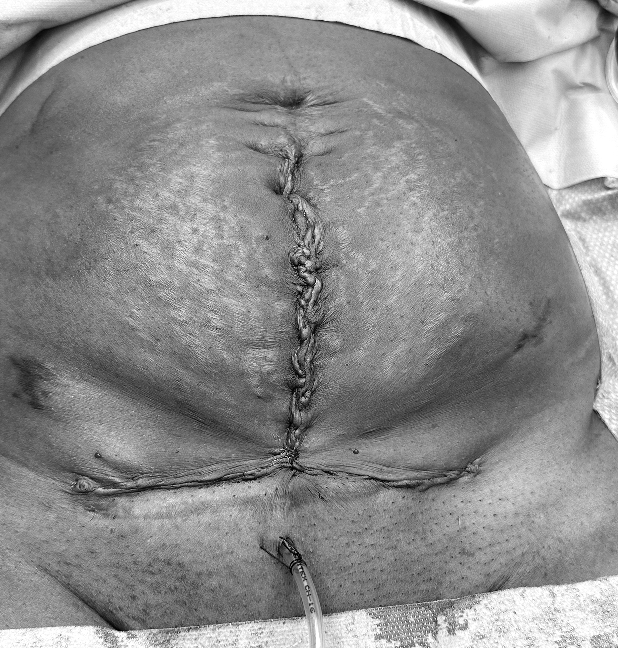
Postoperative view after excision (Case 2).
Illustrative Cases
Informed consent was obtained from both patients for the purpose of publishing this article and video.
Case 1
Case 1 was a 28-year-old female patient who underwent a cesarean section with a Pfannenstiel incision 6 years prior to presenting with cyclical pain around the scar. This patient reported a throbbing cyclical discomfort that had increased progressively over the past year, along with dysmenorrhea. She had no other past medical history, and her body mass index (BMI) was 26. The differential diagnoses included CSE, incisional hernia, granuloma, hematoma, lipoma and desmoid tumor. An MRI investigation revealed multiple rounded foci centrally within the previous cesarean-section scar, indicating CSE. The size of the endometriosis spanned 4.2 x 2.6 x 5.0 cm in total, with the dominant rounded cystic focus measuring 1.8 cm in its maximal dimension. Additionally, the right ovary appeared to be pulled toward the right lateral aspect of the uterus.
This patient was reviewed by the plastic surgery team and she underwent excision of her scar endometriosis and laparoscopic excision of her pelvic endometriosis as a joint procedure. During the laparoscopy, there were deposits of endometriosis noted on the left pelvic side wall and left uterosacral ligament; the deposits were excised. The patient had an uneventful recovery and was discharged the next day. Histopathology confirmed the presence of endometriosis. She reported complete resolution of symptoms at her 6-month follow up.
Case 2
A 44-year-old female underwent a cesarean section through a midline laparotomy, which was complicated by a postoperative wound infection. This was followed by 3 more cesarean sections, using Pfannenstiel incisions. She reported experiencing constant pain at the site of a CSE nodule, which worsened during her menstrual periods. Her BMI was 32. An MRI confirmed the clinical diagnosis of divarication of the recti muscles measuring at least 5 cm from left to right, along with a 1.5 x 1.0 x 0.7–cm focus of endometriosis within this defect, just to right of the midline. She underwent excision of scar endometriosis and abdominal-wall reconstruction as a joint procedure. This patient's recovery was similarly uneventful. Her redivac drain was removed after yielding <30 mL of drainage in 24 hours, and she was discharged on the third day.
Discussion
This article and its accompanying Supplementary video present the step-by-step surgical management of 2 cases of CSE.
CSE is considered iatrogenic. Clinicians should maintain high levels of suspicion when managing cases presenting with palpable painful nodules around the cesarean-section incision site.
The recommended surgical technique for CSE involves wide local excision of the lesion with an adequate margin of at least 1 cm to reduce the risk of recurrence.2,7 However, no studies have addressed if size of the surgical margin has an impact on the rate of recurrence. The rate of recurrence ranges from 4.3% to 29%. 3
In some cases, CSE may involve the rectus abdominis muscle and/or fascia and its resection necessitates en bloc resection of the underlying myofascial elements. It is imperative for surgeons to anticipate the potential risk of hernia formation proactively. Patients must be informed of the potential need for mesh repair or abdominal-wall reconstruction, and possible mesh-related complications such as mesh extrusion and mesh infection should be communicated to patients as well. 8 The optimal anatomical placement of the mesh is controversial and there are no comparative studies. If subsequent cesarean sections are anticipated, mesh placement should be avoided. A multidisciplinary approach and the involvement of a plastic surgeon in a joint surgical procedure may be necessary to achieve optimal results, as demonstrated in the accompanying cases video. Advanced plastic surgery techniques, such as abdominal-wall component separation, locoregional flaps, or free flaps may be required for more-extensive lesions. 9
A history of dysmenorrhea, dyspareunia, and chronic pelvic pain suggests the presence of pelvic endometriosis; the overall risk of coexistence of pelvic endometriosis with CSE is estimated to be 13%. 3 Patients should be informed and counseled about the option of undergoing laparoscopy for diagnosis and treatment. Preoperative assessment with MRI provides additional information on the depth of the lesion and potential fascial involvement. 10
The pathophysiology of CSE is likely to involve complex mechanisms; however, the most widely accepted theory is direct implantation. During cesarean section, endometrial cells are believed to implant on the abdominal scar. 3 The surgical procedure itself, along with the associated stress response, can induce immune suppression, while the surgical wound provides a microenvironment that is rich in growth factors and inflammatory mediators, creating a conducive milieu for the survival and proliferation of these endometrial cells, 11 ultimately leading to the development of CSE. Cases of CSE-associated clear-cell carcinoma have been reported; this is a clinically aggressive disease with a high mortality rate. However, the precise incidence remains uncertain. 12
Various strategies have been proposed for preventing CSE, including avoiding swabbing the endometrial cavity, using separate needles for uterine and abdominal closure, and closing the peritoneum.3,5 However, robust evidence supporting the effectiveness of these strategies is lacking. Moreover, the underlying reasons remain unclear why certain women are at higher risk of developing CSE while most women having cesarean sections do not encounter this complication. The current body of evidence on scar endometriosis is primarily derived from case reports, limiting understanding of the condition. Larger prospective studies are needed to provide more-comprehensive insights into the pathophysiology, optimal management strategies, and long-term outcomes—including reproductive outcomes—of CSE.
Conclusions
CSE is a challenging condition that requires a high index of suspicion and comprehensive evaluation. This phenomenon is likely to become more prevalent in view of the rising cesarean-section rate worldwide. Surgical excision with an appropriate margin remains the most-effective treatment for CSE. This article with its accompanying video illustrates the surgical technique for excising a CSE mass that involves the underlying myofascial layer. A joint procedure with a plastic surgeon or a general surgeon and mesh placement may be necessary for complex cases.
Footnotes
Authors' Contributions
Suruchi Pandey conceptualized this project and supervised the work. Mickey Buckingham performed the investigation and wrote the original draft of this article. Gino Vissers, Orestis Tsonis, and Suruchi Pandey wrote the final version and reviewed and edited it.
Author Disclosure Statement
No financial conflicts of interest exist.
Funding Information
No funding was provided for this project.
References
Supplementary Material
Please find the following supplemental material available below.
For Open Access articles published under a Creative Commons License, all supplemental material carries the same license as the article it is associated with.
For non-Open Access articles published, all supplemental material carries a non-exclusive license, and permission requests for re-use of supplemental material or any part of supplemental material shall be sent directly to the copyright owner as specified in the copyright notice associated with the article.