
Abstract
Abstract
Jiang, Sihua, Feipeng Jin, De Li, Xingmei Zhang, Yun Yang, Dachun Yang, Kun Li, Yongjian Yang, and Shuangtao Ma. Intermittent hypobaric hypoxia promotes atherosclerotic plaque instability in ApoE-deficient mice. High Alt Med Biol 14:175–180, 2013.—
Results:
IHH-exposed mice did not significantly differ from control mice in plasma lipid levels, including triglyceride, total cholesterol, low-density lipoprotein, and high-density lipoprotein. The hematoxylin and eosin-stained sections of the aortic root showed similar plaque size between the groups. However, IHH-treated mice exhibited significantly decreased plaque collagen content, a feature of atherosclerotic plaque stability. Additionally, matrix metalloproteinase (MMP)-9 protein expression was significantly increased, whereas tissue inhibitor of MMP (TIMP)-2 expression was decreased after exposure to IHH.
Introduction
High altitude settings are characterized by a peculiar environmental factor: hypoxia. It has been reported that intermittent normobaric hypoxia experienced by patients with obstructive sleep apnea may enhance the development of atherosclerosis. Previous studies have suggested that intermittent normobaric hypoxia accelerates atherosclerotic plaque growth in ApoE-deficient (ApoE-/-) mice without affecting the plaque composition (Jun et al., 2010). Additionally, normobaric hypoxia was reported to cause atherosclerosis in C57BL/6 mice that were fed a high cholesterol diet (Savransky et al., 2007). However, the intermittent hypoxia that replicates obstructive sleep apnea may dramatically differ from a high altitude air environment. Thus, the role of hypobaric hypoxia, which mimics the ambient air at high altitude, in the development of atherosclerosis has not been elucidated. We hypothesized that intermittent hypobaric hypoxia (IHH) could affect atherosclerotic plaque growth and stability.
Collagen is an important component of the extracellular matrix in the atherosclerotic plaque. It has been shown that matrix collagen contributes to the integrity of the fibrous cap and the stability of the plaque (Nadkarni et al., 2009). The matrix metalloproteinases (MMPs), which are responsible for the proteolysis of matrix collagen, have emerged as important regulators of plaque instability (Beaudeux et al., 2004; Watanabe and Ikeda, 2004). The tissue inhibitors of metalloproteinase (TIMPs) are the endogenous inhibitors of MMPs. It was previously reported that hypoxia may affect several kinds of MMP in different tissues or cells. We therefore hypothesized that IHH could influence plaque stability by regulating the balance of MMPs and TIMPs.
In the present study, ApoE-/- mice were fed a normal diet and exposed to control conditions or IHH for 8 weeks. Their lipid profiles, atherosclerotic plaque size, and surrogate markers of plaque stability were measured.
Materials and Methods
Animal care
Twenty 8-week-old male ApoE-/- mice were purchased from the Model Animal Research Center of Nanjing University (Nanjing, China). The animals were housed under a 12 h/12 h day/night cycle and standard conditions of temperature (22±1°C) and humidity (50%–60%) with ad libitum food and water. The mice were fed a normal chow diet. The experimental procedures were approved by the Hospital Animal Care and Use Committee.
Hypobaric hypoxia
A hypobaric chamber with pneumatic pumps was used to establish the hypobaric hypoxia condition replicating an altitude of 4000 meters (Pi
Serum lipids
Fasting blood samples were obtained and triglycerides (TG), total cholesterol (TC), LDL cholesterol (LDL-C), and HDL cholesterol (HDL-C) were measured by colorimetric assays using a commercially available kit (Jiancheng Bioengineering Institute, Nanjing, China) according to the manufacturer's instructions.
Atherosclerosis analysis
Atherosclerotic lesions of the thoraco-abdominal aorta were analyzed by Oil-red-O staining. Atherosclerotic lesions in aortic roots were examined in cross-sections of aortic origin. Six consecutive 5 μm thick sections were cut from the aorta where the valve cusp was visible. The sections were stained with hematoxylin (90 s) and eosin (2 s) and photographed on an Olympus BX41 microscope (Olympus Corp., Tokyo, Japan). The atherosclerotic lesion area was quantified using Nikon NIS-Elements Research Software (Ma et al., 2011). The average plaque area was used for statistical analysis.
Collagen analysis
Paraffin-embedded aortic sinus sections were stained with Masson's Trichrome using a fast Masson dye kit (Jiancheng Bioengineering Institute, Nanjing, China) following the manufacturer's instructions. The percentage of positively stained area was calculated using Nikon NIS-Elements research software.
Immunohistochemistry
Thoracic aortas were fixed in 4% paraformaldehyde for 12 h, embedded in paraffin, and then cut into sections (5 μm). The sections were incubated with anti-MMP-2, anti-MMP-3, anti-MMP-9, anti-MMP-14, anti-TIMP-1, and anti-TIMP-2 antibodies (diluted in 1:50 dilution, Boster Bioengineering Co., Wuhan, China). Specific binding was detected with biotinylated goat anti-rabbit IgG secondary antibody-horseradish peroxidase complexes using an ABC kit (Boster Bioengineering Co.). The antigen–antibody complex was subsequently visualized with 3', 3'-diaminobenzidine solution. Sections were viewed under a light microscope. The proportion of the positively stained area was calculated using Nikon NIS-Elements research software.
Statistical analysis
Continuous data are presented as a mean±SEM. Comparisons between groups were determined by a one-way ANOVA with a post-hoc Student t-test (SPSS Inc., Chicago, IL). Probabilities of p<0.05 were considered statistically significant.
Results
Food intake, body weight, and lipid profiles
The daily food intake for each mouse was recorded during the course of the study. The amount of food consumed was similar between the control and IHH groups (Fig. 1A). The baseline body weight was similar between the two groups. At the end of the experiment, mice exposed to IHH had lower body weight than the controls (p<0.01) (Fig. 1B). Exposure to IHH did not affect the plasma lipid profiles, including TG, TC, LDL-C, and HDL-C (Fig. 1C–F).

The effects of IHH on the metabolic character of ApoE-/- mice. The daily food intake
Atherosclerotic plaque size
The atherosclerotic plaque size in the thoraco-abdominal aorta was similar between the control and IHH groups (Fig. 2A, B). Additionally, there was no difference in the mean plaque size within the aortic root between the two groups (Fig. 2C, D).
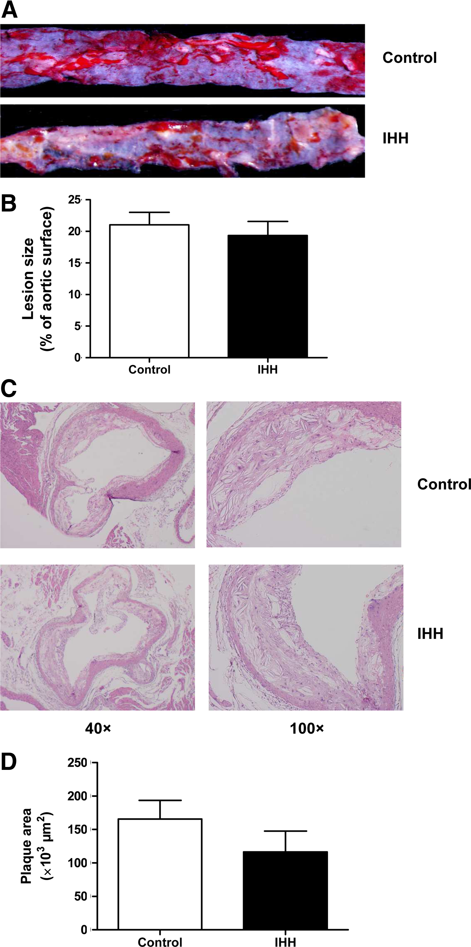
The effects of IHH on atherosclerotic plaque size in ApoE-/- mice.
Plaque instability
As hypothesized, collagen deposition was significantly decreased in the IHH group compared to the control group (p<0.01) (Fig. 3A, B).

The effects of IHH on plaque instability in ApoE-/- mice.
MMP/TIMP expression
The expression of MMP-2, MMP-3, MMP-9, MMP-14, TIMP-1, and TIMP-2 protein in the thoracic aorta was measured by immunohistochemical staining. MMP-3 and TIMP-1 were not obviously visible after staining. Exposure to IHH significantly increased the expression of MMP-9 (p<0.01), decreased the expression of TIMP-2 (p<0.01), and had no significant effect on MMP-2 and MMP-14 in aortic sections (Fig. 4A, B).
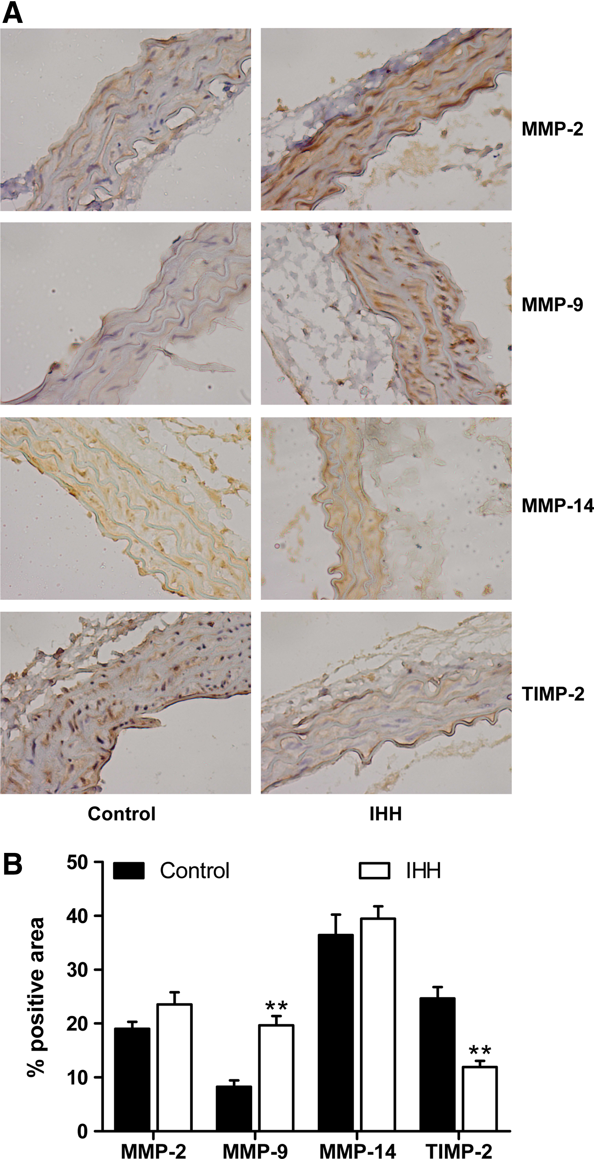
The effects of IHH on MMP-2/-9/-14 and TIMP-2 expression in aortas from ApoE-/- mice.
Discussion
Hypoxia has been implicated in the development of atherosclerosis (Hulten and Levin, 2009; Mayr et al., 2008). Most previous studies have focused primarily on the harmful effects of normobaric hypoxia on patients with obstructive sleep apnea (Arnaud et al., 2009; Drager et al., 2005; Levy et al., 2009). Chronic intermittent hypoxia causes atherosclerotic lesions in C57BL/6 wild-type mice, which are usually resistant to atherosclerosis (Savransky et al., 2007). Additionally, intermittent hypoxia accelerates atherosclerotic plaque growth in ApoE-/- mice (Jun et al., 2010; Nakano et al., 2005). Although the aforementioned conditions dramatically differ from the air environment at high altitude, it is reasonable that exposure to IHH could affect the development of atherosclerosis. Unfortunately, the present study demonstrated that IHH had no effect on the plaque size in ApoE-/- mice. It is herein proposed that hypobaric conditions could attenuate the hypoxia-induced promotion of atherosclerosis.
There is a growing body of evidence that the ascent to high altitude is associated with a high risk of sudden cardiac death (Bartsch and Gibbs 2007; Burtscher and Ponchia 2010; Rimoldi et al., 2010). However, a causal link between altitude environment and sudden death has not been established. The main finding of this study was that 8-week IHH exposure resulted in plaque instability, as expressed by decreased collagen deposition. This result indicates that a high altitude environment mainly characterized as IHH may induce acute coronary syndrome and sudden cardiac death by increasing the vulnerability of the atherosclerotic plaque. A previous study demonstrated that hypoxia alone could not affect plaque composition, including fibrotic caps and collagen deposition (Jun et al., 2010). Taken together, the effects on plaque instability may mostly be attributed to hypobaric conditions.
Chronic hypoxia induces adaptive metabolic changes in several different types of cells (Lodi et al., 2011; Plunkett et al., 1996). We found that exposure to IHH significantly decreased body weights of mice but did not affect the food intake of mice. The observed weight loss was consistent with previous studies (Jun et al., 2010; Savransky et al., 2007). The reduction in body weight may be attributed to the metabolic switch caused by hypoxia. As body weight control has a protective role in atherosclerosis, this might counteract hypoxia-induced plaque growth. In prior studies of mice exposed to normobaric hypoxia, atherogenic lipid profiles were significantly enhanced (Arnaud et al., 2009; Li et al., 2007; Nakano et al., 2005; Roche et al., 2009; Savransky et al., 2008). However, the present study demonstrated no effect on serum lipids after 8 weeks under IHH conditions. We hypothesize that the IHH-induced decrease in body weight may offset the changes in lipids.
Elevated blood pressure is an important factor in the development of atherosclerosis. Previous studies have demonstrated that hypoxia has many effects on blood pressure in rodents, including acute decreases in blood pressure (Campen et al., 2004) superimposed on chronic increases in blood pressure (Fletcher, 2000). However, another study showed that chronic intermittent hypoxia did not affect the blood pressure of Chilean mine workers (Farias et al., 2006). The present study demonstrated that the tail-cuff blood pressure was similar between the two groups (data not shown). However, the tail-cuff blood pressure was measured only once at the end of the present study. Moreover, the ambulatory blood pressure was not evaluated in the present study, and whether plaque instability is attributed to changes in blood pressure remains unclear.
Mechanisms by which hypoxia causes plaque instability have not been well established (Martin et al., 1991). MMPs and TIMPs have been viewed as the regulators of plaque instability (Murillo et al., 2009). We found that the plaque instability induced by IHH was concomitant with the increased MMP-9 and decreased TIMP-2 expression. A previous study demonstrated that hypoxia elevated the level of MMP-9 in cultured microvascular endothelial cells from the human brain (Kolev et al., 2003). Moreover, hypoxia lowers TIMP-2 protein secretion by human endothelial cells (Lahat et al., 2011). Taken together, the increased MMP-9 levels and decreased TIMP-2 levels induced by hypoxia may stimulate the degradation of collagen and consequently cause plaque instability. The results of the present study indicate that the imbalance of MMP and TIMP might be responsible for the linkage between IHH and plaque vulnerability. Therefore, restoring the balance between MMPs and TIMPs may be a novel strategy to prevent hypobaric hypoxia-induced plaque instability.
In conclusion, the present results demonstrate that exposure to IHH promotes atherosclerotic plaque instability without affecting plaque size in ApoE-/- mice. Moreover, the IHH-induced vulnerability to plaques is associated with the imbalance between MMP-9 and TIMP-2. The current study also provides a model of hypobaric hypoxia for the study of atherosclerosis at high altitude. The present findings may contribute to mechanistic insights into altitude-associated sudden cardiac death.
Footnotes
Acknowledgments
We thank Yan Luo and Yihua Chen (Department of Pathology, General Hospital of PLA Chengdu Military Area Command) for skillful technical assistance. We acknowledge Song Hu (Chengdu Medical College) for animal care.
Author Disclosure Statement
No competing financial interests exist.
This study was supported by grants from the General Hospital of PLA Chengdu Military Area Command (No. 2011YG-B32 and No. 2011YG-B22).