
Abstract
In this article, we detail a comprehensive laboratory evaluation of an immunoassay for the rapid detection of abrin using the Meso Scale Diagnostics Sector PR2 Model 1800. For the assay evaluation, we used inclusivity and exclusivity panels comprised of extracts of 11 Abrus precatorius cultivars and 35 near-neighbor plants, 65 lectins, 26 white powders, 11 closely related toxins and proteins, and a pool of 30 BioWatch filter extracts. The results show that the Meso Scale Diagnostics abrin detection assay exhibits good sensitivity and specificity with a limit of detection of 4 ng/mL. However, the dynamic range of the assay for the quantitation of abrin was limited. We observed a hook effect at higher abrin concentrations, which can lead to potential false negative results. A modification of the assay protocol that incorporates extra wash steps can decrease the hook effect and the potential for false negative results.
Introduction
Abrin is a potent water-soluble phytotoxin found in the seeds of Abrus precatorius, a vine common to many tropical areas throughout the world. 1 A precatorius seeds and plants are also known as rosary pea, jequirty, crab's eye, John Crow bead, precatory bean, prayer bead, Buddhist rosary bead, love bean, Indian bead, Seminole bead, lucky ben, akar saga, giddee giddee, and jumbie bead.1,2 The seeds have been used for jewelry, including bracelets and necklaces, as well as for rosary beads. A precatorius seeds, roots, and leaves have a long history in ethnobotany and have been used as an analgesic, aphrodisiac, abortifacient, anticonvulsant, laxative, sedative, insecticide, and herbal remedy, the latter to treat chronic eye diseases, fevers, coughs, worms, sexually transmitted diseases, and other conditions. 3 All parts of A precatorius plants are toxic, but the seeds contain the highest concentration of the toxin abrin (≈0.08%). 1
Abrin is a heterodimeric type 2 ribosome-inactivating protein (RIP) consisting of an A-chain of 251 amino acids (relative molecular mass 30,000 Da) linked by a single disulfide bridge to a B-chain consisting of 268 amino acids (relative molecular mass 33,000 Da). The B-chain is a lectin that binds to cell surface glycoproteins containing D-galactopyranose moieties. Once bound, the heterodimer is transported intact across the cell membrane by receptor-mediated endocytosis and then to the endoplasmic reticulum by the retrograde pathway.4-7 The intersubunit disulfide bond must be reduced before the A-chain can be translocated to the cytosol where it interacts with the ribosome. The A-chain of abrin is an N-glycosidase that cleaves the C-N bond of adenine at position 4324 in the eukaryotic 28S ribosomal RNA of the large ribosomal subunit. 8 This prevents the formation of a critical stem-loop configuration, thereby preventing elongation factor 1 and 2 from binding to the ribosome, resulting in the inhibition of protein synthesis, which leads to apoptosis and cell death.9-10 Other type 2 RIPs include phytotoxins such as ricin (from the seeds of Ricinus communis), modeccin (from the fruits and roots of Adenia digitata), volkensin (from the roots of Adenia volkensii), and viscumin (from mistletoe, Viscum album). 11
Abrin occurs in 3 isoforms, designated I, II, and III, which can be separated by lactamyl-sepharose affinity chromatography 12 ; other researchers have reported the existence of a fourth isoform. 13 Isoforms I, II, and III have different mouse intraperitoneal median lethal dose values of 22, 2.4, and 10 μg/kg body weight, respectively. 12 Abrin is significantly more toxic than ricin. Based on the minimal lethal intravenous dose in mice (0.7 μg/kg), abrin is approximately 4 times more potent than ricin (2.7 μg/kg). 14 The human lethal dose of abrin is estimated to be between 0.1 and 1.0 μg/kg by ingestion. 15 However, abrin can enter humans by several routes. Individuals with abrin intoxication can present with gastrointestinal hemorrhaging following ingestion, muscle necrosis following injection, or acute pulmonary disease following inhalation. 16 Abrin can also enter the body through a break in the skin or it can be absorbed through intact skin if dissolved in certain solvents. Another protein found in seeds of A precatorius is an agglutinin, which is capable of agglutinating red blood cells, but is nontoxic. 15
Although most reported cases of abrin intoxication involve children who have ingested seeds of A precatorius, the consequences of seed ingestion are generally not severe owing to the hard shell that protects the toxin. 17 However, if the integrity of the shell has been compromised (eg, by chewing or drilling holes, as is done for beadwork or jewelry), abrin can affect the digestive tract even as digestive enzymes act to break down the toxin.2,18 Symptoms may not appear until 1 to 3 days following ingestion and can include nausea, vomiting, diarrhea, abdominal cramps, hematemesis, and melena. 18 Acute renal failure and hepatotoxicity may also occur. 18 The clinical course of toxicity can be more than 10 days. The mortality rate is reported to be 5%, with death occurring up to 14 days after ingestion. 18 There is no specific treatment for abrin intoxication resulting from ingestion. Treatment is supportive with intravenous fluids, correction of electrolyte imbalance, and blood transfusions, if required. 18 Nonspecific treatment may involve inducing emesis, gastric lavage, activated charcoal, and whole bowel irrigation.18,19 Gut decontamination (ie, gastric lavage or administration of activated charcoal) may be considered if presentation is within 1 hour of ingestion. 15
Plant toxins such as abrin and ricin are generally easy to produce in large quantities at minimal cost in a low-technology environment. 20 However, when compared with spores of Bacillus anthracis, they are not an efficient weapon for mass casualty events. While the dispersion of phytotoxins over a large area is possible but logistically impractical, abrin represents a viable threat in limited air release scenarios. 21
The purpose of this study was to fulfill the critical need to promote public safety, public health, and food biodefense (potential aerosol release leading to inhalation exposure or the adulteration of food or feed is a potential bioterrorism concern). To support this critical need, we conducted a comprehensive evaluation of the Meso Scale Diagnostics (MSD) Sector PR2 Model 1800 for the rapid detection of abrin. The platform uses electrochemiluminescence (ECL) labels that are conjugated to detection antibodies. When stimulated by electricity in the appropriate chemical environment, SULFO-TAG (STAG) labels emit light, which enables ultrasensitive detection of key proteins and molecules.
The evaluation was conducted using comprehensive inclusivity and exclusivity panels of A precatorius and near-neighbor plant materials, along with other toxins/related materials, lectins, white powders, environmental background material, and BioWatch filter extracts based on the Public Health Actionable Assay standards. 22 To ensure an unbiased evaluation, blinded samples were prepared and provided to perform the testing and evaluation. The primary purpose of this study was to evaluate the robustness, sensitivity, specificity, and repeatability of this assay to understand the probability of false positive (ie, assay is positive but the target analyte of interest is not present) and false negative (ie, assay is negative but the target analyte of interest is present in amounts at or above the limit of detection) results. Although confirmatory or secondary testing is possible on positive test results, it is not practical to confirm negative test results. Thus, the goal is to provide test results with an agreed upon level of certainty so that appropriate and effective decisions can be made in a timely manner to protect the public, avoid unnecessary disruption of civil society, and reduce the economic impact.
Materials and Methods
The MSD Sector PR2 Model 1800 uses ECL technology to carry out highly sensitive multiplexed immunoassays for the presence of bacterial, viral, and toxin biothreat agents. The PR2 Model 1800 instrument is a plate reader that measures ECL generated in specialized 96-well MULTI-SPOT microplates that include integrated screen-printed electrodes located in the bottom of the wells. The electrodes serve as both the solid-phase support for capture reagents used in solid-phase binding assays and as the source of electrical energy for inducing ECL. ECL measurements carried out in these plates use detector antibodies that are labeled with an ECL-active organometallic ruthenium complex (STAG MSD). These labels generate light when oxidized at an electrode surface in the presence of a tertiary amine such as tripropylamine. During the ECL measurement, the plate reader applies a voltage to electrodes in the wells of the MULTI-ARRAY plates and measures the resulting ECL at 620 nm with an array of photodiodes to quantify the target analytes of interest in the sample.
The test and evaluation of the MSD assay for the rapid detection of abrin was performed using MSD MULTI-SPOT abrin/ricin plates. Each well is designed for the multiplex detection of 7 analytes with 2 quality control spots, positive control high (PC-H) and positive control low (PC-L). The analytes are abrin (spot 3), ricin (spot 5), and bovine serum albumin (spots 2, 4, 6, 7, 8) (Figure 1). The 5 bovine serum albumin spots are nonrelevant in this assay; MSD simply offers the option for additional target detection in the future. The PC-H spot is coated with a trademarked capture antibody for the PC-H analyte. The PC-L spot is an uncoated blank surface to monitor nonspecific binding. According to the manufacturer, the PC-H control spot should be between 5,000 and 20,000 counts and the nonspecific binding PC-L control spot should be less than 500 counts. Low PC-H counts could indicate either an error in the assay protocol, instrument operation, or a significant matrix effect.

MULTI-SPOT plate well design. Each well is designed to be used for multiplex detection of 7 analytes with 2 quality control spots (PC-H and PC-L). The analytes are abrin (spot 3), ricin (spot 5), and BSA (spots 2, 4, 6, 7, 8). Abbreviations: BSA, bovine serum albumin; PC-H, positive control high; PC-L, positive control low.
MSD Standard Protocol
The workflow of the MSD Abrin/Ricin Assay is depicted in Figure 2. A 50 μL volume of STAG-labeled anti-abrin and anti-ricin detector antibodies (MSD) and 50 μL of PC-H analyte were added to 2.4 mL of BioD Diluent Buffer (MSD) to prepare a working stock; 20 μL of the working stock solution was added to each well of the plate. Test samples (100 μL) in MSD BioD Extraction Buffer were then added to each well. The plate was covered with an adhesive seal and incubated on a plate shaker (Benchmark Scientific Incu-Mixer MP) for 1 hour at 500 rpm at 37°C. Each well was then washed 3 times with phosphate-buffered saline (PBS) (500 μL/well) using a plate washer (BioTek ELx405). MSD Read Buffer T reagent (1X concentration) with surfactant (125 μL) was added to each well and the ECL signal was immediately read using the MSD Sector PR2 Model 1800.
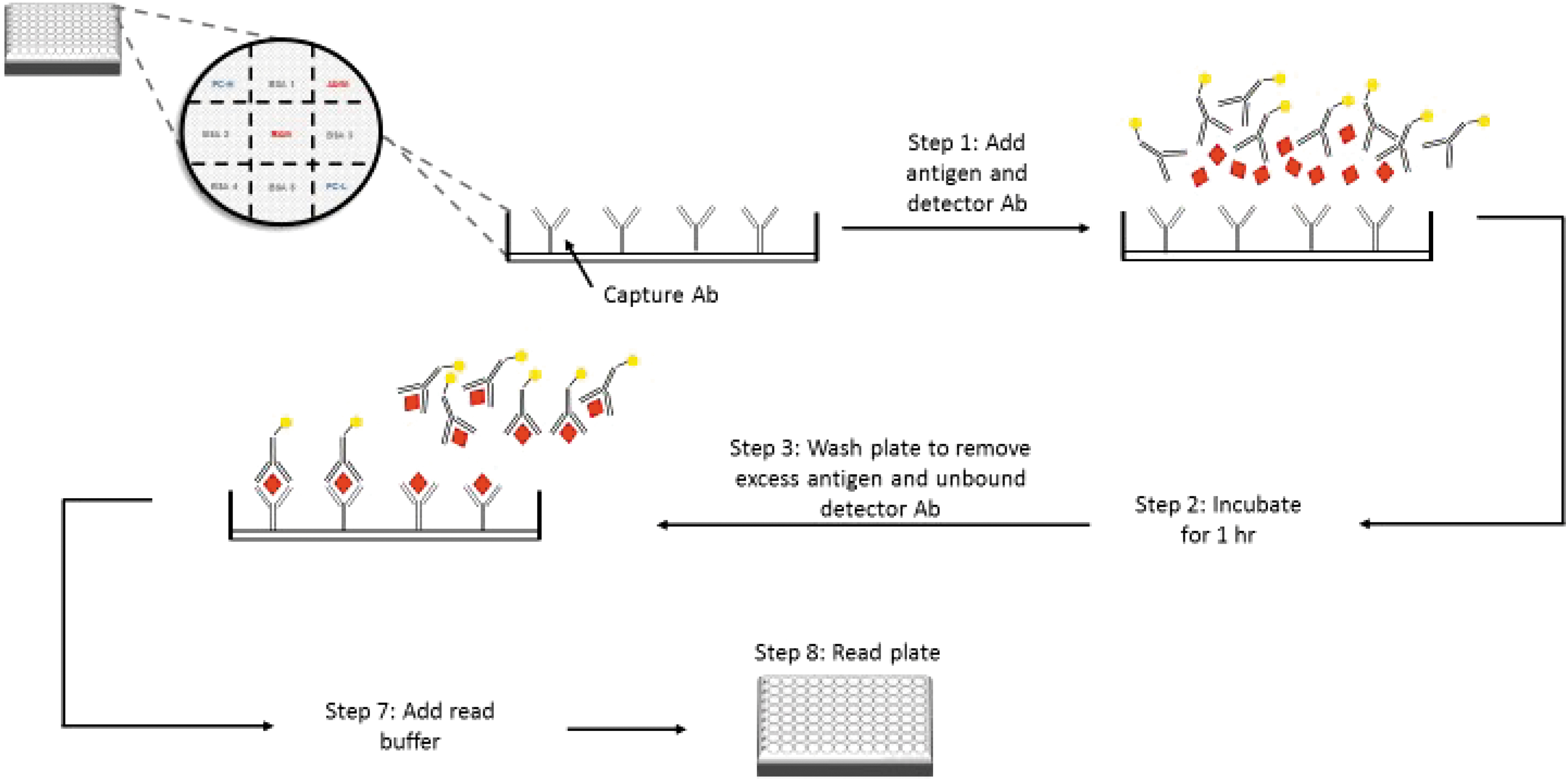
Workflow of the MSD Abrin/Ricin ECL assay. The original assay chemistry defined by the manufacture lacked steps 4 through 6, which are shown in Figure 3. Abbreviation: Ab, antibody.
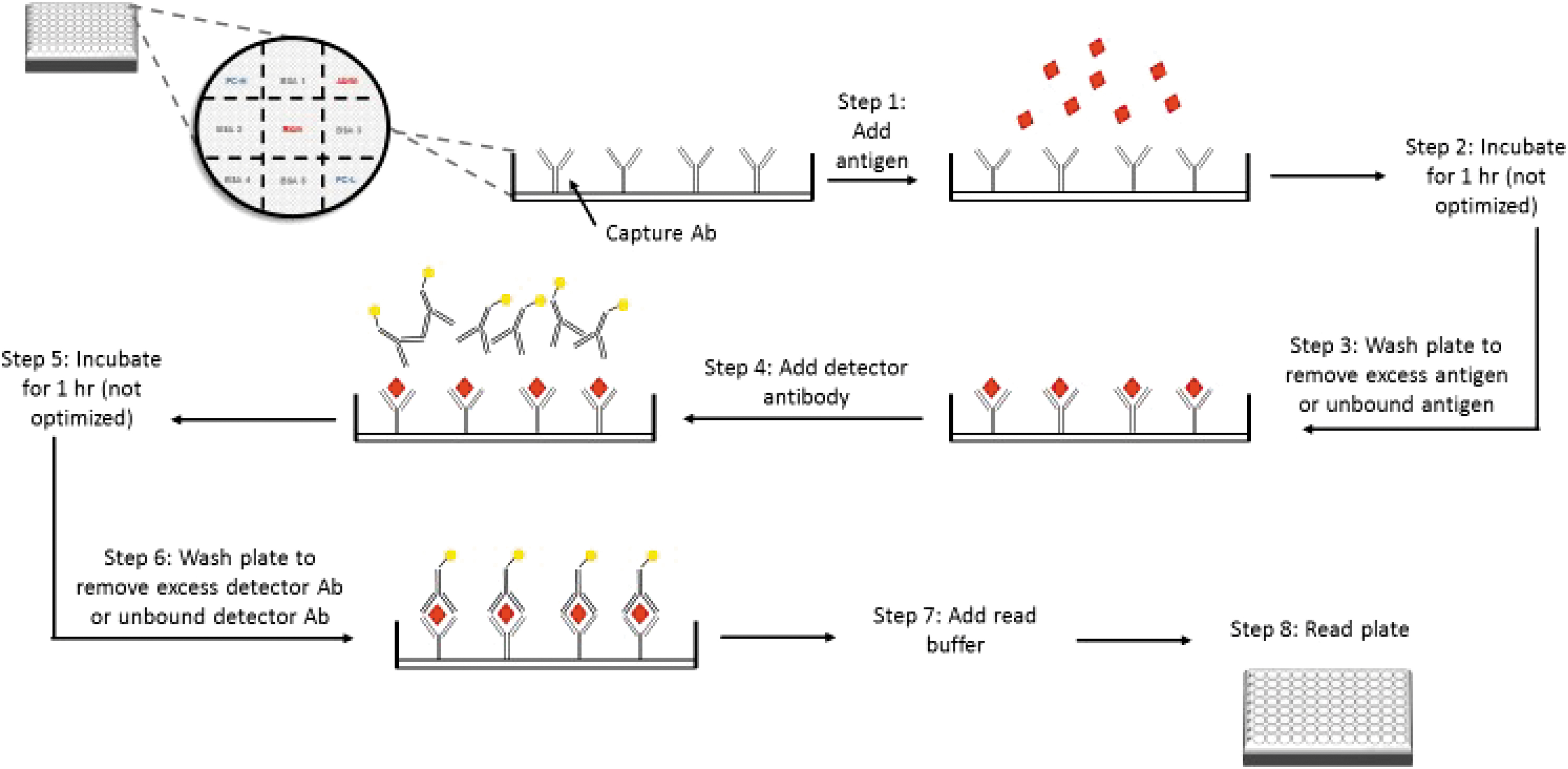
Modified assay protocol for the detection of abrin toxin. The original assay chemistry defined by the manufacture lacked steps 4 through 6. We modified the assay by including steps 4 through 6 to eliminate hook effect and also signal bleed over. Abbreviation: Ab, antibody.
Modified Protocol
A modified protocol (Figure 3) was developed in an attempt to overcome the hook effect. Briefly, a 50 μL volume of PC-H analyte was added to 2.4 mL of BioD Diluent Buffer to prepare a working stock; 20 μL of the working stock solution was added to each well of the plate. Test samples (100 μL) in MSD BioD Extraction Buffer were then added to each well. The plate was covered with an adhesive seal and incubated for 1 hour on a plate shaker at 500 rpm at 37°C. After the adhesive seal was removed, each well was washed 3 times with PBS (500 μL/well) using a plate washer. Twenty μL of a stock solution of detector antibodies (50 μL of STAG-labeled anti-abrin and anti-ricin detector antibodies added to 2.4 mL of BioD Diluent Buffer) and 100 μL of MSD BioD Extraction Buffer was added to each well. The plate was re-covered with an adhesive seal and incubated on a plate shaker for 1 hour at 500 rpm at 37°C. The adhesive seal was removed, and each well was washed 3 times with 500 μL of PBS. MSD Read Buffer with surfactant (125 μL) was added to each well and the ECL signal was read immediately.
Cutoff Values
Cutoff values were calculated using the ECL counts from the negative panels (near neighbors, white powder, biowatch filter extracts, lectins, and informational panels) plus 3 times the standard deviation (SD). The abrin cutoff was determined to be 780 without the inclusion of data from the ricin inclusivity panel.
Plant Sources
Seeds from 11 A precatorius cultivars were obtained from the following sources. Banana Tree Brown and Banana Tree Red were obtained from the Banana Tree Seed Company (Easton, PA). EG 13625 24840 and EG 13625 24530 were obtained from B & T World Seeds (Aigues-Vives, France) and provided by E. A. Garber from the US Food and Drug Administration Center for Food Safety and Applied Nutrition. Seeds with the following designations were obtained from the Desert Legume Program (Tucson, AZ): DES, AUS, CBFL, MelFL, and ZMB. Seeds designated ONA were purchased from Onalee's Seeds (Madeira Beach, FL). Seeds designated TRO were purchased from Tropilab, Inc. (St. Petersburg, FL). Seeds from near neighbors of A precatorius and other plants were obtained from various sources. Abrus laevigatus, Adriana quadripartita, Bryonia dioica, Canavalia gladiata, Canavalia rosea, Canavalia virosa, Cinnamomum camphora, Cucurbita moschata, Dianthus caryophyllus, Jubernardia globifera, Luffa acutangular, Luffa cylindrica (aegyptiaca), Lychnis chalcedonica, Momordica charantia, Phytolacca americana, Phytolacca dioica, Plukenetia volubilis, Sambucus ebulus, Sambucus nigra, Saponaria officinalis, Senna occidentalis, Trewia nudiflora, and Trichosanthes kirilowii were obtained from B & T World Seeds. Fatsia japonica and Saponaria officinalis were obtained from Plant World Seeds (Newton Abbot, Devon, UK). Macaranga grandifolia was obtained from Top Tropicals (Ft. Myers, FL). Seeds of Mallotus philippensis and Mercurialis annua were obtained from the US Department of Agriculture, Agricultural Research Services (Pullman, WA). Leaves from Acalypha rhomboidei, Viscum album, and Manihot esculenta were obtained from the US Botanic Garden (Washington, DC). Abrus schimperi subs. Africanus, Galactia striata, and Galactia wrightii were obtained from the Desert Legume Program. Iris hollandica bulbs were purchased from American Meadows (Williston, VT).
Crude Extracts
Seeds of A precatorius were weighed and placed in a biological safety cabinet. Using proper precautions, the seed coats were cracked with pliers and then the seeds were soaked in PBS for 6 to 12 hours at 4°C in the dark (1.7 mL PBS/g of seeds). During this time, the seeds expanded and absorbed all the liquid. The expanded seeds were transferred to a mortar and pestle. After grinding, the material was transferred to a 50 mL conical centrifuge tube and PBS plus 0.1% Tween-20 (vol/vol) (PBST) was added in the ratio of 5 mL/g of seed. The tube was vortexed and then incubated on a rocker platform for 16 to 19 hours at 4°C in the dark. After this incubation period, the tube was centrifuged for 4 minutes at 3000 rpm (room temperature) in a Sorvall GSA Rotor. The middle aqueous layer was removed, aliquoted into cryovials and stored at -80°C until used. Crude extracts of other seeds and plant materials were prepared in a similar manner, aliquoted into cryovials, and stored at -80°C until used.
Lectins
The following lectins were purchased from EY Laboratories, Inc. (San Mateo, CA): Agaricus bisporus, Aleuria aurantia, Allium sativum, Amaranthus caudatus, Arachis hypogaea, Artocarpus integrifolia, Arum maculatum, Bauhinia purpurea, Bryonia dioica, Canavalia ensiformis, Caragana arborescens, Cicer arietinum, Colchicum autumnale, Cytisus scoparius, Datura stramonium, Dolichos biflorus, Euonymus europaeus, Galanthus nivalis, Glycine max, Griffonia (Bandeiraea) simplicifolia lectin I, G (B) simplicifolia lectin II, G simplicifolia, Hippeastrum hybrid, Iberis amara, Iris hybrid, Jacalin, Laburnum alpinum, Lens culinaris, Lotus tetragonolobus, Lycopersicon esculentum, Maackia amurensis lectin I, Maclura pomifera, Mangifera indica, Narcissus pseudonarcissus, peanut agglutinin, Phaseolus lunatus, Phaseolus vulgaris, P vulgaris agglutinin, P vulgaris erythroagglutinin, Phytolacca americana, Pisum sativum, Psophocarpus tetragonolobus, P tetragonolobus lectin I, P tetragonolobus lectin II, Robinia pseudoacacia, Salvia horminum, Salvia sclarea, Sambucus nigra agglutinin I, S nigra agglutinin II, Solanum tuberosum, Sophora japonica, soybean agglutinin, Trichosanthes kirilowii, Trifolium repens, Tulipa sp., Ulex europaeus agglutinin I, U europaeus agglutinin II, Urtica dioica, Vicia faba, Vicia graminea, Vica villosa, Vigna radiata, Wisteria floribunda agglutinin, W floribunda lectin, and wheat germ agglutinin.
Toxins and Proteins Informational Panel
Purified abrin, formalin-inactivated ricin toxoid, formalin-inactivated abrin toxoid, and purified Shiga toxin were purchased from Toxin Technology, Inc. (Sarasota, FL). Purified ricin (RCA60), ricin A-chain, ricin B-chain, and ricin agglutinin (RCA120) were purchased from Vector Laboratories (Burlingame, CA). Deglycosylated ricin A-chain and the vaccine candidate rRTA1-33/44-198 were obtained from Martha Hale, US Army Medical Research Institute of Infectious Diseases (Ft. Detrick, Frederick, MD). RiVax, a candidate ricin vaccine consisting of a recombinant ricin A-chain containing residues 1-267 with 2 substitutions, V76M and Y80A, to reduce toxicity, 23 was obtained from P. Legler, US Navy. Abrus agglutinin was obtained from E. A. Garber from the US Food and Drug Administration Center for Food Safety and Applied Nutrition.
White Powders
Powdered milk, powdered coffee creamer, powdered sugar, talcum powder, baking powder, cornstarch, and popcorn salt were purchased from Raley's grocery store (Pleasanton, CA). Rice flour was purchased from 99 Ranch Market (Pleasanton, CA). Wheat flour and soy flour were purchased from Van's Health Foods (Livermore, CA). Baking soda, baby powder, chalk dust, and powdered infant formula (iron fortified and low-iron formulation) were purchased from Target (Livermore, CA). Powdered toothpaste was purchased from Walmart Pharmacy (Livermore, CA). Brewer's yeast was obtained from GNC (Livermore, CA). Drywall dust was obtained from Home Depot (Livermore, CA). Gamma aminobutyric acid, L-glutamic acid, kaolin, chitin, chitosan, magnesium sulfate, and boric acid were purchased from Sigma-Aldrich Corp. (St. Louis, MO). Bacillus thuringiensis (Dipel) powder was purchased from SummerWinds Nursery (Palo Alto, CA).
Protein Determination
Protein concentrations were determined using the Bradford Assay Reagent (Pierce Chemical, Rockford, IL) with a standard curve prepared with bovine serum albumin (EM Sciences, Cole-Parmer, Vernon Hills, IL).
BioWatch Filters
Thirty BioWatch filters, after being subjected to environmental aerosol collection for 24 hours, were extracted by shaking with PBST for 1 hour and the extracts pooled. The protein concentration of the pooled extract was determined and adjusted to 3 μg/mL with PBST containing 0.1% bovine serum albumin (wt/vol) (PBSTB) before testing.
Test and Evaluation Study Plan
This evaluation of the MSD abrin assay was conducted in 7 phases:
Phase 1: Range Finding and Repeatability Study
A range finding study was performed to determine both the dynamic range and the limit of detection of the assay. A mixture of abrin isoforms I, II, and III (Toxin Technology) was used for the range and repeatability studies, which were performed by a single operator in triplicate, with abrin concentrations ranging from 9.56 fg/mL to 100 μg/mL in MSD BioD Extraction Buffer using the standard protocol. For the repeatability study, stock solutions of abrin were prepared at 2, 4, and 8 ng/mL. A single operator tested 20 replicates of each concentration.
Phase 2: Inclusivity Panel
To understand the ability of the MSD assay to detect abrin in diverse cultivars from different geographical regions, seeds from 11 cultivars of A precatorius were extracted as previously described and analyzed for the presence of abrin. The extracts were diluted in PBST to a final protein concentration of 13.2 μg/mL and then shipped to the test site. Before testing, the sample tubes were vortexed and centrifuged for 3 minutes at 3,000 rpm in a microfuge. The extracts were diluted in MSD BioD Extraction Buffer and tested at 1 μg/mL. Three different operators tested each extract in triplicate on different days.
Phase 3: Exclusivity (Near-Neighbor) Panel
Crude extracts were prepared from the seeds or leaves of 35 near neighbors of A precatorius (see Plant Sources). The extracts were diluted in PBST to a protein concentration of 20 μg/mL and shipped to the test site, where they were subsequently vortexed and centrifuged at 3,000 rpm for 3 minutes. The supernatants were then diluted in MSD BioD Extraction Buffer to a final protein concentration of 3 μg/mL and tested. Three different operators tested each extract in triplicate on different days.
Phase 4: Lectin Panel
Stock solutions of 65 lectins (see Lectins) were prepared in PBSTB to yield a concentration of 10 μg lectin/mL and shipped to the test site, where they were subsequently vortexed and centrifuged at 3,000 rpm for 3 minutes. The supernatants were diluted in MSD BioD Extraction Buffer to 1.0 μg lectin/mL and tested. Three different operators tested each lectin in triplicate.
Phase 5: Toxins and Proteins Informational Panel
To help understand the limitations of the MSD Abrin Assay, we tested its performance against closely related toxins and toxin components. These included Shiga toxin, abrin toxoid, abrus agglutinin, RCA60, RCA120, ricin A-chain, ricin B-chain, ricin toxoid, deglycosylated ricin A-chain, and 2 ricin vaccine candidates. All proteins were prepared as 2 μg/mL solutions in PBSTB and shipped to the test site, where they were vortexed and centrifuged for 3 minutes at 3,000 rpm, and the supernatant diluted 10-fold in MSD BioD Extraction Buffer before analysis. Three different operators tested each protein in triplicate on different days.
Phase 6: White Powder Panel
A stakeholder panel consisting of representatives from the US Centers for Disease Control and Prevention, the US Department of Defense, the Environmental Protection Agency, the Federal Bureau of Investigation, state public health laboratories, as well as representatives from the commercial sector identified the 26 white powders (see White Powders) most commonly encountered by first responders and US Centers for Disease Control and Prevention Laboratory Response Network laboratories. These materials were evaluated for their ability to affect the performance of the assay. Five mg of each of the 26 white powders were placed in a tube, suspended in 1 mL of MSD BioD Extraction Buffer, and vortexed for 10 seconds. The suspension was allowed to settle for at least 5 minutes, and then 100 μL aliquots were removed and tested. A single operator tested each powder in triplicate.
As described earlier, 5 mg of each of the 26 white powders were suspended in 0.9 mL of MSD BioD Extraction Buffer. Then, 0.1 mL of a crude extract of the Banana Tree Red cultivar of A precatorius (prepared in PBSTB at a concentration of 100 μg extract protein/mL) was added to each tube. After vortexing for 10 seconds, the suspension was allowed to settle for 5 minutes, after which 3 different operators tested 100 μL aliquots of the supernatant in triplicate on different days.
Phase 7: BioWatch Filter Extract
A pooled BioWatch filter extract containing 13.2 μg protein/mL was prepared as previously described from 30 BioWatch filters previously used in several US cities. 16 The pooled extract was shipped to the test site, where it was vortexed and centrifuged for 3 minutes at 3,000 rpm. The supernatant was diluted with MSD BioD Extraction Buffer to a final protein concentration of 3 μg/mL and tested by 3 different operators in triplicate on different days. A similar preparation was spiked with a crude extract of the Banana Tree Red cultivar of A precatorius (final extract protein concentration of 1 μg/mL) and tested as in Phase 6.
Results
The MSD MULTI-ARRAY Abrin/Ricin Assay is designed to detect these phytotoxins individually or when present together. While this evaluation focuses primarily on the abrin component of this assay, some ricin-related data were collected to determine how the test might perform as a multiplex assay.

Comparison of standard and modified protocols for the detection of abrin. Abbreviation: MSD, Meso Scale Diagnostics.
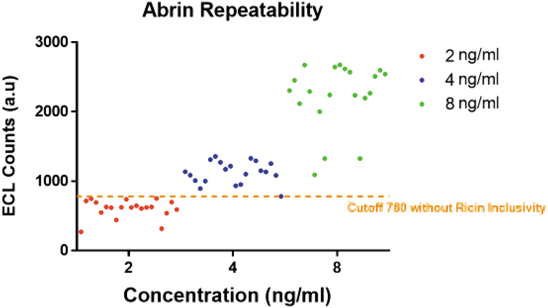
Repeatability test results performed for abrin concentrations 2, 4, and 8 ng/mL, using a cutoff of 790. Abbreviation: ECL, electrochemiluminescence.
The data obtained in the inclusivity study provided an opportunity to compare the variability between the counts of triplicate tests by a single operator and the variability in the counts between 3 operators. Replicates differed by an average of 9.93% (variance 28,349, SD 5324) while differences between operators differed by an average of 21.21% (variance 133,214, SD 11,542).
Summary of Informational Panel Results
Using a cutoff of 857 counts.
Abbreviation: SD, standard deviation.
Discussion
Several methods have been developed to detect abrin in food, beverages, and environmental sample matrices. Detection in food and/or environmental sample matrices may require extraction or elimination of interfering factors before analysis. 26 Methods include real-time polymerase chain reaction with a limit of detection of approximately 1.2 copies of abrin DNA in crude preparations of abrin. 27 However, this method can detect only the presence of targeted DNA sequences and not the presence of the toxin protein and whether it is active. A method involving immuno-extraction followed by quantitative mass spectrometry has been developed that can be used with complex matrices. 28 Mass spectrometry has also been used to measure the activity of type 2 RIPs such as ricin. 29 However, antibody-based assays in various formats remain the gold standard for the detection and identification of abrin in both food and environmental samples.16,17,30-32
ECL technology for antigen and antibody detection is similar to enzyme-linked immunosorbent assays except that the detector antibody is directly labeled with a chemiluminescent label, such as ruthenium, whereas in enzyme-linked immunosorbent assays it is typically labeled with horseradish peroxidase or alkaline phosphatase.33,34 Because of its small size, ruthenium can be easily conjugated to any protein ligand without affecting immunoreactivity or solubility of the protein. The MSD ECL technology employs 96-well assay plates that include integrated electrodes located at the bottom of the wells. When an electric field is applied to the electrode, a rapid electron transfer reaction between the substrate (tripropylamine) and the ruthenium atom occurs, resulting in the production of photons, which in turn are sensed by the photon detector. The measurement of a single sample can be repeated numerous times in the reader, because the electron transfer photon-release reaction regenerates the ruthenium resulting in an amplified signal and potentially greater sensitivity. Detection limits of 200 fmol/L are feasible with ECL, with a linear dynamic range spanning 6 orders of magnitude. 35 ECL assays have been used for the sensitive detection of threat agents and toxins in various matrices.17,33,34,36,37 In general, these assays are simple, rapid, and sensitive; however, assay sensitivities may vary depending on the sample matrices encountered and assay performance (ie, sensitivity and specificity) may depend on the capture and detection antibodies. 38
We conducted a 7-phase evaluation of the performance of a custom prepared MSD Abrin/Ricin assay. The range finding study revealed a linear dynamic range spanning 4 orders of magnitude (0.5 to 1,000 ng abrin/mL); however, a hook effect was observed at abrin concentrations higher than 1,000 ng/mL. This could be overcome by a modification of the protocol or by serially diluting unknown samples before analysis. However, at abrin concentrations higher than 50 ng/mL, the ricin spot exhibited high ECL counts, which would be interpreted as a positive result for ricin in an unknown sample. Thus, confirmation of a result indicating the presence of both abrin and ricin may require reanalysis of diluted samples.
A repeatability study was conducted at abrin concentrations of 2, 4, and 8 ng/mL. Twenty replicates at each concentration were run. The abrin cutoff value was determined to be at 780 counts without the inclusion of data from the ricin inclusivity panel and 857 with the inclusion of the ricin inclusivity panel data. Using the cutoff of 780, the limit of detection was determined to be 4 ng/mL, where we observed 20 out of 20 positive results. The standard deviations ranged from 13.8% to 24% of the mean values of the concentrations tested. The repeatability study was performed by multiple operators on different days, which would magnify errors due to pipetting, washing, and sample mixing.
Because of the widespread geographical distribution of A precatorius, we wanted to determine whether the MSD abrin assay would detect the presence of abrin in extracts from geographically diverse cultivars. Eleven geographically diverse cultivars were tested at 1 μg extract protein/mL. If one assumes that 2.4% of the extract protein is abrin plus abrus agglutinin, 39 then 1 μg of extract protein contains approximately 24 ng abrin plus abrus agglutinin. The MSD assay identified abrin in 10/11 cultivars tested. Abrin was not detected in extracts of A precatorius DES West Africa. This extract was positive for abrin plus abrus agglutinin when tested with an LFA. 16 One explanation for this discrepancy is that the West Africa cultivar may produce an isoform of abrin (I, II, or III) that is not recognized by the capture or detector antibody. In support of this possibility is the observation that the abrin control, consisting of isoforms I, II, and III, routinely gives lower ECL counts (ie, <33%) than the ricin control when tested at equivalent concentrations.
The MSD Abrin/Ricin Assay was also tested against extracts from 35 near neighbors and other plant materials and 65 lectins, including wheat germ agglutinin, which had been shown previously to cause false positive results in LFAs.16,24,25 Crude extracts of near neighbors were tested at a protein concentration of 3 μg/mL, while the lectins were tested at 1 μg/mL. All 65 lectins, including wheat germ agglutinin, yielded negative results (585/585) at the concentration tested. Of the 35 near neighbors and other plant materials, all yielded negative test results (315 out of 315). In a previous evaluation of an abrin LFA, positive results were obtained with an extract of A laevigatus 16 ; however, negative results were obtained with the MSD ECL assay. Negative results were also obtained with an extract of A schimperi subsp. Africanus. Other Abrus species such as A pulchellus produce a type 2 RIP.40,41 The A-chain of pulchellin, which is present in the seeds of A pulchellus, exhibits a high (>86%) amino acid sequence identity with the A-chain of abrin-c. 41 A laevigatus also produces a protein that cross-reacts with the abrin antibodies that are used in some assays. 16 The antibody used in the MSD assay does not appear to cross-react with these proteins.
The MSD Abrin/Ricin ECL Assay was also tested against 11 related proteins and toxins including abrin toxoid, abrus agglutinin, ricin toxoid, ricin and ricin subunits, and ricin vaccine candidates at a concentration of 1 μg/mL. Abrus agglutinin, which shares a high degree of homology with abrin, and abrin toxoid produced results that would be interpreted as positive. However, the ECL counts for abrus agglutinin and abrin toxoid were only 5.9% and 3.2% of the abrin control, respectively, suggesting limited cross-reactivity. No reactivity was observed with any of the other proteins or toxins. In contrast, ricin toxoid, RCA60, RCA120, ricin A-chain, deglycosylated ricin A-chain, and the 2 ricin vaccine candidates produced positive results; ricin B-chain was negative. Thus, 1 or both ricin antibodies in this assay were directed against epitopes on the A-chain. ECL counts ranged from 39% to 161% (average 81%) of the ricin control.
As part of this study, we evaluated the ability of this assay to detect abrin in commonly encountered powders spiked with an extract of the Banana Tree Red cultivar of A precatorius. The ECL assay gave positive results with 26/26 spiked powders; no significant inhibitory effects were noted. We also evaluated the ability of this assay to detect abrin in a pooled BioWatch filter extract. The extract alone did not appear to inhibit the ECL assay; however, the presence of the extract reduced abrin-specific counts by 40% to 50%. The reasons for this reduction are unknown currently. However, at the present time, the MSD Abrin/Ricin Assay is not used by first responders in the field to identify these phytotoxins in powders and other environmental samples. It is likely that this assay would be better suited to a laboratory environment to confirm a positive field test or to detect abrin and ricin in food and other matrices.
Conclusion
The MSD Sector PR2 Model 1800 instrument is robust and user-friendly. The Abrin/Ricin ECL Assay can be completed within 4 to 6 hours, including sample preparation time. Based on our evaluation, the Abrin ECL Assay exhibits good sensitivity and specificity. However, some negative aspects were identified during this evaluation. The dynamic range of the assay for quantitation is limited. At high abrin concentrations there is a hook effect, which may be mitigated by an additional wash step or serial dilution of the sample before analysis. The assay has the potential to include other analytes and thus may be a more cost-effective alternative if analytes such as botulinum toxin, staphylococcal enterotoxin B, saxitoxin, and tetrodotoxin are included. There were also problems with the internal controls. For example, the PC-L counts were elevated as the target analyte concentration increased suggesting signal bleed over, which could be misinterpreted as an invalid run based on the manufacturer's guidelines (Figure 6).
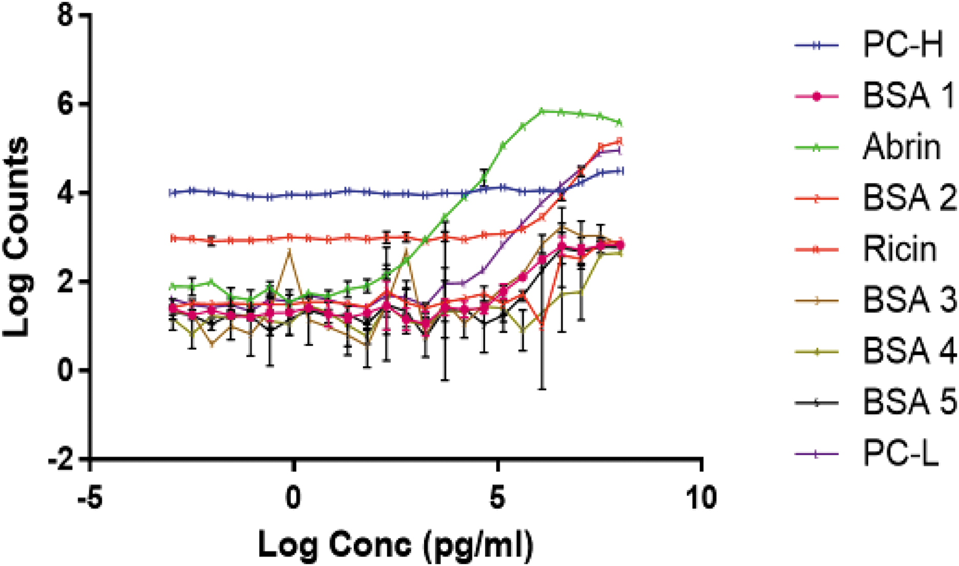
Range finding study results performed for abrin concentration ranging from 9.56 fg/mL to 100 μg/mL in MSD BioD Diluent Extraction Buffer using the standards MSD protocol. Abbreviations: BSA, bovine serum albumin; PC-H, process control high; PC-L, process control low.
Footnotes
Acknowledgments
This research was funded by the Department of Homeland Security Science and Technology Directorate IAA 70RSAT18KPM000127 and US Food and Drug Administration research participation program agreement administered by the Oak Ridge Institute for Science and Education.