
Abstract
We investigated the safety and neuroregenerative potential of an adeno-associated virus (AAV2) containing human glial cell line-derived neurotrophic factor (GDNF) in an MPTP primate model of Parkinson's disease. Dopaminergic function was evaluated by positron emission tomography with 6-[18F]fluoro-
Introduction
Another clinical trial that delivered GDNF by putaminal infusion also failed to show therapeutic effects beyond that shown in a control group (Lang et al., 2006). However, significant increases in positron emission tomography (PET) measures of striatal fluorine-labeled dihydroxyphenylalanine (F-dopa) uptake were observed, suggesting a subtherapeutic functional effect of GDNF. In addition to the lack of therapeutic effects, several patients developed neutralizing anti-GDNF antibodies, raising safety concerns perhaps related to the delivery method. Further concerns were raised after the observation of multifocal cerebellar Purkinje cell loss in monkeys after continuous putaminal infusion of GDNF over a 6-month period (Hovland et al., 2007). Although some PD patients experienced benefit after putaminal infusions of GDNF (Gill et al., 2003; Patel et al., 2005; Slevin et al., 2005, 2006), the overall failure of randomized trials and the safety concerns have prompted the exploration of alternative methods for administering viral vectors in nonhuman primates.
A lentiviral GDNF vector system evinced protective effects of GDNF in both aged nonhuman primates (NHPs) and in young 1-methyl-4-phenyl-1,2,3,6-tetrahydropyridine (MPTP)-lesioned NHPs (Kordower et al., 2000). This study showed protection of dopaminergic innervation of the striatum, improved motor performance, and gene expression for 8 months. Although these findings were promising, lentiviral vectors have not been as well characterized as adeno-associated viral vectors for CNS applications. A study with an adeno-associated serotype 2 viral (AAV2) GDNF vector in marmosets found a protective effect against striatal delivery of 6-hydroxydopamine (6-OHDA) (Eslamboli, 2005). It must be emphasized that such studies did not explore the efficacy of GDNF in the context of a fully lesioned nigra, but only under conditions in which toxin was administered nearly contemporaneously with GDNF, raising the possibility that efficacy was due to protection of existing nigral neurons rather than to regeneration of dopaminergic function (Eberling et al., 1997). This is an important issue, particularly in the context of the failure of a phase 2 clinical trial of neurturin (NTN) delivered by striatal injections of an AAV2 encoding human NTN. The preclinical rationale for this study was based primarily on the neuroprotective effect of AAV2-NTN against 6-OHDA in rats (Gasmi et al., 2007) and MPTP in macaques (Kordower et al., 2006). However, Parkinsonian subjects recruited for the phase 1 and 2 clinical trials of AAV2-NTN (CERE-120) were enrolled at a stage of the disease (stage 3 on the Hoehn and Yahr scale) in which substantial loss of nigral innervation of the striatum had been present for at least 5 years (Marks et al., 2008). On this basis, we regard the failure of this phase 2 study as evidence that any neurotrophic therapy for PD must demonstrate the ability of the agent to stimulate regeneration in a stably lesioned animal. Accordingly, we investigated the safety and neuroregenerative potential of AAV2-GDNF in a stable MPTP primate model of PD. The present report represents an initial analysis of in vivo responses to AAV2-GDNF treatment of MPTP-lesioned NHP over the first 12 months of a 2-year study. Because our PD model is characterized by an extensive nigral lesion in the ipsilateral hemisphere and mild lesioning in the contralateral hemisphere, we were able to model early and advanced PD in the same animal. Our findings suggest that GDNF has the capacity to induce dopaminergic regeneration regardless of the degree of nigral injury, and thus augurs well for broad clinical application of AAV2-GDNF in the treatment of PD.
Materials and Methods
Experimental subjects
The protocol was approved by the Institutional Animal Care and Use Committee at the University of California, San Francisco (San Francisco, CA) and the Animal Welfare and Research Committee at the Lawrence Berkeley National Laboratory. Eleven male rhesus monkeys (8–14 kg) and 1 female rhesus monkey (7 kg) were lesioned with 1-methy-4-phenyl-1,2,3,6-tetrahydropyridine (MPTP) as previously described (Bankiewicz et al., 1986, 2000; Eberling et al., 1998). Briefly, MPTP lesioning consisted of one or two right intracarotid artery infusions of 2.0–4.0 mg of MPTP-HCl followed by additional intravenous administrations of 0.2- to 0.5-mg/kg doses of MPTP-HCl. This method of lesioning produces nearly a complete dopaminergic lesion on the side of carotid artery infusion (ipsilateral side) and a partial lesion on the other side of the brain (contralateral side). Intravenous dosing with MPTP continued until the animal showed bilateral parkinsonian signs and a clinical rating scale (CRS) score between 21 and 26, as described below. Animals were not scheduled for surgery until they had achieved a stable behavioral disability rating in more than 10 assessments over about a 4-month period.
Clinical rating scale
All ratings were performed by a single investigator blinded to the experimental conditions. The modified Parkinson's CRS was developed by the Bankiewicz laboratory, and closely approximates those reported in the literature (Imbert et al., 2000). The scale evaluates 14 parkinsonian features, each of which receives a score from 0 to 3 in order of increasing severity (0, normal; 1, mild; 2, moderate; 3, severe). Individual scores are summed to arrive at a final score. Features evaluated include tremor (right and left sides), locomotion, “freezing,” fine motor skills (right and left hand), bradykinesia (right and left sides), hypokinesia, balance, posture, startle response, and gross motor skills (right and left hands). Normal animals score in the range of 0 to 4, and severely parkinsonian monkeys score over 20. CRS assessments were performed after MPTP lesioning to determine a stable baseline before treatment. Animals were assessed after AAV treatment both with and without
Magnetic resonance imaging
Baseline magnetic resonance imaging (MRI) was performed to enable accurate placement of the infusion cannula in the target structures. During the MRI procedure, NHPs were sedated with ketamine (Ketaset, 7 mg/kg, intramuscular) and xylazine (Rompun, 3 mg/kg, intramuscular). Each animal was positioned in the MRI-compatible stereotactic frame. Ear- and eye-bar positions were recorded, and an intravenous line was installed. Coronal images (1 mm) were collated on a GE Signa 1.5-T system. Images were T1-weighted and taken in three planes with a repetition time of 700 msec, an echo time of 20 msec, and a flip angle setting of 30°. A scanning time of approximately 20 min was employed, with a 15-cm field of view, a 192 matrix, and a 2NEX (number of averages per signal unit). Each scan lasted approximately 20 min. The coronal MRI images were used to determine the three-dimensional structure of the caudate nucleus and the putamen, and surgical coordinates were generated from magnified coronal images (×1.5).
AAV2-GDNF vector construction
The human GDNF cDNA was cloned into an AAV2 shuttle plasmid, and a recombinant AAV2 carrying GDNF under the control of the cytomegalovirus promoter was generated by a triple-transfection technique with subsequent purification by CsCl gradient centrifugation (Matsushita et al., 1998; Wright et al., 2003). AAV2-GDNF stock was concentrated to 1.1 × 10¹³ vector genomes per ml (vg/ml) as determined by quantitative PCR, and then diluted immediately before use to 3.3 × 10¹² vector genomes (VG)/ml in phosphate-buffered saline (PBS)–0.001% (v/v) Pluronic F-68.
Stereotactic surgery and AAV2 infusion
Monkeys received bilateral infusions of AAV2-GDNF (n = 6) or PBS (n = 6) into the putamen at anterior and posterior levels by convection-enhanced delivery (CED). Infusions were performed with a step-design fused silica cannula (Krauze et al., 2005) with an outside diameter (at the tip) of 0.3 mm by a sequential, ascending infusion rate method (0.2, 0.5, 0.8, 1, 1.5, and 2 μl/min for 10 min and 2.5 μl/min for 6 min) in which 75 μl/site was delivered into two sites in each putamen. After infusion, the cannula was left in place for 5 min before it was retracted, and the surgical site was then closed within anatomical layers. This method of delivery has been extensively used and refined by our group over more than a decade (Bankiewicz et al., 2000, 2006b; Forsayeth et al., 2006; Hadaczek et al., 2009).
Positron emission tomography methods and analysis
We have published extensively on the use of 6-[18F]fluoro-
Data were reconstructed via an ordered subset expectation maximization (OSEM) algorithm with weighted attenuation and scatter correction. PET data were quantified by means of a multiple-time graphical analysis (“Patlak plot”) with the time–activity curve for a region, the cerebellum, in which the tracer is nonspecifically bound as the input function. The Patlak model was fit with dynamic data from each region between 24 and 90 min. Parametric images of FMT influx (K i) from both time points were generated and coregistered in order to identify functional changes after treatment. FMT influx in identical regions within the putamen was thereby measured before and after treatment for each monkey. Comparisons were made between baseline and posttreatment K i values by paired t tests. Pearson product–moment correlation coefficients were used to assess relationships between K i values and CRS scores.
Results
PET scans performed both before and after treatment displayed a considerable imbalance in FMT signal between the right and left putamen resulting, respectively, from the full and partial lesioning with MPTP (Fig. 1). AAV2-GDNF-treated monkeys generally displayed a clear bilateral increase in the intensity and distribution of FMT signal 6 months after treatment when compared with the baseline scans, whereas no apparent change in FMT uptake was evident in PBS-treated control monkeys (Fig. 1). Quantification of the PET signals showed a ∼4-fold difference in K i values between the right (0.013 min−1) and left (0.003 min−1) putamen at baseline (Fig. 2A). In comparison, a similar sized cohort of unlesioned animals (n = 4) had a mean K i of 0.023 ±0.001 min−1. Six months after treatment the FMT uptake in AAV2-GDNF-infused monkeys was significantly (p < 0.05) increased bilaterally over baseline levels, with both hemispheres showing a similar increase in K i of ∼0.002 min−1. However, given the large baseline PET difference between hemispheres, the percentage increase in FMT uptake after AAV2-GDNF treatment was considerably greater in the fully lesioned right putamen (54 ± 19%) than in the partially lesioned left putamen (18 ± 5%; Fig. 2B). Control animals did not show a significant change in FMT uptake for either right or left putamen. One control animal was notable in its sharp behavioral improvement after surgery despite the absence of an increase in PET signal.
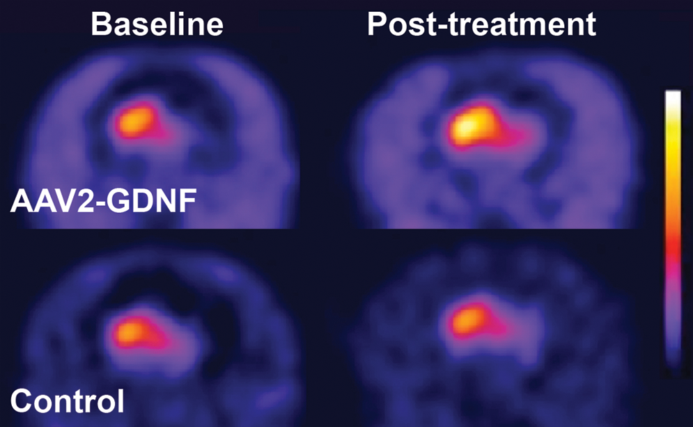
FMT-PET images before and after treatment. Representative parametric PET images show increased FMT uptake bilaterally for a GDNF-treated monkey but minimal change in FMT uptake for a control monkey.

FMT-PET uptake (K
i) in the putamen. (
The extent of motor deficits was assessed at various times throughout the study both with and without
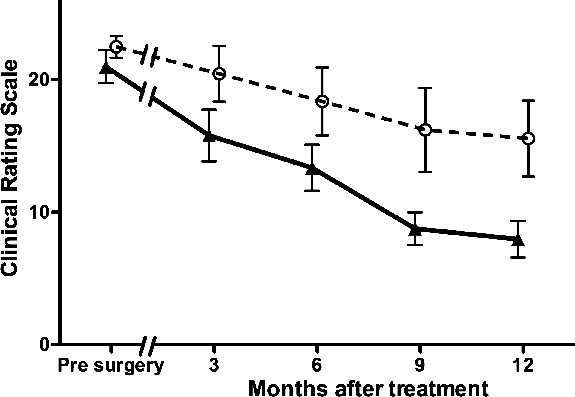
Clinical rating scale (CRS) scores. AAV2-GDNF-treated monkeys (triangles) showed rapid improvement in CRS score after treatment relative to controls (circles). A significant reduction in CRS score was observed to occur during the 9 months after treatment and was maintained at the longest time point of 12 months (two-way ANOVA, AAV2-GDNF vs. controls, p < 0.001). Error bars indicate the SEM.
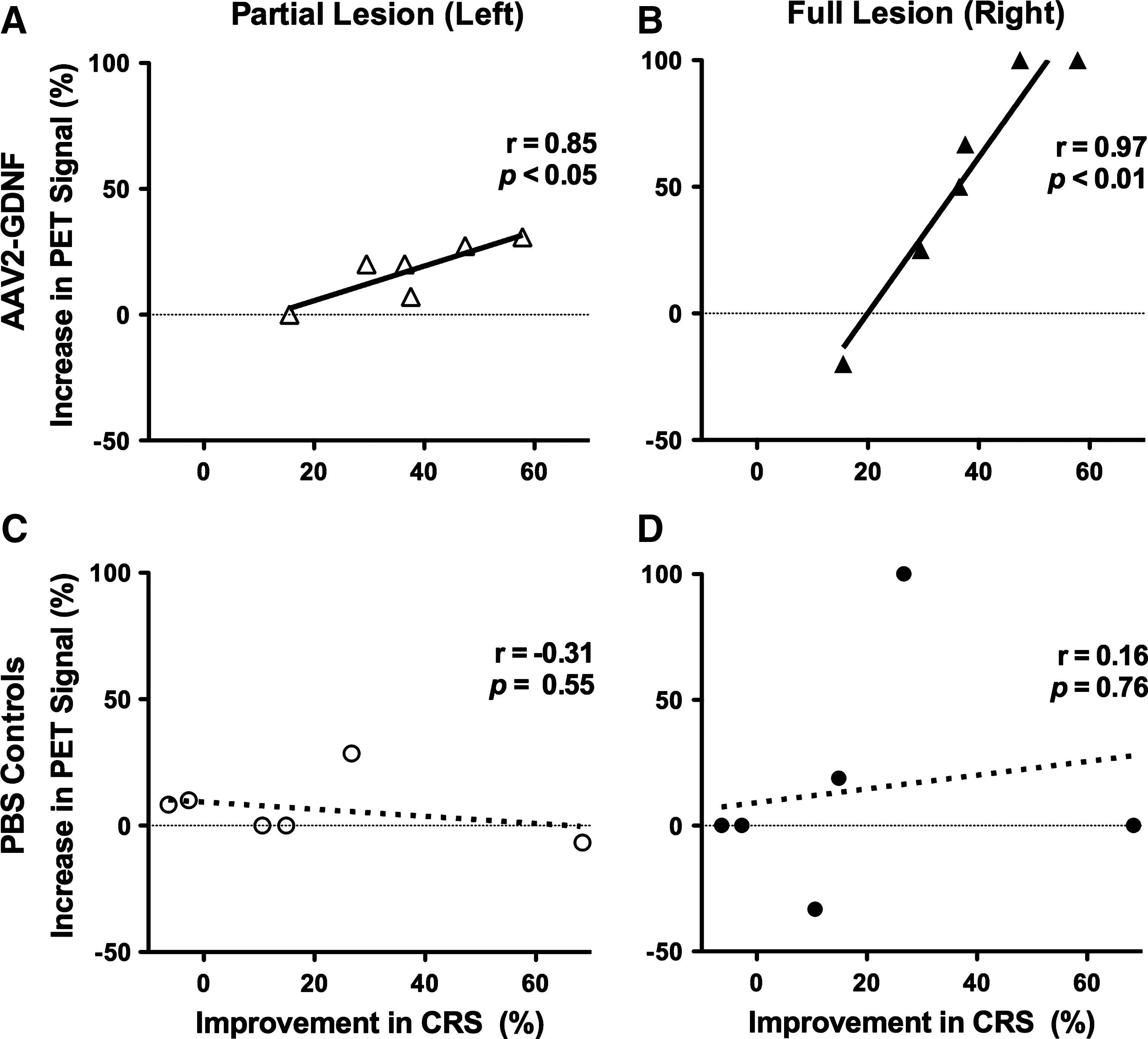
Bilateral increases in FMT uptake correlate with reductions in clinical rating scale (CRS) scores. Shown are independent correlations between the percentage change in CRS score 6 months after treatment and the percentage increase in FMT-PET signal for the left and right putamen of AAV2-GDNF (
Comprehensive biochemical and histological analyses of the brains will be undertaken when the 24-month end-point monkeys are killed and the results will be published separately. Preliminary data at the 6- and 12-month end points showed bilateral expression of GDNF protein within the AAV2-GDNF-infused putaminal regions and an enhancement of tyrosine hydroxylase (TH) expression in both the partially lesioned (left) and fully lesioned (right) caudate–putamen when compared with the control PBS-infused monkeys (Fig. 5). No evidence of MPTP lesioning could be detected by TH immunostaining on the partially lesioned hemisphere of AAV2-GDNF-treated monkeys. Enhanced TH-positive staining in the fully lesioned hemisphere was most evident in the medial–ventral region of the putamen with the MPTP-induced lesioning of the nigrostriatal projection still clearly evident over most of the caudate–putamen. Further analysis of these brains, including histological assessment of the substantia nigra, may provide further insight into the basis of the enhancement and/or regeneration of TH-expressing neurons.
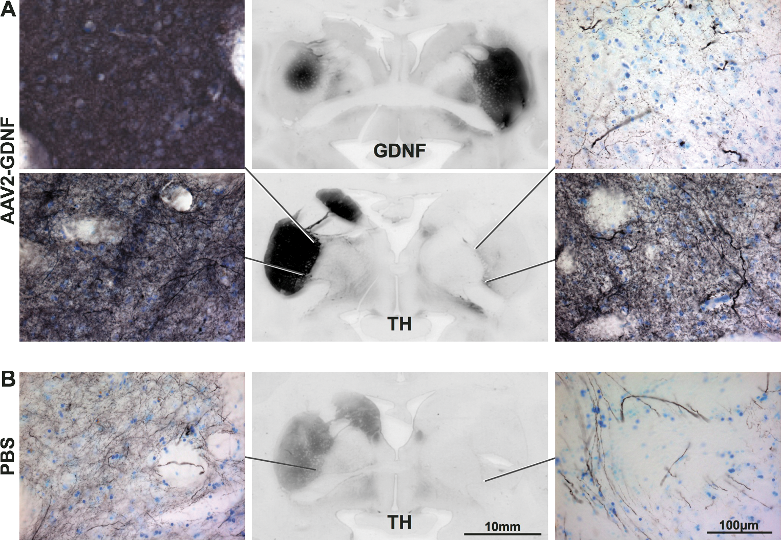
Enhanced expression of tyrosine hydroxylase (TH) after AAV2-GDNF delivery. Immunostaining for TH demonstrated almost complete destruction of TH-positive fibers in the fully MPTP lesioned (right) caudate–putamen compared with the partially lesioned (left) hemisphere. Twelve months after bilateral AAV2-GDNF delivery (
Discussion
We used an overlesioned hemiparkinsonian MPTP primate model to evaluate the safety and efficacy of AAV2-GDNF for PD. In this model, the nigrostriatal pathway is modestly lesioned in one hemisphere and severely lesioned in the other, resembling both early- and late-stage PD, respectively (Eberling et al., 1998). We chose this model to evaluate the effectiveness of GDNF at different stages of the degenerative process. FMT uptake was significantly increased by comparable magnitudes in the putamen in both hemispheres of AAV2-GDNF-treated monkeys, indicating increased dopaminergic activity in the nigrostriatal pathways. In addition, AAV2-GDNF-treated monkeys showed clinical improvement without the development of adverse effects, such as dyskinesia. The control monkeys did not show significant changes in FMT uptake with only slight recovery of clinical measures, as is often seen with the MPTP lesion model.
We reported the results of a parallel study in aged nonhuman primates treated with AAV2-GDNF and showed that this vector directed broadly distributed GDNF expression in the striatum, improved several clinically relevant measures of nigrostriatal function, and displayed no apparent adverse effects (Johnston et al., 2009). The use of aged and MPTP-treated NHP for the clinical development of novel therapeutics for PD addresses different critical issues for potential clinical application. The use of aged primates (>20 years) addresses issues related to the neuroregenerative potential of the aged brain. This is clearly significant because the mean age of onset for PD is about 60. Aged NHP undergo many of the changes seen in aged humans, including the loss of the dopaminergic phenotype in the substantia nigra, and improvements in dopaminergic function have been reported after treatment with growth factors (Kordower et al., 2000; Maswood et al., 2002; Grondin et al., 2003). The MPTP model is important because the clinical syndrome produced closely mimicks that seen in humans with PD, and therefore addresses questions regarding clinical efficacy (Fiandaca et al., 2008). In this respect, our studies resemble similar studies in aged and MPTP-treated, monkeys conducted before the phase 1 trial of CERE-120 (AAV2-NTN) (Kordower et al., 2006; Herzog et al., 2007; Marks et al., 2008; Palfi, 2008).
The present study differs from previous reports by demonstrating the neuroregenerative potential of AAV2-GDNF in a stable primate model of PD. We previously showed temporal changes in nigrostriatal degeneration by evaluating monkeys at various time points after MPTP administration (Eberling et al., 1997). These findings guided subsequent work aimed at neuroprotection by administering growth factors and neurotoxins closely in time in order to prevent the degeneration that occurs several days after a neurotoxic insult to the dopaminergic system (Kordower et al., 2000; Eslamboli et al., 2005). This neuroprotective strategy is problematic because it assumes that the cause of idiopathic PD is a neurotoxic insult. The use of the overlesioned primate model, in which the dopaminergic deficit was well established (>5 months before the administration of AAV2-GDNF), enabled the simultaneous evaluation of the neuroregenerative capacity of severely and mildly injured nigrostriatal pathways. The increased FMT uptake in both hemispheres suggests that AAV2-GDNF may be effective in addressing early and later stages of the disease process when applied to clinical populations. It is important to note in this context that the placebo-controlled phase 2 efficacy study of CERE-120 was reported to have failed to meet its end points (see
In summary, interim in vivo findings in stably lesioned NHPs presented here demonstrate that AAV2-GDNF delivered by CED into the putamen exhibits significant neuroregenerative capacity in both mildly and severely lesioned nigrostriatal pathways. This model more closely recapitulates the situation encountered in PD patients in whom more than 60% of the nigra is lost before symptoms appear. This initial report will be followed by detailed postmortem analyses that, together with our study in aged NHPs (Johnston et al., 2009), will form the basis of an Investigational New Drug application for AAV2-GDNF.
Footnotes
Acknowledgments
The authors thank Avigen for preparing the AAV2-GDNF. This work was supported under a U54 Cooperative Translational Research Program from NIH-NINDS.
Author Disclosure Statement
No competing financial interests exist.