
Abstract
Gene therapy may hold promise as a therapeutic approach for the treatment of intractable ocular diseases, including retinitis pigmentosa (RP). Gene transfer vectors that are able to show long-lasting transgene expression in vivo are highly desirable to treat RP; however, there is a dearth of information regarding long-term transgene expression in the eyes of large animals. We previously reported that the simian immunodeficiency virus from African green monkeys (SIVagm)-based lentiviral vector showed efficient, stable, and safe retinal gene transfer, resulting in significant prevention of retinal degeneration by gene transfer of a neurotrophic factor, human pigment epithelium-derived factor (hPEDF), in rodents. Before applying this strategy in a clinical setting, we here assessed the long-lasting transgene expression of our third-generation SIVagm-based lentiviral vectors in the retinal tissue of nonhuman primates. Approximately 20–50 μl of SIV-EGFP (enhanced green fluorescent protein) or SIV-hPEDF was injected into the subretinal space via a glass capillary tube. To detect EGFP expression in the retina, we used a fluorescence fundus camera at various time points after gene transfer. Human PEDF expression was assessed by immunohistochemical analysis, Western blot assay, and enzyme-linked immunosorbent assay. The retinas demonstrated frequent EGFP expression that was preserved for at least 4 years without significant decline. The expression of hPEDF was stable, and occurred mainly in the retinal pigment epithelium. The secreted protein was detected in vitreous and aqueous humor. We thus propose that SIVagm-mediated stable gene transfer might be significantly useful for ocular gene transfer in a clinical setting.
Introduction
Using recombinant adeno-associated virus (rAAV) vectors, many reports have demonstrated stable transgene expression in retinal cells, including photoreceptor cells and retinal pigment epithelium (RPE), of nonhuman primate eyes (Bennett et al., 1999; Lotery et al., 2003; Le Meur et al., 2005). On the other hand, there has been only one report assessing the short-term features of lentiviral vector-mediated retinal gene transfer in primates (Lotery et al., 2002). In that previous study, recombinant feline immunodeficiency virus (FIV)-based lentiviral vectors efficiently transduced a variety of retinal cells in nonhuman primates at between 20 and 50 days after subretinal injection. However, long-term retinal gene expression with a lentiviral vector has never been demonstrated in nonhuman primates.
Lentiviral vectors hold promise as a potential treatment modality for ocular diseases. However, the possible risks associated with the use of current human immunodeficiency virus (HIV)-based lentiviral vectors slow their clinical application. We developed a novel lentiviral vector derived from the nonpathogenic simian immunodeficiency virus from African green monkeys (SIVagm), to minimize these safety issues (Nakajima et al., 2000). Our third-generation SIVagm-based lentiviral vectors may have the same gene transfer tropism of conventional HIV-based lentiviral vectors, because they are pseudotyped with vesicular stomatitis virus glycoprotein G (VSV-G). We previously demonstrated efficient and stable retinal gene transfer mediated by SIVagm-based lentiviral vector in rodent retinas, as well as a therapeutic outcome in an animal model of retinal degeneration using the recombinant SIVagm vectors carrying human pigment epithelium-derived factor (hPEDF) (Ikeda et al., 2003; Miyazaki et al., 2003, 2008; Murakami et al., 2008).
Our current study attempted to determine the expression efficiency of our third-generation SIVagm-mediated retinal gene in nonhuman primates. We observed stable gene expression (over 3 years) in nonhuman primate retinas via subretinal injection of SIVagm vectors. We thus propose that SIVagm-mediated stable gene transfer might be a useful method for ocular gene transfer in a clinical setting. We are currently conducting a long-term safety study in nonhuman primates. In addition, we are preparing for a clinical application of the gene therapy to treat retinitis pigmentosa (RP).
Materials and Methods
Simian immunodeficiency virus from African green monkeys (SIVagm)-based lentiviral vectors
The third-generation recombinant SIVagm-based lentiviral vectors were produced as previously described (Ohmori et al., 2006; Miyazaki et al., 2008). Briefly, human embryonic kidney (HEK) 293T cells were transfected with the packaging vector, the gene transfer vectors encoding the human pigment epithelium-derived factor (hPEDF) or enhanced green fluorescent protein (EGFP) driven by the cytomegalovirus (CMV) promoter, the Rev expression vector, and the envelope vector, pVSVG (Clontech Laboratories, Mountain View, CA), using lipofection. Twelve hours later, the culture medium was replaced to start harvesting viral particles. Harvesting was undertaken at 48 hr, and viral particles were concentrated by ultracentrifugation. The U3 region in the 3′ and 5′ long terminal repeats (3′ and 5′ LTRs) of SIVagm was deleted to induce self-inactivation. The viral titer was determined by transduction of the HEK 293T cell line as expressed as transducing units per milliliter (TU/ml), and these viruses were kept at −80°C until just before use. Vector stocks were confirmed to be free from endotoxin, and without extraordinary cytotoxicity by simultaneous transfection testing using HEK 293T cells and human RPE cells (ARPE-19) obtained from the American Type Culture Collection (Manassas, VA).
Retinal gene transfer in nonhuman primates
All nonhuman primates were cared for in accordance with the Association for Research in Vision and Ophthalmology (ARVO, Rockville, MD) guidelines for the use of animals in ophthalmic and vision research. This study was conducted according to the Rules for Animal Care and Management of the Tsukuba Primate Research Center (Honjo, 1985) and the Guiding Principles for Animal Experiments Using Nonhuman Primates formulated by the Primate Society of Japan (Primate Society of Japan, 1986). The protocols of the experimental procedures were approved by the Animal Welfare and Animal Care Committee of the National Institute of Biomedical Innovation (Osaka, Japan).
Anesthetization was achieved with ketamine (6 mg/kg) and xylazine (1.2 mg/kg). For all subretinal injections, we used an operating microscope to monitor related events. First, approximately 20 μl of balanced salt solution (BSS: 122.2 mM NaCl, 5.08 mM KCl, 1.05 mM CaCl2, 0.98 mM MgCl2, 25.0 mM NaHCO3, 3.0 mM Na2HPO4, 5.11 mM dextrose; pH 7.4) was injected into the subretinal space through the pars plana via a glass capillary tube. The anterior chamber was then tapped with 26-gauge needles to remove the aqueous humor. Finally, approximately 20–50 μl of SIV-hPEDF solution (low titer, 2.5 × 10⁷ TU/ml; high titer, 2.5 × 10⁸ TU/ml; or maximal titer, 1.0 × 10⁹ TU/ml) or BSS was injected into the subretinal space from the previous retinal hole. The total solution volume injected into the subretinal space was approximately 40–70 μl.
In the gene transfer efficiency study, five Macaca fascicularis were enrolled and SIV-EGFP (low titer, n = 2, #17 right eye [#17R] and #20 left eye [#20L]), SIV-hPEDF (low titer [#6R], high titer [#5L], and maximal titer [#4R], n = 1, respectively) or BSS (n = 1, #6L) was injected. In the acute safety study, nine Macaca fascicularis were enrolled and SIV-hPEDF (low titer [#7R, #25R, and #28R], high titer [#2R, #14R, and #27L], and maximal titer [#10R, #15L, and #23R]) was injected. In the long-term safety study, seven Macaca fascicularis were enrolled and SIV-hPEDF (low titer [#22R, #24R, #32L, and #33R] and high titer [#29L, #30L, and #31R]) was injected.
Detection of EGFP with fundus camera
Ophthalmoscopy and fundus photography were performed at various time points after gene transfer (days 14, 30, 90, 180, 270, 360, 540, 720, 900, 1080, and 1450). To detect EGFP expression in the retina, we used a portable fundus camera (RC-II; Kowa, Tokyo, Japan) with interference filters (Z6, Z7; Spectrotech, Lincoln, MA). The animals were anesthetized, their pupils were dilated with 2.5% phenylephrine (Santen, Osaka, Japan), and they were placed on a bed.
Immunohistochemistry
An immunohistochemical analysis was performed to evaluate therapeutic transgene expression. The eyes were enucleated and fixed with ice-cooled 4% paraformaldehyde in 0.1 M phosphate-buffered saline (PBS) for 1 day at 4°C. They were then mounted in paraffin, and 5-μm-thick sections were made and allowed to dry overnight. The specimens were incubated overnight at 4°C with mouse monoclonal anti-human PEDF IgG antibody (Chemicon International/Millipore, Temecula, CA) diluted 1:400. Signals were then developed with an EnVision/AP (Dako, Carpinteria, CA) and a Vector red alkaline phosphate substrated kit (Vector Laboratories, Burlingame, CA). The sections were counterstained with hematoxylin, if necessary.
Western blot analysis
Vitreous body and aqueous humor samples were obtained from the enucleated eyes in the acute safety animals 90 days after treatment. Each protein from vitreous body or aqueous humor sample was separated on a sodium dodecyl sulfate–10% polyacrylamide gel, and proteins were transblotted. After blocking with 3.0% nonfat dried milk, the membrane was reacted with a mouse monoclonal anti-human PEDF IgG antibody (Chemicon International/Millipore) diluted 1:1000. Immunoreactivity for human PEDF was visualized by ECL Plus (Amersham Biosciences, Buckinghamshire, UK). For the positive control, a recombinant human PEDF (Chemicon International/Millipore) and human vitreous humor sample were used.
Enzyme-linked immunosorbent assay
To measure the secreted human PEDF levels in the aqueous humor samples in the long-term safety study animals at various time points (days 360, 540, 720, and 1080), commercially available enzyme-linked immunosorbent assay (ELISA) systems (Chemicon International/Millipore) were used according to the manufacturer's instructions. The concentration of each protein was standardized by the concentration of total protein. Monkey PEDF could not be detected in this ELISA system.
Results
SIV-mediated stable GFP gene expression in nonhuman primate retinas
Previously, we demonstrated gene expression of more than 1 year in rodent retinas, using our simian immunodeficiency virus from African green monkeys (SIVagm)-based lentiviral vectors. In the present study, we examined whether stable gene expression could be achieved in the retinas of nonhuman primates. Gene expression in the same eyes was evaluated with enhanced green fluorescent protein (EGFP) as the reporter transgene. We injected a low titer of SIV-EGFP (2.5 × 10⁷ transduction units [TU]/ml), which was previously shown to achieve stable transgene expression without damage to the retinal tissue in rodents, into the subretinal space of the nonhuman primate eyes.
As shown in Fig. 1, extensive EGFP fluorescence was found in the vector-injected retinas of both treated animals (#17R and #20L), and the amount and frequency of fluorescence were maintained for 4 years after gene transfer. No EGFP fluorescence was detected in the control eye (#6L) treated with BSS (data not shown). Depigmentation and/or hyperpigmentation of the retinal pigment epithelium were observed at the site of injection of vector solution, by color fundus photography on day 1450 (Fig. 1c and f, arrows). We currently have no histological data, but we will gather this information during our continued examination of these animals.

Expression of enhanced green fluorescent protein (EGFP) in the retina of nonhuman primates [(
SIVagm-mediated functional gene expression in nonhuman primate retinas
Next, we tried to assess gene expression and histopathological change in nonhuman primate retinas, using a functional gene encoding human PEDF. hPEDF is one of the neurotrophic factors and a possible agent for our clinical trial of gene therapy in retinitis pigmentosa (RP). We injected three different titers of SIV-hPEDF vector solution into the subretinal space: a low titer (2.5 × 10⁷ TU/ml), a high titer (2.5 × 10⁸ TU/ml), and a maximal titer (1.0 × 10⁹ TU/ml).
As shown in Fig. 2a–c, an abundant amount of hPEDF protein (red staining) was detected in the RPE of the injected area 50 days after injection of the maximal titer of SIV-hPEDF. Artifactual retinal detachment in preparation for paraffin sectioning was observed (Fig. 2a and c). In the rod and cone layer of the retina of the injected area, an abundant amount of secreted hPEDF protein accumulated, just as in human retinal tissue (Fig. 2f and g), and no hPEDF was detected in the peripheral retina (Fig. 2d). No hPEDF signal was detected in retina treated with BSS (Fig. 2e). No apparent retinal structural destruction or severe inflammatory reaction, such as that previously seen after administering a high titer of SIVagm-based lentiviral vector to rodent retinas (Ikeda et al., 2003), was observed after injection of the maximal titer of SIV-hPEDF vector solution, or after low- or high-titer vector administration.
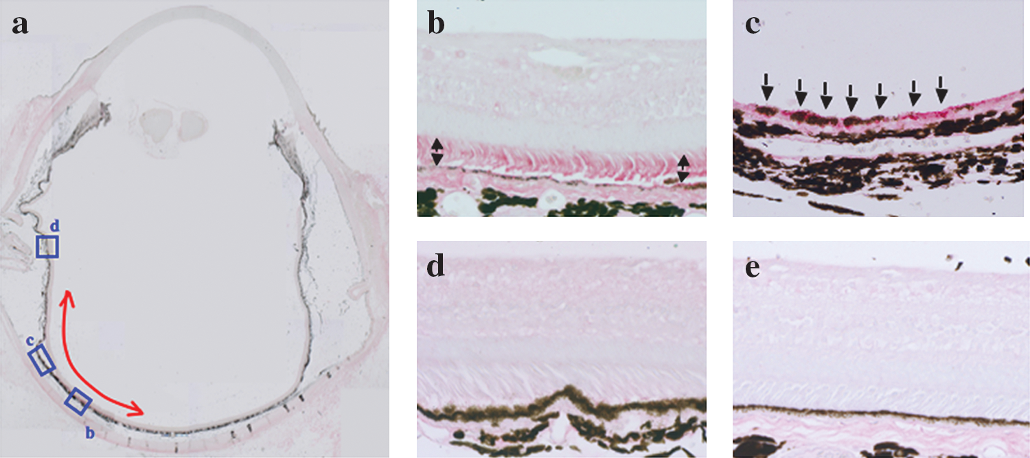
Immunohistochemical stainings against human pigment epithelium-derived factor (hPEDF). (
Secretion of human PEDF protein into vitreous and aqueous humor
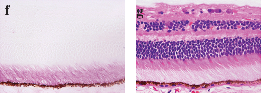
PEDF is well known as a secretory protein and is abundantly present in the vitreous and aqueous humor. In Western blot analysis using monoclonal antibody against human PEDF, the samples from buffer- or SIV-EGFP-injected eyes did not show a positive reaction, indicating there was no cross-reaction between human and simian PEDF (Fig. 3). We detected human PEDF, which was secreted from RPE transduced with the SIV-hPEDF vector, both in the vitreous and aqueous humor samples obtained from the acute safety study animals 90 days after vector injection (Fig. 3).
Western blot analyses of human PEDF protein in (
Long-lasting secretion of human PEDF protein into aqueous humor
We are currently conducting experiments to assess the long-term safety of administration of SIV-hPEDF, including the possibility of carcinogenesis, using nonhuman primates. Aqueous humor samples have been collected at regular intervals from the animals enrolled in this long-term safety study. After 3 years of gathering these data, we have continued to detect an abundant level of hPEDF in these samples (Table 1). About 1000–2000 ng of hPEDF protein per milliliter was detected in the aqueous humor samples of the high-titer SIV-hPEDF-treated eyes (#29L, #30L, and #31R). Moreover, we could not detect hPEDF protein in the aqueous humor samples of untreated control eyes (#22L, #24L, #29R, #30R, #32R, and #33L) by the commercial available ELISA system (Chemicon International/Millipore).
Abbreviations: hPEDF, human pigment epithelium-derived factor; L, left; nd, not done; R, right.
Concentration expressed as nanograms per milliliter.
Discussion
In this study, we demonstrated long-term transgene expression of at least 4 years without significant decline, following reporter gene transfer by an SIVagm-based lentiviral vector in nonhuman primate retinas. Moreover, therapeutic gene (hPEDF) expression was also detected in aqueous humor 1080 days after gene transfer.
Previously, we demonstrated stable and efficient retinal gene transfer in rodents via subretinal injection of an SIVagm-based lentiviral vector (Ikeda et al., 2003), and a morphologically and functionally neuroprotective effect of hPEDF gene transfer in a rodent model of photoreceptor degenerative diseases (Miyazaki et al., 2003). Further evaluation in large animals, such as nonhuman primates, must be performed before clinical application, because of the anatomical difference between the retinas of primates and those of other animals, such as rodents. In the case of lentiviral vectors, there has been only one report demonstrating efficient gene transfer to nonhuman primate retinas (Lotery et al., 2002), despite the many reports of efficient transfer by recombinant adeno-associated virus (rAAV) vectors (Bennett et al., 1999; Lotery et al., 2003; Le Meur et al., 2005). Furthermore, the previous report using lentiviral vectors assessed only the short-term efficiency (Lotery et al., 2002). In the present study, on the other hand, we demonstrated that our SIVagm-based lentiviral vector has the power to achieve long-term (at least for 1450 days) transgene expression in nonhuman primate retinas (Fig. 1), just as in our previous study in rodents (Ikeda et al., 2003).
We next assessed therapeutic gene expression in nonhuman primates. This was done in preparation for a future clinical trial, because almost all previous works have assessed only the expression of reporter genes, such as EGFP and β-galactosidase (Bennett et al., 1999; Lotery et al., 2002, 2003; Le Meur et al., 2005). In the present analysis, transgene expression was immunohistochemically observed only in the retinal pigment epithelium (RPE) 50 days after injection (Fig. 2a–c), in agreement with our previous results in rodent retinas (Ikeda et al., 2003; Miyazaki et al., 2003). However, the previous report showed that recombinant FIV vectors could transduce a variety of retinal cells via subretinal injection (Lotery et al., 2002). The exact reason why our SIVagm-based lentiviral vectors could not transduce other retinal cells is unclear, but the interphotoreceptor matrix (IPM) is well known to be a physical barrier blocking lentiviral vector-mediated gene transfer into photoreceptor cells (Gruter et al., 2005). One possible explanation is that the total amount of vector particles (average volume, 66 μl; titer, 5.0 × 10⁸ to 5.0 × 10⁹ IU/ml) injected into subretinal spaces in the previous study was greater than that used here (volume, 20–50 μl; titer, 1.0 × 10⁹ TU/ml). Moreover, we should consider the sensitivity of our antibody to detect the human PEDF protein. Thus, further studies will be needed to clarify gene transfer properties, and we will check histological sections of EGFP gene-transferred retinal tissue after a 5-year observation period.
Immunohistochemically, human PEDF protein (red staining) was seen in the rod and cone layer of the retina, at the injected area (Fig. 2a and b). This finding suggests that human PEDF secreted from the RPE probably accumulated, because it is well known that PEDF is deposited on the extracellular matrix between the retinal pigment epithelium and the neural retina (Fig. 2f) (Tombran-Tink et al., 1995; Wu et al., 1995; Becerra et al., 2004, 2008). Western blot assay showed that hPEDF protein was detected into both vitreous and aqueous humor 90 days after vector injection (Fig. 3). Amazingly, even 1080 days after administration, a large volume of secreted hPEDF protein was detected in the aqueous humor of other nonhuman primates in the long-term safety study (Table 1). These findings may indicate that a large amount of therapeutic protein is secreted from the RPE over a long period of time. The median aqueous humor PEDF concentration in RP patients (0.24 μg/ml) is lower than in normal cataract patients (0.86 μg/ml) (Ogata et al., 2004). At least 1 μg of hPEDF per milliliter was contained in the aqueous humor of eyes treated with high-titer SIV-hPEDF, suggesting SIV-hPEDF administration could recover hPEDF up to normal level.
Becerra and colleagues previously reported that the same monoclonal antibody to hPEDF (Chemicon International/Millipore) immunoreacted with monkey PEDF in a Western blot assay and immunohistochemically (Becerra et al., 2004). However, we could not detect monkey PEDF (Fig. 2d and e, Fig. 3, and Table 1). As shown in Fig. 2f and g, our immunohistochemistry worked well in human retinal tissue. Moreover, samples from buffer- or SIV-EGFP-injected eyes and untreated control eyes did not show a positive reaction by immunohistochemistry, Western blot assay, or ELISA (Chemicon International/Millipore). The exact reason for this discrepancy is unclear at present.
In summary, our current study using nonhuman primates suggests that the SIVagm-based lentiviral vector may be a useful tool for long-term transgene delivery to retinal tissue and may thus be clinically applicable for chronic progressive ocular diseases. We are currently performing a preclinical safety study to estimate local and systemic acute toxicity and carcinogenesis over the long term. The safety study is being conducted with nonhuman primates given a subretinal injection of our third-generation SIV-hPEDF, in preparation for a clinical trial in patients with retinitis pigmentosa.
Footnotes
Acknowledgments
The authors thank H. Fujii for assistance with the experiments. KN International provided language assistance. This work was supported in part by a Grant-in-Aid (to Y.I., Y.Y., K.S., and T.I.) from the Japanese Ministry of Education, Culture, Sports, Science, and Technology (#15209057, #16390118, #17689047, and #19209012) and by a Grant of Promotion of Basic Science Research in Medical Frontier of the National Institute of Biomedical (Y.Y., K.S., and T.I. project no. 21).
Author Disclosure Statement
Yoshikazu Yonemitsu is a member of the Scientific Advisory Board of DNAVEC Corporation.