
Abstract
The use of nonionic polymeric micelles orally to protect and deliver plasmid DNA in vivo was investigated. Parathyroid hormone (PTH)(1–34) gene (179 bp) was inserted into a human cytomegalovirus promoter (PCMV) and E. coli competent cells were used to amplify the cDNA. Polymeric micelle formations (100 μl) formed from PCMV-PTH(1–34) cDNA (7.2 μg/μl) and 6% (w/v) polyethylene oxide-polypropylene oxide-polyethylene oxide (PEO-PPO-PEO) was administered at 8-hr intervals for 48 hr and then at 8-hr intervals for 24 hr weekly for 3 weeks. Parathyroidectomized rats receiving 150 μl of EDTA (10 mM) before each dose of formation served as the study group; rats receiving drinking water, EDTA (10 mM), PCMV-PTH(1–34) cDNA and PCMV-PTH(1–34) cDNA plus EDTA at the same amount and time intervals served as the control groups. Serum levels of calcium and PTH(1–34) were measured weekly for 4 weeks. Immunohistochemical stain for PTH(1–34), reverse transcriptase polymerase chain reaction for PTH(1–34) mRNA and the relative density of PTH(1–34) mRNA were performed at 2 and 4 weeks after oral gene therapy in different organs. One third to three of five rats in the control groups died after parathyroidectomy. Serum levels of calcium and PTH(1–34) were higher in the study than in the control groups. In the study group, positive stain of PTH(1–34) and PTH(1–34) mRNA could be found in those organs. Relative densities of PTH(1–34) mRNA were higher in the study than in the drinking water group in different organs. Oral gene therapy can maintain calcium and PTH(1–34) levels in parathyroidectomized rats.
Introduction
Although permanent hypoparathyroidism after thyroid or parathyroid surgery rarely occurs in general practice, it is a troubling problem. Long-term vitamin D3 and calcium therapy is inconvenient and causes hypersecretion of gastric juice, constipation, and milk-alkali syndrome (Tal and Powers, 1996). Long-term parathyroid hormone (PTH) therapy is rarely applied because PTH has a short half-life (2–4 min) and must be injected daily (Winer et al., 1996, 2003; Shoback, 2008). Gene therapy for hypoparathyroidism through gene transfer to skeleton cells or stem cells has been reported previously (Zhou et al., 2005; Wen et al., 2006; Zhu et al., 2006). However, gene delivery through the gastrointestinal tract not only has many potential applications, but is also less invasive and easier to perform (Alton et al., 1993).
The purpose of this study was to investigate the feasibility of using nonionic polymeric micelles orally to protect and deliver plasmid DNA containing human cytomegalovirus promoter (PCMV)-parathyroid hormone (PTH)(1–34) in vivo. We assessed the expression of a PTH(1–34) gene in gastrointestinal tissues, the expression after gene distribution to other organs through the circulation, as well as serum PTH(1–34) levels of parathyroidectomized rats given oral gene therapy (21 days).
Materials and Methods
Nonionic polymeric micelles of polyethylene oxide-polypropylene oxide-polyethylene oxide (PEO-PPO-PEO) copolymer with an average molecular mass of 8400 Da, was obtained from BASF (Ludwigshafen, Germany). EDTA was obtained from Sigma-Aldrich (St. Louis, MO). All other chemicals used in this study were of analytical grade without further purification.
Animals
Male Sprague-Dawley rats were used throughout the experiments. The animals were purchased from the National Science Council (Taipei, Taiwan) animal center and housed in a controlled environment at 24 ± 1°C, humidity of 55 ±5%, with a circadian light rhythm of 12 hr.
Standard chow and water ad libitum were available to the rats. The study was performed according to the guidelines provided by the experimental animal laboratory and approved by the animal care and use committee.
Five groups were designed as follows: parathyroidectomy treated with oral PCMV-PTH(1–34) cDNA in PEO-PPO-PEO polymeric micelle formations plus EDTA as the study group (n = 9); parathyroidectomy treated with oral drinking water (n = 9); parathyroidectomy treated with oral EDTA (n = 9); parathyroidectomy treated with oral PCMV-PTH(1–34) cDNA; and parathyroidectomy treated with oral PCMV-PTH(1–34) cDNA plus EDTA.
Plasmid DNA
The plasmid carrying full-length human PTH(1–34) under the control of CMV promoter (Mission Biotech, Taipei, Taiwan) was cloned. The PTH(1–34) gene (179 bp) was inserted into PCMV (4219 bp) via the EcoRI and BamHI cloning sites, and competent Escherichia coli cells were used to amplify the cDNA. The cDNA concentration was measured by ultraviolet absorption at 260 nm. The stability of plasmid DNA in cDNA/PEO-PPO-PEO polymeric micelle formulation was determined by electrophoresis immediately after preparation, after 2 days of storage at room temperature, and after three freeze-thaw cycles (Liaw et al., 2001).
Preparation of DNA/PEO-PPO-PEO polymeric micelles
All DNA [PCMV-PTH(1–34)]/PEO-PPO-PEO polymeric micelles were freshly prepared in collaboration with J. Liaw and S.-F. Chang at Taipei Medical University (Taipei, Taiwan).
Polymeric micelles were formed with 6% (w/w) PEO-PPO-PEO in water. A pyrene fluorescence probe was used to determine the formation of micelles (Chang et al., 2004). PCMV-PTH(1–34) cDNA (7.2 μg/μl) was gently mixed with PEO-PPO-PEO polymeric micelles in a vial for 2 hr at 25°C.
Oral gene transfer in vivo
After successful parathyroidectomy, Sprague-Dawley rats were fasted for 24 hr before the experiments but allowed free access to water. Polymeric micelle formations (100 μl) formed from PCMV-PTH(1–34) cDNA (7.2 μg/μl) and 6% (w/v) PEO-PPO-PEO were administered at 8-hr intervals (6 a.m., 2 p.m., and 10 p.m.) for 48 hr as the first course and for 24 hr on days 7, 14, and 21. Rats receiving 150 μl of EDTA (10 mM) 10 min before and concurrent with administration of PCMV-PTH(1–34) cDNA/PEO-PPO-PEO polymeric micelles served as the study group. Rats receiving 150 μl of drinking water, rats receiving 150 μl of EDTA (10 mM), rats receiving 100 μl of PCMV-PTH(1–34) cDNA, and rats receiving 100 μl of CMV-PTH(1–34) cDNA plus 150 μl of EDTA (10 mM) at the same intervals served as the control groups.
Parathyroidectomy
Under sodium pentothal anesthesia, a longitudinal skin incision was made at the anterior neck. The right and left parathyroid glands were exposed and removed under microscopic examination ( × 16). After this procedure the rats were returned to the animal center, until the studies were arranged.
Serum levels of PTH(1–34), calcium, and phosphorus
Blood samples were obtained for PTH(1–34), calcium, and phosphorus determinations, before oral gene therapy (on day 0, before the first oral gene therapy) and on day 7 (after the second gene therapy), day 14 (after the third gene therapy), day 21 (after the fourth gene therapy), and day 28.
Levels of PTH(1–34) were measured by enzyme-linked immunosorbent assay (ELISA) with a human PTH(1–34) kit (Peninsula Laboratories, San Carlos, CA). The minimal detectable concentration was 0.04–0.06 ng/ml, with a detectable range of 0–25 ng/ml. Serum levels of calcium and phosphorus were measured by a cresophthalein complexion and phosphomolybdate UV method. For the determination of PTH(1–34) gene expression, rats were killed 14 or 28 days after oral gene therapy for the detection of mRNA by reverse transcriptase-polymerase chain reaction (RT-PCR).
Detection of PTH(1–34) mRNA in tissues by RT-PCR
Two and 4 weeks after the first oral dosing of PCMV-PTH(1–34) cDNA/PEO-PPO-PEO polymeric micelles, total RNA was extracted from duodenum, liver, spleen, heart, lung, and kidney with TRIzol reagent (Invitrogen Life Technologies, Carlsbad, CA) according to the manufacturer's instructions. cDNA was prepared with SuperScript II reverse transcriptase (Invitrogen Life Technologies) and stored at −20°C. A 179-bp segment of the PTH(1–34) gene was amplified by polymerase chain reaction (PCR) as described subsequently. Sequences of the primer pair for amplification of the transferred gene were as follows: 5′-AGTTTACTCAGCATCAGCTACTA-3′ and 5′TTCTTAACAGATTTCCCATCCGA-3′ (Mission Biotech). For PCR, the thermocycler was set at 94°C for 5 min, 94°C for 30 sec, 51.6°C for 30 sec, 72°C for 30 sec, 72°C for 7 sec, and 4°C to end. The products of PCR were separated by electrophoresis on 2% agarose gels, and stained with ethidium bromide to visualize and quantify the bands by computerized densitometric scanning, using BioDoc-It and VisiDoc-It (UVP, Upland, CA) and LabWorks (version 4.6) software (UVP).
Immunohistochemical staining with PTH(1–34) antibody
After formalin fixation, paraffin-embedded tissue sections were deparaffinized in xylene and dehydrated in a graded ethanol series. Endogenous peroxide was quenched with 3% hydrogen peroxide in methanol. After incubation of the tissue with rabbit anti-human parathyroid hormone(1–34) serum (diluted 1:600; Phoenix Pharmaceuticals, Belmont, CA) horseradish peroxidase (Zymed Laboratories/Invitrogen, San Francisco, CA) was added, and after a wash step chromogen was then added. The peroxide then catalyzed the substrata and converted the chromogen diaminobenzidine to a brown-colored deposit.
Statistical analysis was performed with the Statistical Product and Service Solution (SPSS) version 10.0 software package (SPSS, Chicago, IL). All data are expressed as means ± SD. The Wilcoxon signed ranks test was used for nonparametric data and repeated measures of analysis of variance was used for between-group analysis. To determine whether a group was significantly different from the others, simultaneous multiple comparison Bonferroni techniques were used. A p value less than 0.05 was considered significant.
Results
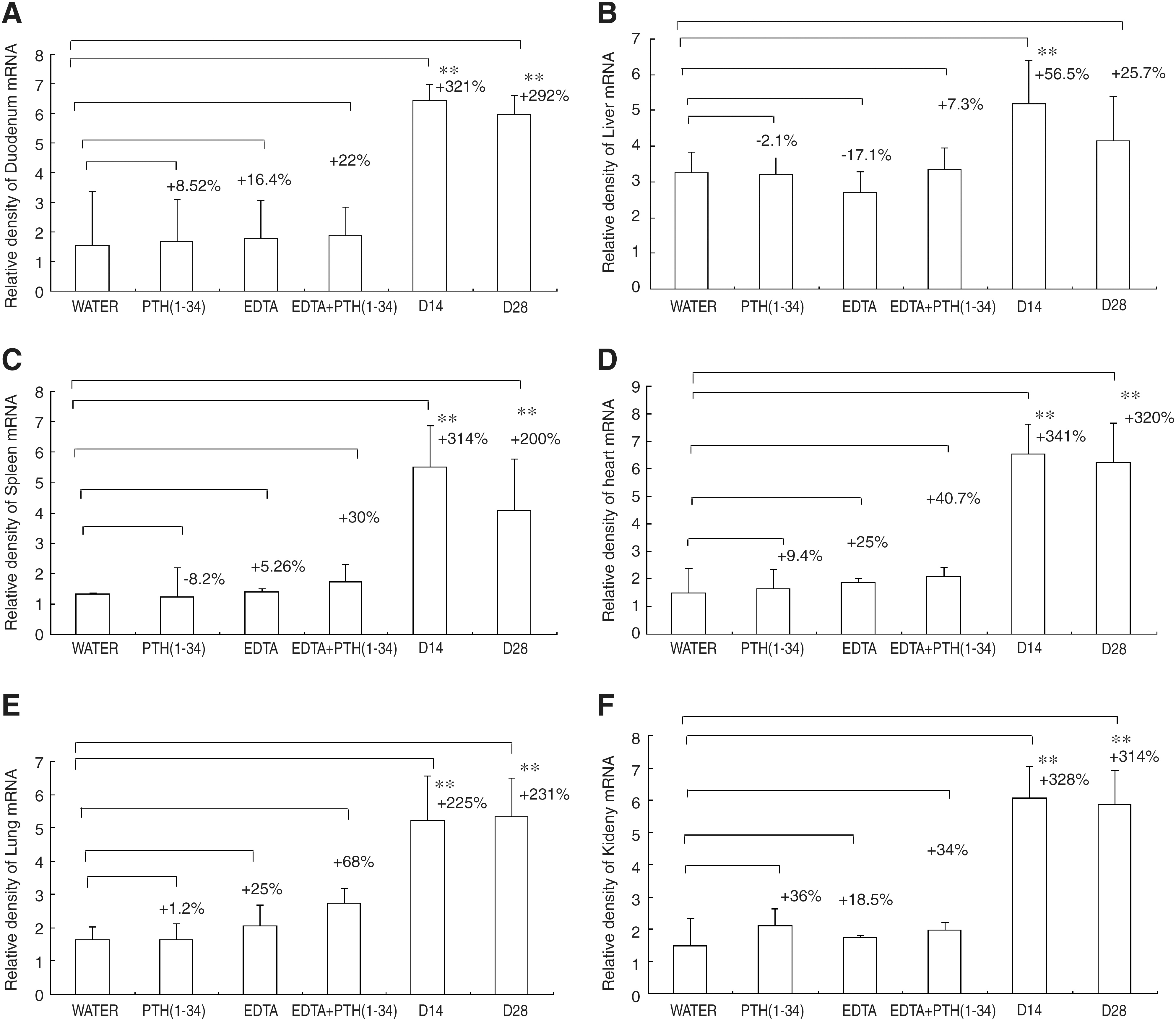
All rats in the study group survived well with normal appetites and activities. Although two of three rats in the PCMV-PTH(1–34) cDNA, EDTA, and PCMV-PTH(1–34) plus EDTA groups survived, their appetites and activities decreased and three of five rats in the drinking water group died of exhaustion 2 to 3 weeks after the first oral gene therapy, due to poor appetite. Serum levels of calcium were significantly higher in the study group than in the control groups (Table 1) (all p < 0.001). Serum levels of phosphorus were significantly different between the study and drinking water groups (Table 2) (p = 0.004). Serum levels of PTH(1–34) were significantly higher in the study group than in the control groups (Table 3) (all p < 0.001). In the study group, both serum calcium and PTH(1–34) levels were higher on days 7, 14, 21, and 28 than at baseline (day 0) (Tables 1 and 3). Immunohistochemical staining was positive for PTH(1–34) in duodenum, liver, spleen, heart, lung, renal glomeruli, and renal tubules on days 14 and 28 in the study group, but not in the control groups (Fig. 1A–C). PTH(1–34) mRNA (179 bp) was detected in duodenum, liver, spleen, heart, lung, and kidney 14 and 28 days after oral gene therapy but was not detected in the control groups (Fig. 2). The relative density of PTH(1–34) mRNA (179 bp) in various organs was higher at 14 and 28 days in the study group than in the drinking water group. There was no significant difference between days 14 and 28 regarding the relative density in the study group (Fig. 3A–F).

Top: Immunohistochemical staining with PTH(1–34) antibody reveals PTH(1–34)-positive cells in the duodenal mucosa in the oral PCMV-PTH(1–34) cDNA/PEO-PPO-PEO plus EDTA (study group), at (
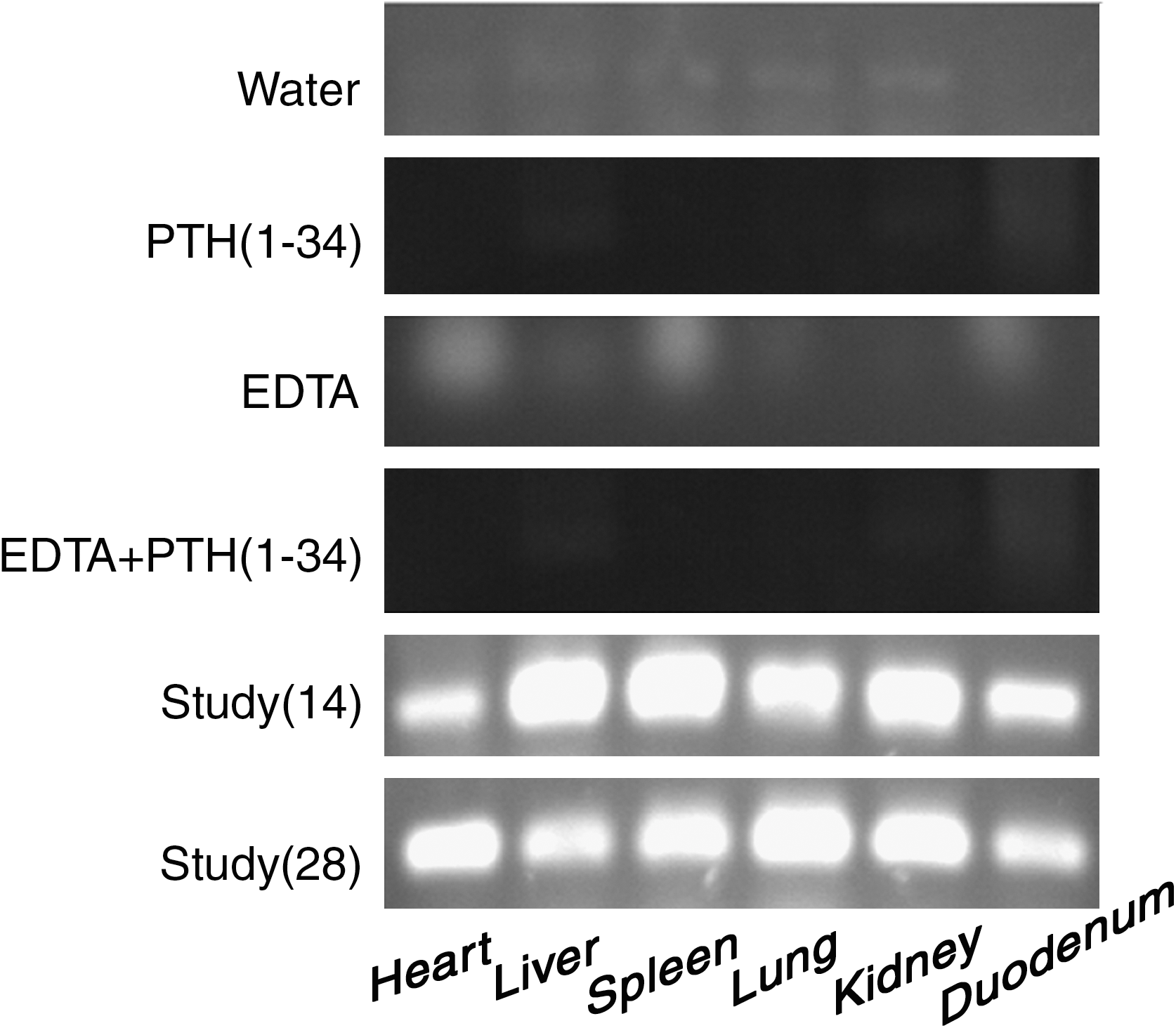
RT-PCR of tissue (179 bp) mRNA in duodenum, liver, spleen, heart, lung, and kidney after 14 and 18 days of weekly oral PCMV-PTH(1–34) cDNA/PEO-PPO-PEO plus EDTA (study group). No mRNA was detected in the drinking water, EDTA, PTH(1–34) cDNA, and PTH(1–34) cDNA plus EDTA groups.
(
Serum Calcium Levels after Parathyroidectomy: Before and 1, 2, 3, and 4 Weeks after Weekly Oral Gene Therapy in Various Groups a
Abbreviations: PCMV, human cytomegalovirus promoter; PEO, polyethylene oxide; PPO, polypropylene oxide; PTH, parathyroid hormone.
All data represent means ± SD.
Using the Wilcoxon signed rank test, all p ≤ 0.011 compared with levels before oral gene therapy.
Using repeated measures analysis of variance, all p < 0.001 compared with PTH (1–34) cDNA/PEO-PPO-PEO plus EDTA group.
Serum Phosphate Levels after Parathyroidectomy: Before and 1, 2, 3, and 4 Weeks after Weekly Oral Gene Therapy in Various Groups a
All data represent means ± SD.
Using repeated measures of variance, p = 0.004 compared with PCMV-PTH(1–34) cDNA/PEO-PPO-PEO plus EDTA group.
PTH(1–34) Levels after Parathyroidectomy: Before and 1, 2, 3, and 4 Weeks after Weekly Oral Gene Therapy in Various Groups a
All data represent means ± SD.
Using the Wilcoxon signed ranks test, all p ≤ 0.036 compared with levels before oral gene therapy.
Using repeated measures analysis of variance, all p < 0.001 compared with PCMV-PTH(1–34) cDNA/PEO-PPO-PEO plus EDTA group.
Discussion
A growing number of polymer therapeutics have been approved for clinical use in the treatment of cancer, infections, and genetic disease (Liaw and Lin, 2000; Kabanov et al., 2002). Polymeric micelles have been evaluated in multiple pharmaceutical applications as drug and gene delivery systems as well as for diagnostic imaging as carriers of various contrasting agents (Yokoyama, 1992; Kwon et al., 1994; Kwon and Katota, 1995; Stolnik et al., 1995; Alakhov and Kabanov, 1998; Kwon and Okano, 1999; Forchillin, 2001; Huang et al., 2008; Zhao et al., 2009). It has been hypothesized that the addition of hydrophilic PEO polymers to liposome or polymers can prevent complexes from binding to protein/serum and from being degraded by digestive enzymes (Kwon et al., 1994; Forchillin, 2001).
The surface charge seems to be a requisite structural factor for colloidal carriers, leading to significant interaction with the mucosa of the intestine. Gene transfer by PEO-PPO-PEO polymeric micelles through the gastrointestinal tract is currently under investigation. Because PEO-PPO-PEO has been approved by the U.S. Food and Drug Administration for use in food, toxicity should not be a factor in its use for gene delivery to muscle and in ophthalmic pharmaceuticals (Pepic et al., 2004; Thuret et al., 2005; Roque et al., 2009).
We used PEO-PPO-PEO polymers to increase the oral bioavailability of plasmid cDNA in the intestine. Improved transfection efficiency was achieved by the use of EDTA, which is known to open tight junctions of the duodenum and increase paracellular transport for the further distribution of cDNA to remote organs such as liver, spleen, lung, heart, and kidney (Chang et al., 2004; Tong et al., 2007).
A previous study (Chang et al., 2004) found that 10 mM EDTA and mixtures containing plasmid DNA (0.26 μg/μl) with 6% PEO-PPO-PEO polymeric micelles yielded a significant increase in the reporter vector for 2 days, compared with a DNA concentration of 0.26 μg/μl alone. They used 39 μg of plasmid DNA six times for each study mouse. In this study, the average weight of Sprague-Dawley rats was 15–20 times higher than that of nude mice. Preliminary results with plasmid DNA at 360 μg/100 μl, administered six times (2160 μg), showed poor calcium and PTH(1–34) levels (data not shown). We therefore used 4300 μg for each study rat, about 720 μg/100 μl each time.
After weekly oral gene therapy, serum calcium levels increased significantly 1 week later and could be maintained until 4 weeks later. Serum calcium levels were higher in the study group than in the control groups. Serum PTH(1–34) levels also were higher in the study group than in the control groups. However, serum phosphate levels were lower in the study group than in the drinking water group.
Immunohistochemistry revealed PTH(1–34)-positive staining in the duodenum, liver, spleen, lung, heart, and kidney. PTH(1–34) mRNA studies demonstrated mRNA (79 bp) in the various organs. Relative densities of PTH(1–34) mRNA in those organs were higher on days 14 and 28 in the study group than in the drinking water group. There were no significant differences in results between days 14 and 28 regarding the relative density of mRNA in the study group. Weekly oral gene therapy maintained calcium levels until 4 weeks, but it cannot have the effect of summation of calcium levels. One third to three of five rats in the control groups died 2 to 3 weeks after parathyroidectomy because of hypocalcemia and exhaustion. However, this did not happen in the study group.
Footnotes
Acknowledgments
This work was supported by grants from the National Science Council of Taiwan (NSC96-2314-B-182A-017-MYZ).
Author Disclosure Statement
No competing finanical interests exist.