
Abstract
β-Thalassemia is an anemia caused by a relative excess of α-hemoglobin (αHb) due to absent or reduced β-hemoglobin (βHb) synthesis. In this study, we explore whether the introduction of α-hemoglobin stabilizing protein (AHSP), a chaperone protein for proper folding and stabilization of free αHb in red blood cells, thus aiding hemoglobin A (HbA) assembly, could relieve the pathogenic state of red blood cells in β-thalassemia. For that, a human ahsp vector was constructed to generate transgenic human ahsp mice in a model of βIVS-2-654-thalassemia by microinjecting the vector into fertilized eggs, resulting in the production of double heterozygous mice (h-ahsp +/βIVS-2-654+). Real-time quantitative RT-PCR and Western blot analysis confirmed AHSP expression in three h-ahsp +/βIVS-2-654+ mice. Hematologic determination showed an improvement in the red blood cell indices of these h-ahsp +/βIVS-2-654+ mice. The red blood cell count and hemoglobin level were elevated to various extents as compared with their diseased siblings. A dramatic reduction in anisocytosis in the peripheral blood of h-ahsp +/βIVS-2-654+ mice was observed (16.2 ± 4.6 vs. 30.0 ± 5.2%). Few erythroid precursors appeared in the liver sinusoids of h-ahsp +/βIVS-2-654+ mice. Splenomegaly with extramedullary hematopoiesis was also ameliorated. Significantly, serum iron concentration was remarkably reduced as compared with that of h-ahsp −/βIVS-2-654+ mice (43.2 ± 14.9 vs. 82.4 ± 12.9 μM), and iron deposition in the liver was decreased in h-ahsp +/βIVS-2-654+ mice. All these results suggested amelioration of the anemia phenotype in h-ahsp +/βIVS-2-654+ mice after introduction of the ahsp gene. We therefore propose that an ahsp transgene could provide an adjuvant method for gene therapy of β-thalassemia.
Introduction
Materials and Methods
Construction of ahsp expression vector
The human ahsp gene with its native promoter (from position −184 to + 884, 1068 bp) was amplified from human genomic DNA by nested polymerase chain reaction (PCR), using specific primers that contained PacI and EcoRI cutting sites in the inner pair of primers. Such a design would allow efficient erythroid expression of its transgene (Gallagher et al., 2005). The sequences of the outer primer pair were 5′-GTAATAGGGCTCAGTAAACG-3′ and 5′-AAGCAGGTCACAGGACAA-3′, and those of the inner primer pair were 5′-CC
The previously described amplified fragment was digested with PacI and EcoRI and then cloned into the corresponding sites of the lentiviral vector FUGW (Lois et al., 2002). Eventually, the vector was cut with PacI and PmeI to obtain a fragment containing only h-ahsp (with its promoter), the woodchuck hepatitis posttranscriptional regulatory element (WRE, to enhance expression), and the HIV-1 3′ mini-long terminal repeat (LTR) sequence (for polyadenylation signaling), which was used to generate h-ahsp transgenic mice by the pronuclear microinjection method. The human ahsp gene vector (FA) is shown schematically in Fig. 1.
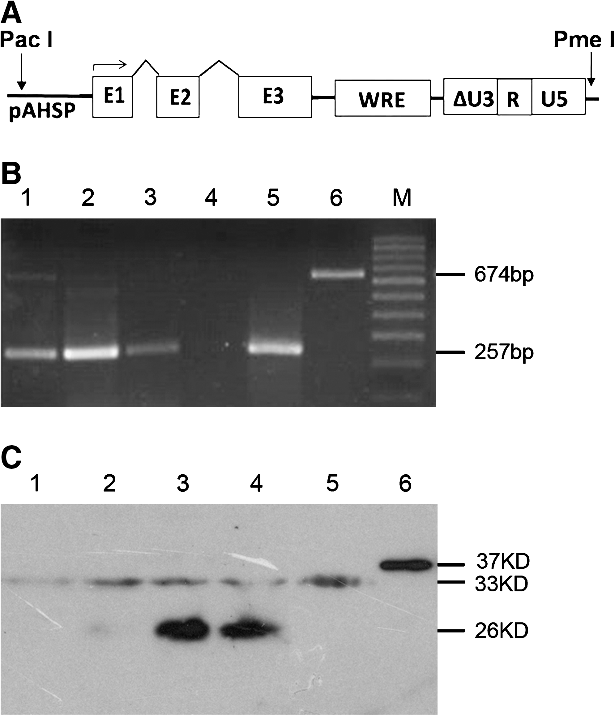
Human ahsp expression cassette and its expression in transgenic mice. (
Mouse strains
βIVS-2-654-Thalassemia mice were obtained from the Jackson Laboratory (Bar Harbor, ME). In this mouse model, the two cis murine adult β-globin genes (i.e., major and minor) have been replaced with a single copy of the human βIVS-2-654-thalassemia gene in one of the two alleles. The same aberrant splicing was observed as in its human counterpart because of a C → T substitution at nucleotide 654 of intron 2 in the β-globin gene. Heterozygous mice produce reduced amounts of mouse β-globin chains and no functional human β-globin, and present an intermediate β-thalassemia phenotype (anemia, splenomegaly, and abnormal hematologic indices) (Lewis et al., 1998). The βIVS-2-654-thalassemia mouse strain used in this study was reviewed and approved by the Review Board of Shanghai Children's Hospital (Shanghai, China).
Generation of transgenic mice
βIVS-2-654-Thalassemia male mice were mated to superovulated wild-type females. Single-cell embryos were collected, injected with ahsp DNA (4 ng/μl) via the pronucleus, and then immediately implanted into the oviduct of pseudo-pregnant wild-type mice and allowed to develop to full term, to provide the founder transgenic mice (F0) (Hogan et al., 1994; Zhang et al., 2006; Xie et al., 2007; Li et al., 2008). F1 and F2 generations were produced by crossing founder (F0) or F1 mice, respectively, with wild-type mice.
DNA analysis
After the pups were weaned (approximately 3 weeks after birth), mouse genomic DNA was isolated from tail tissue. PCR was then performed to determine human ahsp integration, and the alleles of human βIVS-2-654 and murine βmajor , using primer pair 1 (5′-ACAGCCTGTTAGAACTGAAG-3′ and 5′-CACAGCCTAAGGACATGAAG-3′), primer pair 2 (5′-AGTGATAATTTCTGGGTTAAGGT-3′ and 5′-AGGGCCTAGCTTGGACTCAG-3′), and primer pair 3 (5′-AGGCAGCTCACAAGAAGAAG-3′ and 5′-TGGAGACTGCTCCCTAGAAT-3′), respectively. Reactions were performed in a total volume of 25 μl including 2.5 μl of 10 × PCR buffer, 2 μl of MgCl2 (25 mM), 2 μl of dNTPs (each 2.5 mM), 0.6 μl of each primer pair (each primer, 10 μM), and 0.2 μl of Taq enzyme (5 U/μl), adding double-distilled H2O to 25 μl. Thirty cycles were performed: denaturing at 94°C for 45 sec, annealing at 60°C for 45 sec, and extension at 72°C for 60 sec.
Human ahsp mRNA analysis
Total RNA was isolated from fresh murine peripheral blood using TRIzol (Invitrogen, Carlsbad, CA), according to the manufacturer's instructions. After the reverse transcriptase (RT) reaction using Oligo-dT (TaKaRa Bio, Shiga, Japan), human ahsp transcript was amplified with specific primers (5′-ACAGCCTGTTAGAACTGAAG-3′ and 5′-GTTCAGCTCTTGCCGAAGCT-3′) for 30 cycles in a PCR machine (Eppendorf, Hamburg, Germany): denaturing at 94°C for 45 sec, followed by annealing and extension at 70°C for 60 sec.
Real-time PCR analysis
Real-time PCR was performed on the previously described RT product. The sequences of primers and probes were as follows: the primer pair used to quantify murine ahsp: 5′-AGGACCTTTCTGAAGTCCAAAGAG-3′ and 5′-TGATGCCCAGACCCTTTAAGTT-3′, and its probe: 5′-FCCCCAAGCAATACACTGCCCTCCTCP-3′; the primer pair used to quantify human ahsp: 5′-CCTTAATTAAGCTCTTGCCTTCTTGCATTT-3′ and 5′-GGAATTCGGTAGAGTGGCAGGAGCA-3′, and its probe: 5′-FCATCAACTATTACAGGCAGCP-3′. Primers (900 nM), probe (250 nM), and premixed RT solution (5 μl) containing Mg2+ (1.5 mM), dNTPs (0.2 mM each), and Ex Taq (0.04 U/μl; TaKaRa Bio) were used, to produce a total of 25 μl of solution for PCR. The PCR program started at 95°C for 5 min followed by 40 cycles of 95°C for 30 sec and then at 59°C for 30 sec. Carboxyfluorescein (FAM) was observed at a wavelength of 510 nm, and data were collected at 59°C. The signal was analyzed with software installed on a real-time PCR apparatus (RG-3000; Corbett Life Science, Sydney, Australia). The ratio of human ahsp mRNA copy number to murine ahsp mRNA copy number was used to determine trans-human ahsp expression efficiency.
For the determination of transgene copy number in the transgenic mice, LTR was quantified with a single copy number of transgene that we previously identified (Li et al., 2008) as the reference. The primers were 5′-ACAGCCGCCTAGCATTTCAT-3′ and 5′-GAGAGCTCCCAGGCTCAGATC-3′, and the probe was 5′-FACATGGCCCGAGAGCTGCATCCP-3′.
Analysis of human AHSP and murine αHb proteins by Western blot
Fresh peripheral blood from mouse tail veins was collected and then lysed. Proteins were separated by 15% sodium dodecyl sulfate–polyacrylamide gel electrophoresis and then transferred to Amersham Hybond-P polyvinylidene difluoride membranes (RPN2020F; GE Healthcare Life Sciences, Piscataway, NJ) by the electronic transfer method. Primary human anti-AHSP monoclonal antibody (H00051327-M17, at a working concentration of 4 μg/ml; Abnova, Heidelberg, Germany) or primary mouse anti-αHb polyclonal antibody after stripping (sc-21005, at a working concentration of 1 μg/ml; Santa Cruz Biotechnology, Santa Cruz, CA) was used for hybridization at 4°C overnight. After the primary incubation was completed, secondary hybridization was performed with peroxidase-conjugated goat anti-mouse IgG (CG-610-1032, at a final concentration of 0.4 μg/ml; ChinaGen, Shenzhen, Guangdong, China), or goat anti-rabbit IgG–horseradish peroxidase (sc-2030, at 0.8 μg/ml for detecting murine αHb; Santa Cruz Biotechnology) at 4°C for 1 hr. AHSP and αHb bands were visualized by enhanced chemiluminescence staining (RPN2132; GE Healthcare Life Sciences).
Hematologic analysis
Mouse peripheral blood smears were prepared from blood samples (1–2 μl collected in heparinized microhematocrit tubes), air dried, and stained with Wright–Giemsa. Whole blood samples from mice, starting from 6 weeks of age, were collected in 40-μl microhematocrit tubes containing 2 μl of 0.5 M EDTA (pH 8.0). The red blood cell (RBC) count, hemoglobin (HGB), mean corpuscular volume (MCV), mean corpuscular hemoglobin (MCH), and reticulocyte counts for each sample were determined with a hematology analyzer (KX-21; Sysmex, Holbrook, NY) equipped with software to analyze murine cells. Serum iron content was determined by a bathophenanthroline method, using a kit provided by Wako (Kyoto, Japan).
Histopathology
Transgenic mice and sibling controls were used for analysis of tissue pathology. Small pieces of liver and spleen were embedded in paraffin wax, cut with a Leica RM 2135 (Leica Microsystems, Houston, TX), and then mounted onto glass slides. The tissue sections were stained with hematoxylin–eosin and subsequently examined by light microscopy. Liver tissue sections were also stained with Pearl's Prussian blue to investigate iron accumulation. Bone marrow smears were stained with Wright–Giemsa stain to assess nucleated cells. The spleen coefficient was calculated as spleen weight divided by body weight.
Statistical analysis
The χ2 test was used to determine the significance of differences (see Fig. 3C), and one-way analysis of variance (ANOVA) was used to analyze the data presented in Figs. 2, 5, and 6. Analytical data were processed with SPSS 11.0 software (SPSS, Chicago, IL).
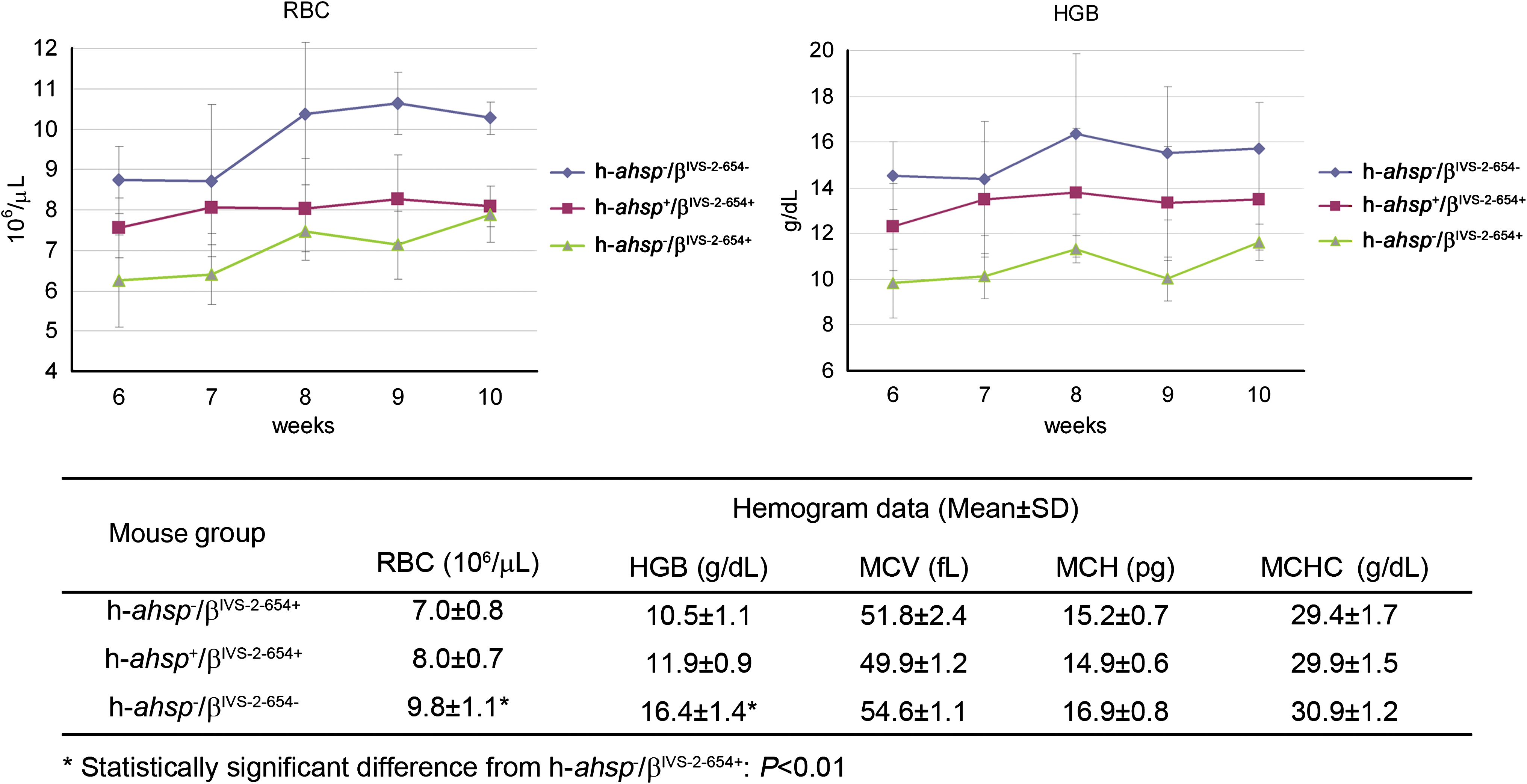
Hematologic improvements in h-ahsp
+/βIVS-2-654+ double heterozygous mice. Top: A comparison of red blood cell (RBC; left) and hemoglobin (HGB; right) indices among mice of the h-ahsp
+/βIVS-2-654+ group, and the positive and negative control groups, was performed for five consecutive weeks, indicating a slight recovery from anemia. Bottom: Hemogram indices for each group of mice are shown. The numbers of mice in each group (h-ahsp−
/βIVS-2-654+, h-ahsp
+/βIVS-2-654+, and h-ahsp−
/βIVS-2-654–) were 4, 3, and 4, respectively. MCH, mean corpuscular hemoglobin; MCHC, mean corpuscular hemoglobin concentration; MCV, mean corpuscular volume. Color images available online at
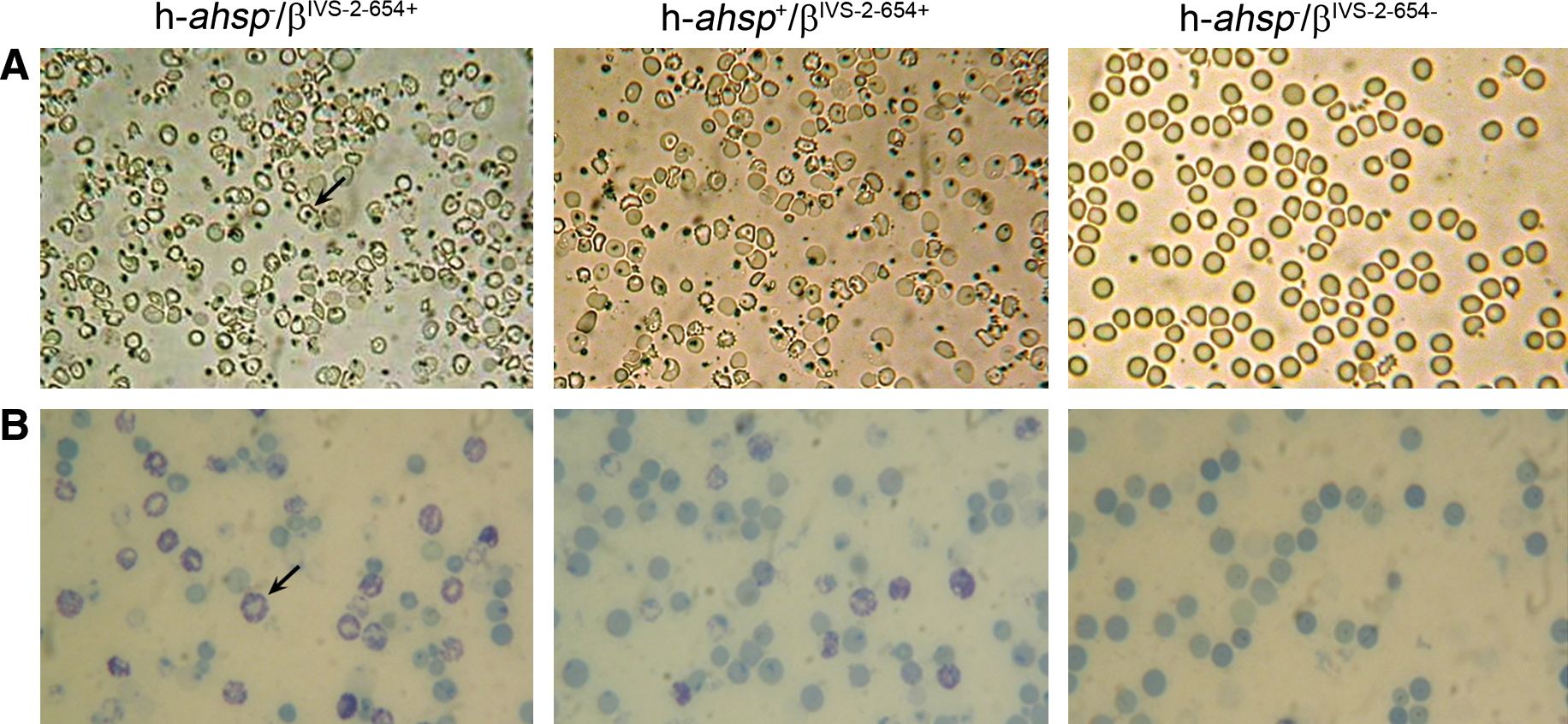
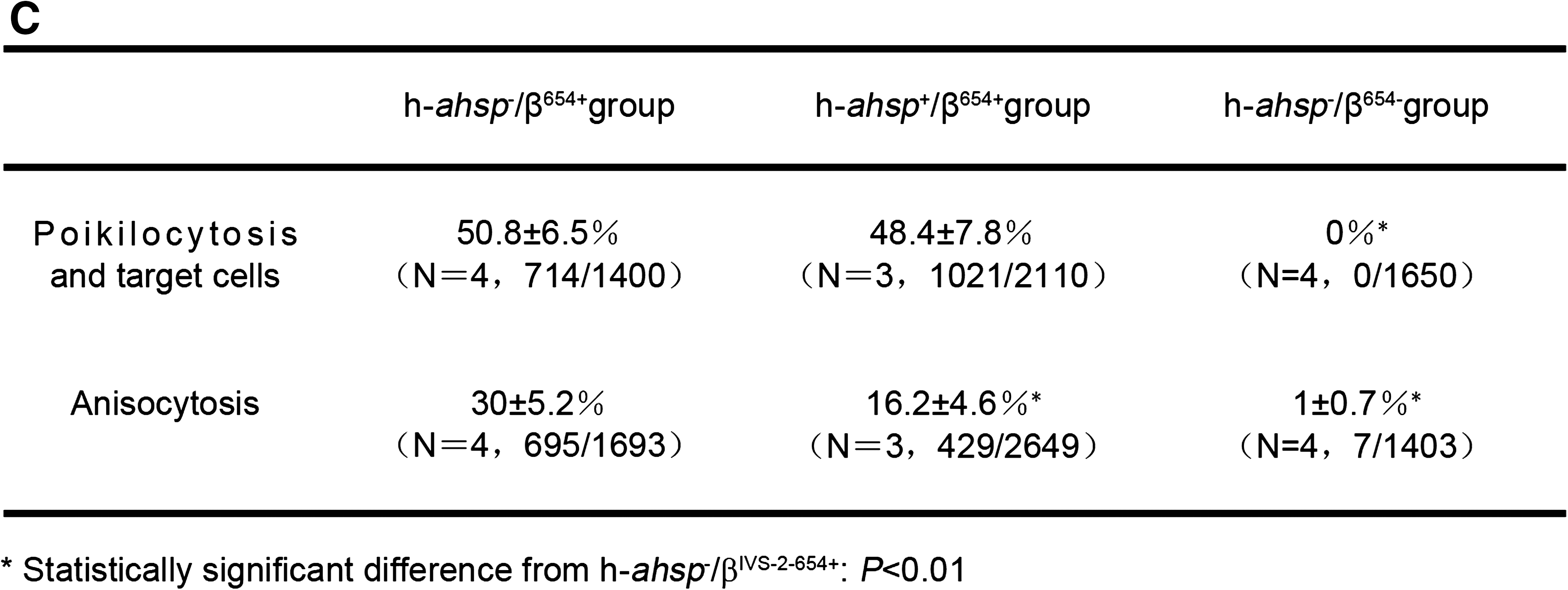
Hematologic parameter changes in h-ahsp
+/βIVS-2-654+ mice. Shown is a comparison of poikilocyte and target cell (
Results
Generation of trans-human ahsp βIVS-2-654-thalassemia mouse model
Erythroid human ahsp gene expressing cassette DNA (Fig. 1A) was injected into the fertilized eggs of βIVS-2-654-thalassemia model mice, followed by implantation and breeding by conventional procedures (Hogan et al., 1994; Zhang et al., 2006). Of the 48 newborn pups, 17 carried the human ahsp gene and 6 were double heterozygotes for human ahsp and βIVS-2-654 (h-ahsp
+/βIVS-2-654+). The genotypes were determined by PCR analysis of genomic DNA from murine tail tips (see Supplementary Fig. 1 at
Erythroid expression of human ahsp in h-ahsp +/βIVS-2-654+ double heterozygous mice
RT-PCR analysis showed human ahsp transcription in the blood cells of the double heterozygous mice (h-ahsp +/βIVS-2-654+) (Fig. 1B). However, real-time RT-PCR analysis indicated that the ratio of human ahsp mRNA to mouse ahsp mRNA was greater than 0.1% in only three mice, accounting for 5.7, 3.8, and 0.6% (average, 3.4%), respectively, with transgene copy numbers of 24, 33, and 1, individually. Western blot analysis showed human AHSP expression in the double heterozygous mice (Fig. 1C). Thus, these three double heterozygous mice were used as the experimental group to compare with the different genotypes of sibling controls (n = 4 and 4) in the following phenotypic studies.
Hematologic improvements in h-ahsp +/βIVS-2-654+ double heterozygous mice
Hematologic parameters were measured once per week for five consecutive weeks (Fig. 2). Mean data indicated that although mice of the double heterozygous group (h-ahsp
+/βIVS-2-654+) had slight improvements in both RBC and HGB levels compared with mice of the h-ahsp−
/βIVS-2-654+ group, the p values were greater than 0.05 and therefore did not reach significant levels. The comparison of peripheral blood smear pictures, stained with Wright–Giemsa (to assess anisocytosis) or without Wright–Giemsa (to assess poikilocytosis and target cells), showed that the h-ahsp
+/βIVS-2-654+ double heterozygous group had less anisocytosis than the h-ahsp−
/βIVS-2-654+-thalassemia group (p < 0.01), although their poikilocytosis and target cell counts were not significantly different (Fig. 3; and see Supplementary Fig. 2 at
Histopathologic recovery of spleen, liver, and bone marrow in h-ahsp +/βIVS-2-654+ double heterozygous mice
To further determine the therapeutic effect of stable human ahsp expression on hematopoiesis, we investigated the extent of extramedullary hematopoiesis (EMH) in h-ahsp
+/βIVS-2-654+ and h-ahsp−
/βIVS-2-654+ control mice. Compared with h-ahsp−
/βIVS-2-654+ mice, h-ahsp
+/βIVS-2-654+ mice showed fewer foci of intrasinusoidal EMH (Fig. 4A; and see Supplementary Fig. 3 at
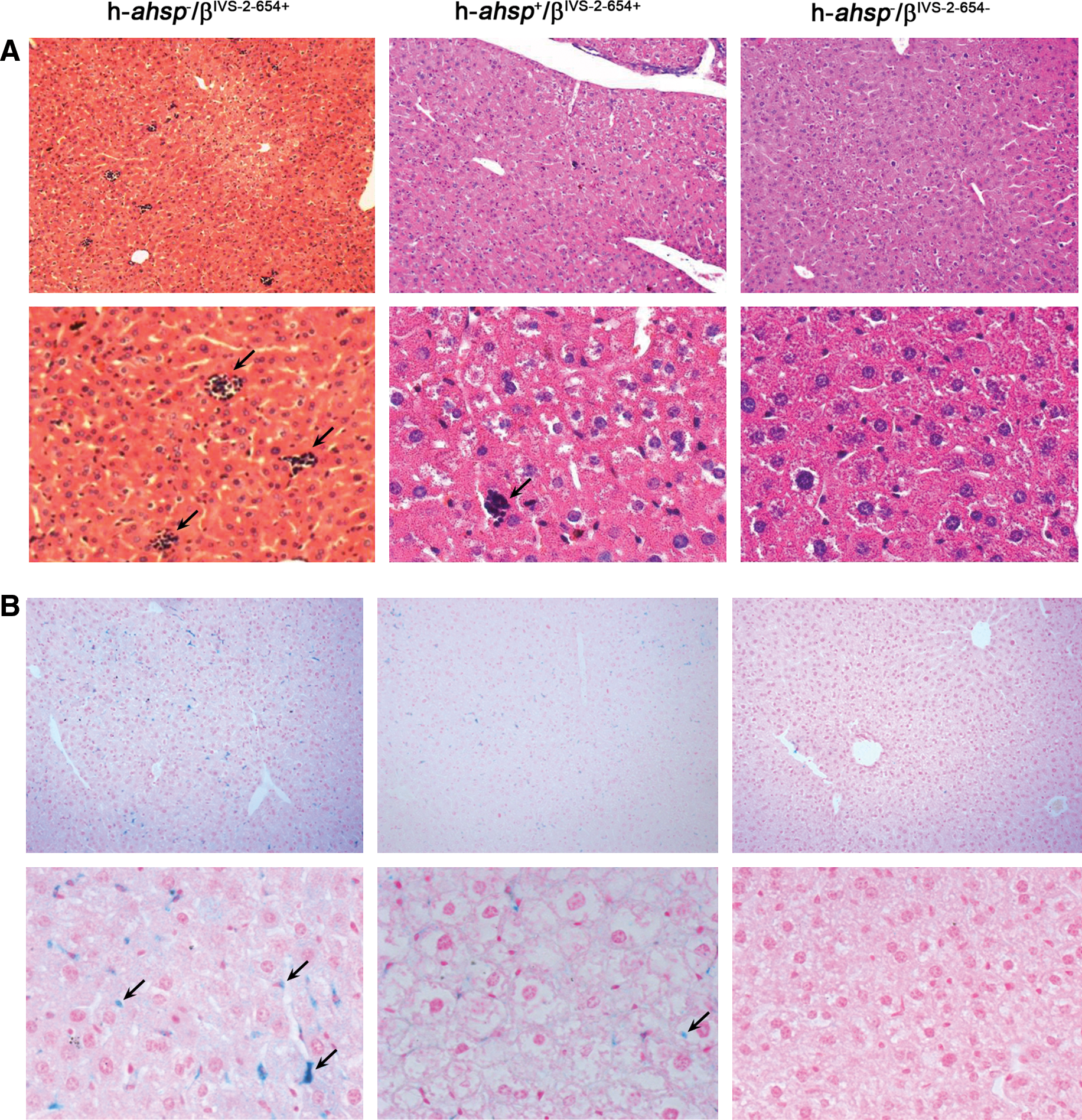
Tissue pathologic changes in h-ahsp
+/βIVS-2-654+ mice. (
Spleen size reduced in h-ahsp +/βIVS-2-654+ double heterozygous mice
Spleens from each group were dissected and weighed. Spleen weight divided by body weight was calculated as a coefficient to reflect its burden. The results showed that spleen weight and size were both reduced in h-ahsp
+/βIVS-2-654+ mice (Fig. 5; and see Supplementary Fig. 5 at
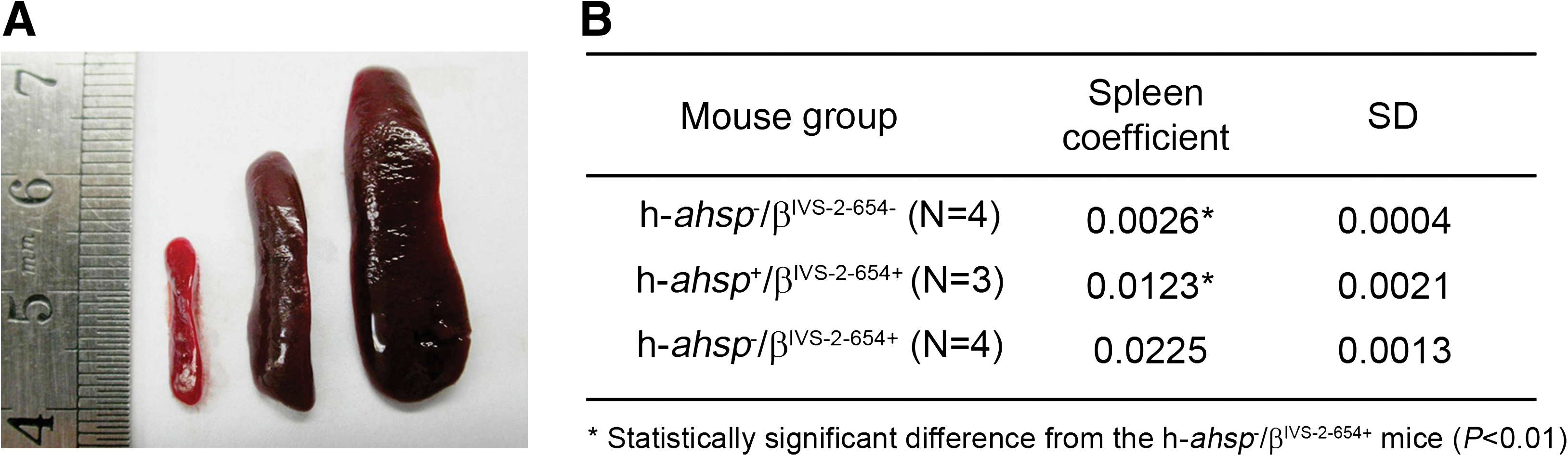
Spleen size was reduced in h-ahsp
+/βIVS-2-654+ double heterozygous mice. (
Serum iron reduced in h-ahsp +/βIVS-2-654+ double heterozygous mice
We found that serum iron concentrations were reduced in three h-ahsp +/βIVS-2-654+ mice (Fig. 6) with a statistically significant difference from the h-ahsp− /βIVS-2-654+ mice (p < 0.05), suggesting an improvement in red cell life span in the diseased mice.
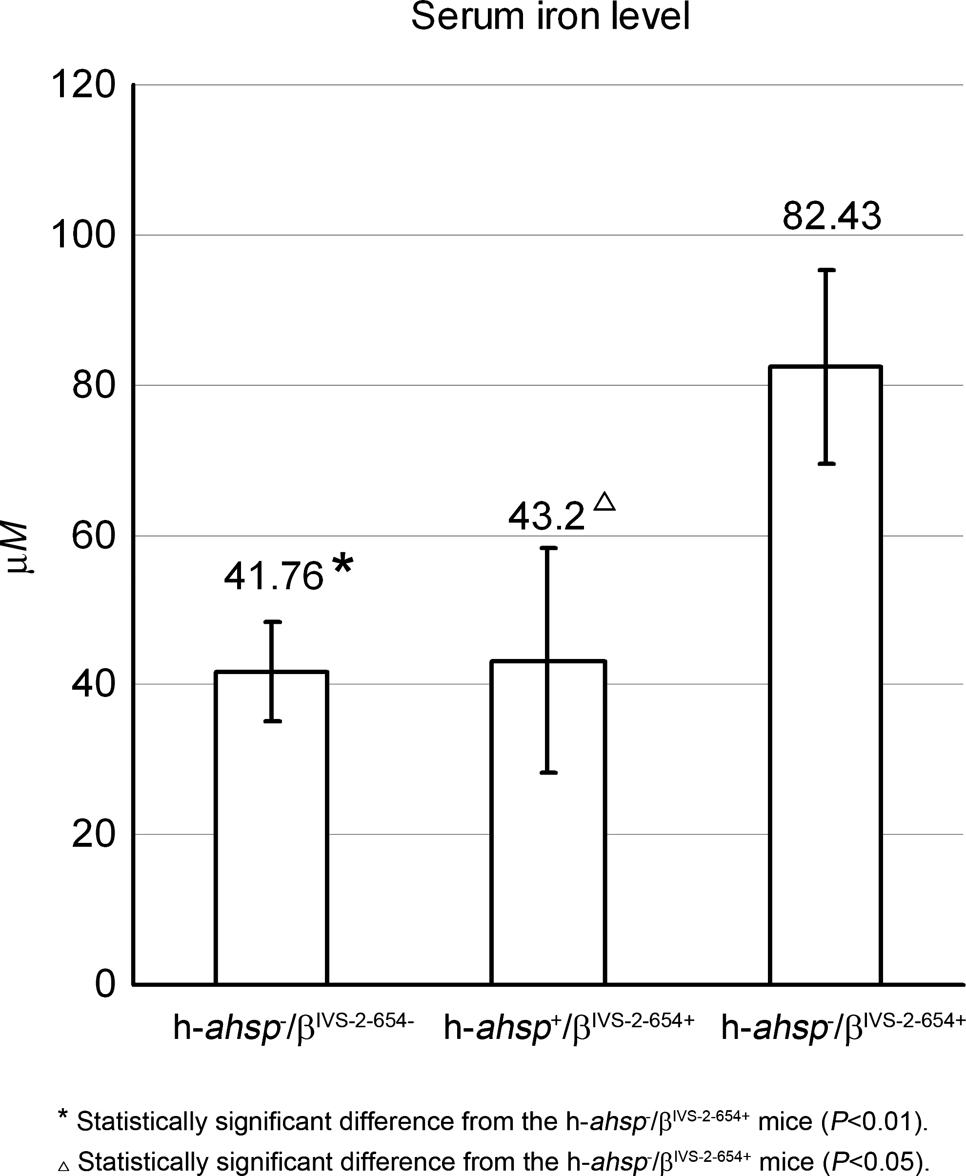
Serum iron levels were reduced in h-ahsp +/βIVS-2-654+ mice. Each column represents the mean serum iron content (μM) for normal mice (n = 4), h-ahsp +/βIVS-2-654+ mice (n = 3), and h-ahsp− /βIVS-2-654+ mice (n = 3).
Discussion
AHSP is a small-molecule protein that is highly expressed in erythroid cells. Human AHSP is a 12-kDa protein formed by 102 amino acids. Its gene is located on human chromosome 16, and contains three exons flanked by two introns, yielding a 0.5-kb transcript (Gell et al., 2002; Kihm et al., 2002). Structurally, it is a globular protein with three α helices, which can interact with αHb in several ways (Gell et al., 2002). The N and C termini of human AHSP are not essential for its interaction with αHb, and are likely to be related to its other functions (Santiveri et al., 2004; Feng et al., 2005).
AHSP appears to have three main functions (Feng et al., 2004; Kong et al., 2004; Zhou et al., 2006; Yu et al., 2007; Pinho et al., 2008): (1) As a chaperone protein, it participates in early HbA synthesis and assembly; (2) AHSP interacts with and stabilizes the excess and free form of αHb to avoid αHb denaturation leading to the generation of reactive oxygen species (ROS); and (3) AHSP renatures denatured αHb, allowing it to take part in HbA synthesis. The preceding features are based on the fact that the binding affinity of αHb for βHb is higher than that for AHSP. Therefore, the αHb–AHSP complex could be easily replaced by αHb–βHb to form the HbA tetramer (Feng et al., 2004). According to the preceding facts, we tested the ability of transgenic ahsp to neutralize the excess αHb in β-thalassemia mice, leading to partial relief of the effects of the denatured αHb in red blood cells.
To allow detection of exogenic ahsp, we used human ahsp, which is highly homologous to mouse ahsp, for the transgenic experiment. Lentiviral vector mediation was initially used to generate transgenic mice, which led to the loss of introns during the pseudo-virus preparation in some cases (our unpublished results). In our experience, in contrast to viral mediation, pronuclear DNA injection would usually give rise to a concatemer in a single site. For efficient expression of human ahsp, we then used its DNA backbone for conventional pronuclear injection to generate h-ahsp +/βIVS-2-654+ double heterozygous mice. RT-PCR and Western blot analysis demonstrated the expression of human AHSP in the blood of the double heterozygous (h-ahsp +/βIVS-2-654+) mice. However, real-time RT-PCR analysis indicated a variation of human ahsp transcripts in the blood cells of h-ahsp +/βIVS-2-654+ mice. The variation in transcripts depended mostly on the location where the transgene (human ahsp) resided. The discrepancy in human ahsp mRNA correlated well with the different effects in different h-ahsp +/βIVS-2-654+ mice. The greater the amount of human ahsp mRNA in the blood cells of h-ahsp +/βIVS-2-654+ mice, the better the effect on the improvement of thalassemia phenotype. The other three double heterozygous mice barely transcribing human ahsp mRNA experienced almost no effect on RBC, HGB, and serum iron levels. Western blotting using either monoclonal or polyclonal antibodies showed 26-kDa and 33-kDa bands instead of the 12-kDa AHSP band, which was similar to the previous size-exclusion chromatography result when α-globin was also present (Kihm et al., 2002). We believe it is due to association with mouse αHb (Fig. 1C). After stripping, the filter was reprobed with anti-mouse αHb antibody to reveal a 26-kDa band, supporting an association of AHSP with αHb.
In comparing the β-thalassemia-related parameters in each group of mice, we found that the average datum of RBC or HGB level in the h-ahsp +/βIVS-2-654+ double heterozygous mice was slightly elevated, although it did not reach the significant level. However, anisocytosis in peripheral blood was obviously decreased, compared with that in control h-ahsp− /βIVS-2-654+ mice. The spleen coefficient was markedly reduced. The histopathology of the spleen and liver was also improved to different extents. Moreover, the proportion of nucleated cells in the bone marrow was considerably decreased in h-ahsp +/βIVS-2-654+ mice (see Supplementary Fig. 7D). Significantly, the serum iron concentration in the blood of h-ahsp +/βIVS-2-654+ mice was also reduced. All these results indicated a partial improvement of the anemia phenotype in h-ahsp+ /βIVS-2-654+ mice. Our current study suggested that an ahsp transgene could provide an adjuvant method for gene therapy for β-thalassemia.
The impact of AHSP on β-thalassemia phenotypes has been demonstrated previously (Cappellini et al., 2004), but a potential role for AHSP in treating β-thalassemia has not been reported. Some studies claimed that AHSP was not a modulator for HbE β-thalassemia, whereas other authors disagreed (Kihm et al., 2002; Kong et al., 2004; Viprakasit et al., 2004). The present study reports the successful generation of h-ahsp +/βIVS-2-654+ double heterozygous mice. Although the approach described in this study only partially relieves the β-thalassemic phenotype in a mouse model, it provides a potential remedy for patients with β-thalassemia. For instance, by such an approach one could stably transduce h-ahsp into autologous hematopoietic stem cells (HSCs), or induced pluripotent stem cells (iPS), followed by safety verification, and then transplant the cells back into patients for the treatment of β-thalassemia, or simply correct the condition prenatally.
In summary, using such mice, we found that AHSP could partially relieve the βIVS-2-654 phenotype, thus supporting the view that AHSP could act as an adjuvant for gene therapy. The results of the present study suggest that AHSP could potentially be used to treat β-thalassemia.
Footnotes
Acknowledgments
The authors are grateful to Professors Yitao Zeng and Shuzhen Huang and to Dr. Wei Li (Shanghai Institute of Medical Genetics, Shanghai Jiao Tong University) for intellectual contributions and helpful discussions. This work was supported by the National High Technology Research and Development Program of China (no. 2007AA021206) and National Basic Research Program of China (nos. 2004CB18806, 2010CBJ29902), the National Natural Science Foundation of China (no. 30870943), and the Shanghai Municipal Natural Science Foundation (no. 08ZR1412100), State and Shanghai Leading Academic Discipline (B204).
Author Disclosure Statement
None of the authors report any potential conflicts of interest.
References
Supplementary Material
Please find the following supplemental material available below.
For Open Access articles published under a Creative Commons License, all supplemental material carries the same license as the article it is associated with.
For non-Open Access articles published, all supplemental material carries a non-exclusive license, and permission requests for re-use of supplemental material or any part of supplemental material shall be sent directly to the copyright owner as specified in the copyright notice associated with the article.