
Abstract
Adeno-associated viruses (AAVs) are small, nonenveloped single-stranded DNA viruses that require helper viruses to facilitate efficient replication. Despite the presence of humoral responses to the wild-type AAV in humans, AAV remains one of the most promising candidates for therapeutic gene transfer to treat many genetic and acquired diseases. Characterization of the IgG subclass responses to AAV and study of the prevalence of both IgG and neutralizing factors to AAV types 1, 2, 5, 6, 8, and 9 in the human population are of importance for the development of new strategies to overcome these immune responses. Natural exposure to AAV types 1, 2, 5, 6, 8, and 9 can result in the production of antibodies from all four IgG subclasses, with a predominant IgG1 response and low IgG2, IgG3, and IgG4 responses. Prevalences of anti-AAV1 and -AAV2 total IgG determined by enzyme-linked immunosorbent assay were higher (67 and 72%) than those of anti-AAV5 (40%), anti-AAV6 (46%), anti-AAV8 (38%), and anti-AAV9 (47%). Furthermore, data showed that cross-reactions are important. The two highest neutralizing factor seroprevalences were observed for AAV2 (59%) and AAV1 (50.5%) and the lowest were observed for AAV8 (19%) and AAV5 (3.2%). Vectors based on AAV5, AAV8, and AAV9 may have an advantage for gene therapy in humans. Furthermore, among individuals seropositive for AAV5, AAV8, and AAV9, about 70–100% present low titers. Better characterization of the preexisting humoral responses to the AAV capsid and cross-reactivity will allow development of new strategies to circumvent AAV acquired immune responses.
Introduction
The development of recombinant AAV vectors in gene therapy is in large part due to the lack of pathogenicity of the wild-type virus, the ability to establish long-term transgene expression, the ability to transduce both dividing and nondividing cells over a broad host range (human, simian, murine, canine, and avian cells), and their low immunogenicity. rAAVs of serotypes 1 and 2 have enjoyed some success in human trials (Kay et al., 2000; Manno et al., 2003; Moss et al., 2004; Nierman et al., 2007; Stroes et al., 2008). However, complications of treatment related to immune responses against the vector have emerged as serious obstacles for successful translations to humans. Indeed, in clinical trials, a cellular immune response against the capsid of AAV1 and AAV2, which may have been responsible for a loss of transgene-expressing cells, has been observed after administration by various routes (Mingozzi et al., 2007a–c, 2009). Nevertheless, several other major observations have showed that the host immune response to AAV capsid is mediated primarily by circulating antibodies, which may prevent repeated administrations (Manno et al., 2006). Data indicate that even low levels of neutralizing antibodies (1:5–1:10) can completely abrogate transduction with high titers of vectors (Jiang et al., 2006; Manno et al., 2006; Scallan et al., 2006).
The first AAV serotypes described were AAV1 to AAV6, and these have since been diversified to include serotypes with different cell tropisms, including AAV8 and AAV9 (Chiorini et al., 1999; Davidson et al., 2000; Gao et al., 2002, 2003). In preclinical models, these serotypes have emerged as interesting candidates for gene therapy (Z. Wang et al., 2005; Nathwani et al., 2007; Toromanoff et al., 2008; Yue et al., 2008; Gregorevic et al., 2009).
For many viruses, studies of the IgG subclasses that arise against viral antigens can provide insight into the nature and duration of the exposure or infection. IgG subclass responses were examined in serum samples of healthy human subjects exposed to AAV2 (Murphy et al., 2009). Analysis indicated a production of antibodies from all four IgG subclasses, with a predominant IgG1 response. A description of binding antibodies specific to these vectors is of importance, but nevertheless neutralizing properties for the antibodies and/or unidentified factors present in the serum must also be studied. Several groups have reported frequencies of antibodies to AAV types 1, 2, 5, and 6 ranging from 30 to 80% in small human cohorts from the United States (Chirmule et al., 1999; Erles et al., 1999; Xiao et al., 1999; Halbert et al., 2000, 2006; Hildinger et al., 2001). Calcedo and colleagues reported the frequencies of AAV1-, 2-, 7-, and 8-specific neutralizing antibodies in humans (Calcedo et al., 2009). They showed that neutralizing antibodies to AAV2 were the most prevalent antibodies in all geographic regions studied, followed by antibodies to AAV1.
Our goal in this study was to determine and compare the prevalence and profiles of serum IgG subclasses and neutralizing factors against AAV types 1, 2, 5, 6, 8, and 9 in a healthy population from France. We found that IgG and neutralizing factors to AAV types 5, 6, 8, and 9 were less prevalent when compared with AAV1 and AAV2. Furthermore, neutralizing factor titers to AAV8 and AAV9 vector types are typically low compared with other vector types. We also show that IgG coprevalence to AAV types 1, 2, 5, 6, 8, and 9 in humans is high for the various combinations, thus probably limiting the use of one of these serotypes as an alternative to another one to circumvent preexisting naturally acquired immunity to AAV in humans.
A better characterization of the preexisting humoral responses to the AAV capsid and cross-reactivity will enable the design of new strategies to circumvent AAV acquired immune responses.
Materials and Methods
Samples
Serum samples were collected from healthy volunteers. A total of 226 donors, recruited in the Île de France community in France, were collected by the French Etablissement Français du Sang (EFS) according to their procedures. All donors were between the ages of 25 and 64 years.
Production of AAV vectors and contaminant proteins
Pseudotyped AAV vectors were generated by packaging AAV2-based recombinant genomes in AAV type 1, 2, 5, 6, 8, and 9 capsids. All the vectors used in the study were produced by a three-plasmid transfection protocol as described elsewhere. Briefly, HEK293 cells were tritransfected with the adenovirus helper plasmid pXX6 (Xiao et al., 1998); a pAAV packaging plasmid expressing the rep and cap genes, pACG2.1 for AAV2 (Li et al., 1997), pLT-RC02 for AAV1 (Riviere et al., 2006), pLT-RC03 for AAV5 (Riviere et al., 2006), pLTRCO6 for AAV6 (Riviere et al., 2006), p5e18-VD2-8 for AAV8 (Gao et al., 2002), and pAAV-2/9 for AAV9 (Gao et al., 2004); and the relevant pAAV2 vector plasmid. Single-stranded AAV vectors were produced with a conventional pGG2 AAV2 vector plasmid expressing luciferase (Snyder et al., 1997) under the transcriptional control of the cytomegalovirus immediate-early (CMV IE) promoter associated with the simian virus 40 (SV40) poly(A) signal. Contaminant proteins were obtained by the same transfection protocol on HEK293 cells but lacking the packaging plasmid. Recombinant vectors and contaminant proteins were purified by double-cesium chloride ultracentrifugation followed by dialysis against sterile phosphate-buffered saline (PBS). Viral genomes were quantified by real-time polymerase chain reaction and vector titers are expressed as viral genomes per milliliter (VG/ml), and contaminant proteins were quantified by the Bradford protein assay.
Antibody subclass enzyme-linked immunosorbent assay
Recombinant AAV particles were diluted in coating buffer (0.1 M carbonate buffer, pH 9.5) to a final concentration of 2 × 10¹⁰ VG/ml. Fifty microliters was added to each well of a 96-well Nunc Maxisorp immunoplate (Thermo Fisher Scientific, Roskilde, Denmark). At the same time, proteins corresponding to contaminants purified during the various steps of rAAV production, but in the absence of viral particle formation, were diluted in the same buffer and seeded, in parallel, in different wells of the same immunoplate, at a concentration of 3.4 μg/ml. This amount of protein corresponds to an amount of contaminant protein equivalent with that seeded in the AAV wells and the signal obtained at the end corresponds to the nonspecific signal (NSS) that was removed from the signal obtained with the same serum in the corresponding AAV wells. In some experiments, to create a standard curve, purified IgG1 antibody was coated in 3-fold dilutions, beginning at a concentration of 3000 ng/ml (Sigma-Aldrich, St. Louis, MO). Plates were then incubated overnight at 4°C. The next day, plates were washed three times with blocking buffer (6% nonfat milk buffer in PBS) and then blocked with blocking buffer for 2 hr at room temperature. Plates were again washed three times with wash buffer (0.05% Tween 20 in PBS) and then incubated with heat-inactivated serum (at 56°C for 30 min) diluted from 1:30 to 1:65,610, for 1 hr at 37°C for IgG, IgG1, and IgG3 and overnight at 4°C for IgG2 and IgG4. After three washes, purified antibodies specific for the IgG, IgG1, and IgG3 subclasses were added and incubated for 1 hr at room temperature and purified antibodies specific for IgG2 and IgG4 were incubated for 2 hr at 37°C. Purified antibodies to IgG1 and IgG2 were purchased from Sigma-Aldrich; horseradish peroxidase (HRP)-conjugated IgG, IgG3, and IgG4 were purchased from Southern Biotech (Birmingham, UK); and HRP-conjugated sheep anti-mouse antibody (SAM) was from GE Healthcare (Amersham, Buckinghamshire, UK). After incubation the plates were washed three times with wash buffer. Unconjugated IgG1 and IgG2 antibodies were incubated with SAM, for 1 hr at room temperature. Last, plates were washed three times with wash buffer and revealed with tetramethylbenzidine (TMB) substrate solution (BD Biosciences, San Jose, CA), 30 min in the dark. The reaction was stopped with H2SO4 solution and measurements were made at 450 nm. The results are expressed as arbitrary optical density (OD) units, using a colorimetric amplification system based on peroxidase.
The AAV-specific signal was reported as the optical density determined in the AAV-coated enzyme-linked immunosorbent assay (ELISA) after removal of the optical density determined in the contaminant protein ELISA, which is a nonspecific signal (ODAAV – ODNSS).
Titration of rAAV serotypes for virus-neutralizing factors
Cell lines differ in their susceptibility to AAV of different serotypes. Therefore, we determined the optimal cell line and multiplicity of infection (MOI) for each AAV serotype tested. HeLa cells were best transduced with rAAV1, rAAV2, rAAV5, rAAV6, or rAAV9, and Huh7 cells (a human hepatoma cell line) with rAAV8. On day 1, 48-well plates were seeded either with 5 × 10⁴ HeLa cells per well or with 5 × 10⁴ Huh7 cells per well for 24 hr. On day 2, recombinant AAV-CMV-Luciferase (AAV-CMV-Luc) was incubated at various MOIs in DMEM–10% FCS for 48 hr at 37°C and 5% CO2. Transduction efficiency was measured for each vector serotype at various MOIs (5000–100,000) and expressed as arbitrary units (relative light units per second per well and normalized per amount of protein per well expressed as optical density; RLU/sec/well/OD). Maximal transduction was reached at an MOI of 5000 VG/cell for rAAV2, rAAV5, and rAAV6; at an MOI of 10,000 VG/cell for rAAV1 and rAAV8; and at an MOI of 100,000 VG/cell for rAAV9.
Neutralizing assay
On day 1, 48-well plates were seeded either with 5 × 10⁴ HeLa cells per well or with 5 × 10⁴ Huh7 cells per well for 24 hr. On day 2, recombinant AAV-CMV-Luciferase (AAV-CMV-Luc) was diluted in Dulbecco's modified Eagle's medium (DMEM; Invitrogen Life Technology, Carlsbad, CA) supplemented with 10% fetal calf serum (FCS; Hyclone, Logan, UT) and incubated with a 10-fold dilution, then 2-fold serial dilutions (1:20 to 1:12,800) of heat-inactivated (at 56°C for 30 min) serum samples for 1 hr at 37°C. Subsequently, the serum–vector mixtures corresponding to 5 × 10³ to 1 × 10⁵ VG/cell, depending on the serotype (as determined previously), were added to cells plated on day 1 and incubated in DMEM–10% FCS for 48 hr at 37°C and 5% CO2. Each mix was performed in duplicate. Cells were then washed in PBS and lysed for 10 min in 0.2% Triton lysis buffer at 4°C. The lysate was transferred to 96-well plates and then the luciferase activity was read with a luminometer (VICTOR2; PerkinElmer Life Sciences, Waltham, MA). Transduction efficiency was measured as relative light units per second per well and normalized per amount of protein per well expressed as optical density (RLU/sec/well/OD).
The neutralizing titer was reported as the highest serum dilution that inhibited the rAAV transduction by ≥50% compared with the control without serum and correlated with the amount of protein quantified in each well after cell lysis by Bradford assay.
Statistical analyses
Results are presented as means ± standard deviation (SD). The Student t test for paired data was used to determine the significance of differences between the two groups. A p value less than 0.05 was considered statistically significant.
Results
Prevalence of serum IgG to AAV in the healthy population
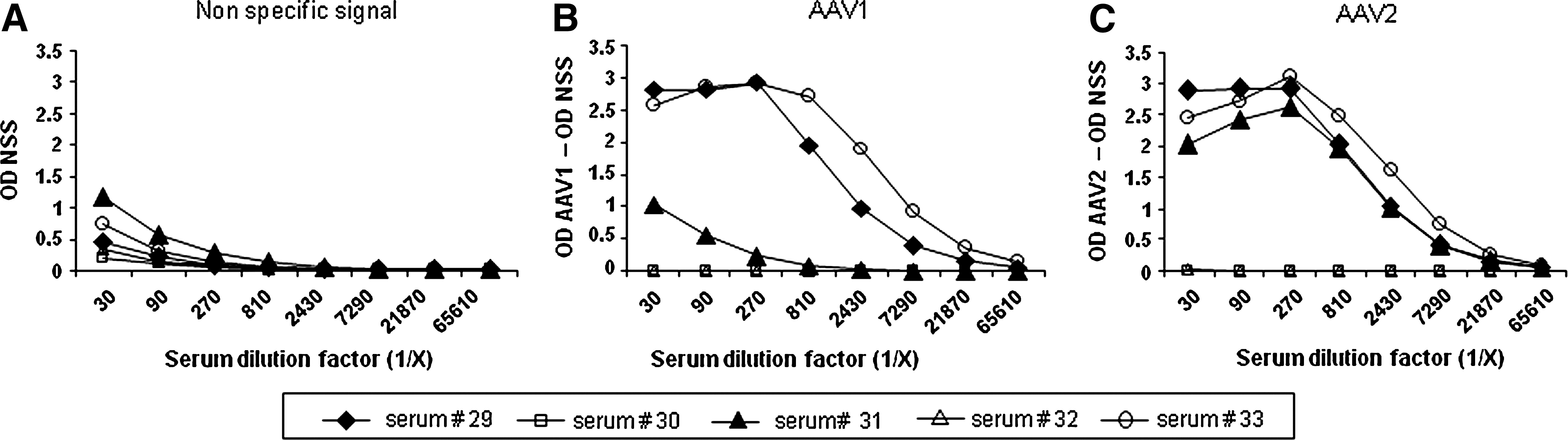
The seroprevalences of total IgG antibodies to AAV serotype 1, 2, 5, 6, 8, or 9 were determined with an AAV-specific ELISA on a large cohort of serum samples from adult healthy donors from the Île de France community in France, which corresponds to a mixed population of different origins (Fig. 1). This prevalence of IgG is a relevant indicator of the frequency of individuals who are not naive for one of the AAV serotypes. We did not observe any age-related effect in this analyzed adult population. Of note, for some human sera, the nonspecific signal (NSS) obtained for proteins copurified with vector particles during the rAAV production process in the absence of viral particle formation were high (Fig. 1A). Therefore, two different coatings were performed in parallel systematically for each serum sample: one coating with AAV vector particles of different serotypes and the other with a corresponding amount of contaminant proteins. The signal obtained in this second case corresponds to the NSS that was removed from the signal obtained with AAV vector particles. Serum samples were incubated for 1 hr with the coated plates, and detection was performed with HRP-conjugated IgG-specific secondary antibodies. Sera were judged positive for AAV-specific IgG when an optical density signal ≥0.5 (cutoff based on mean optical density of 58 negative donors + 3 SD) was observed at a dilution ≥1:30. Specific binding of serum IgG to AAV capsid proteins was evaluated after removal of the NSS (Fig. 1A) from the signal obtained on ELISA coated with one of the rAAV serotypes, as illustrated in Fig. 1B and C for AAV1 and AAV2. Serum samples 30 and 32 were negative for IgG specific for AAV1 and AAV2 and serum samples 29, 31, and 33 were positive for IgG specific for both vector protein capsids. The highest seroprevalences were observed with AAV1 and AAV2, with 67 and 72% of IgG-seropositive donors, respectively (Fig. 1D). Seroprevalences to AAV5, AAV6, and AAV9 were slightly less than 50%. Interestingly, compared with other serotypes a significantly lower seroprevalence (less than 40%) was observed for AAV8 (Fig. 1D). Of note, 27% of the tested population was seronegative to any of these serotypes (data not shown).
Seroprevalence of total IgG to AAV types 1, 2, 5, 6, 8, and 9 in the healthy population. Results of ELISAs measuring (

AAV-specific IgG subclass response in the healthy population
The relative levels of IgG subclass responses to AAV capsid of various serotypes in healthy human subjects were determined by ELISA. Serum samples from 16 to 21 donors positive for IgG specific to the various rAAV serotypes were analyzed (Fig. 2). Plates coated either with rAAV or with contaminant proteins were incubated for 1 hr or overnight with serum samples and detection was performed with HRP-conjugated IgG subclass-specific secondary antibodies. As described previously, the specific signal for each IgG subclass was obtained after removal of the corresponding NSS. Results showed that profiles of IgG subclass responses to AAV type 1, 2, 5, 6, 8, and 9 capsids were almost similar (Fig. 2). Indeed, the dominant IgG subclass response was IgG1 (Fig. 2), and this response was significantly higher with AAV6 compared with the AAV1 and AAV2 serotypes (Fig. 2 and Table 1). IgG2 responses were low, but they were significantly higher with AAV8 compared with other serotypes and they appeared significantly higher for AAV1 than for AAV2, AAV6, and AAV9 capsid proteins (Fig. 2A, B, and D–F). Similarly, IgG2 responses appeared significantly higher for AAV5 capsid proteins compared with AAV2, 6, and 9 (Fig. 2B–D and F). Despite low IgG3 and IgG4 responses; two of the same donors had significant levels of IgG3 specific to AAV6 (Fig. 2D) and IgG4 specific of AAV2, AAV5, and AAV6 (Fig. 2B–D) and one other donor had significant levels of IgG3 specific to AAV2 (Fig. 2B).
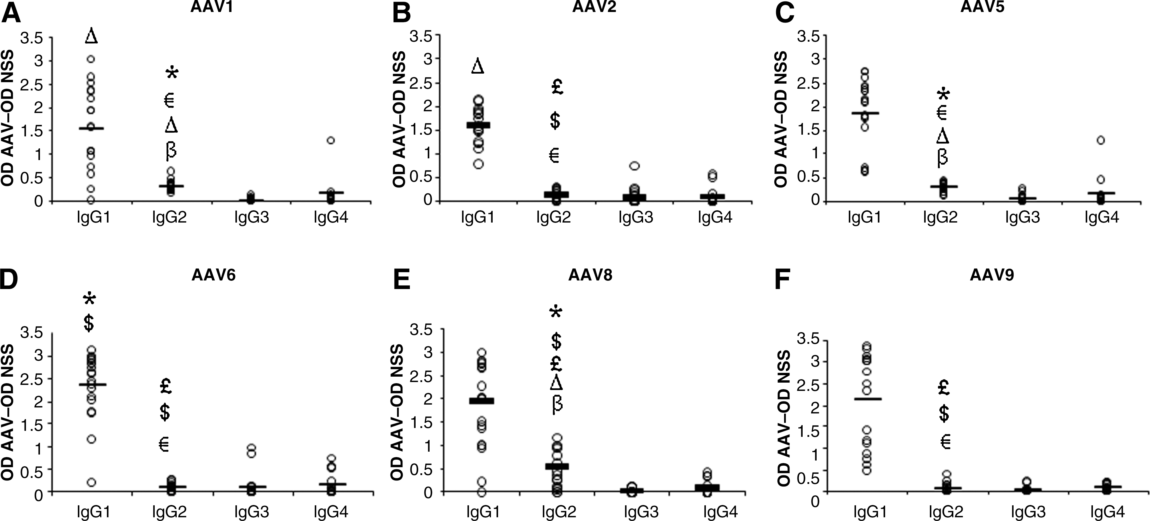
IgG subclass profiles to AAV types 1, 2, 5, 6, 8, and 9 in the healthy population. Comparison of IgG subclass antibody profiles to (
Values shown represent antibody concentrations, in picograms per milliliter.
Coprevalence of AAV-specific IgG
An interesting question for gene therapy protocols based on AAV is the coprevalence of IgG antibodies to capsid proteins of two different AAV types within individuals. Sera from 9 to 146 donors were analyzed for each serotype (Table 2). As shown, 93% of the individuals positive for IgG to AAV2 capsid were also positive for AAV1 capsid and, reciprocally, 100% of the individuals positive for IgG to AAV1 were also positive for AAV2 capsid. Of note, all donors positive for AAV5, AAV6, AAV8, or AAV9 types were positive for AAV1 and two capsid types. Interestingly, all donors positive for AAV5 were also positive for AAV6 capsid. Surprisingly, only 73% of donors positive for AAV1 were positive for AAV6 capsid, whereas they differ by six amino acids, and only 59% of donors positive for AAV2 were positive for AAV6 capsid. The lower coprevalences were observed among AAV2-positive donors because less than 60% were positive for AAV type 5, 6, 8, or 9 capsid.

Prevalence of AAV serum neutralizing factors
Healthy donors were analyzed for the prevalence of neutralizing factors against rAAV types 1, 2, 5, 6, 8, and 9 (Fig. 3A). Sera were judged positive for neutralizing capacity when a 1:20 dilution of serum inhibited vector transduction by 50% or more (Fig. 3A). Serum samples from 152 donors were analyzed for the presence of neutralizing factors. The seroprevalence of neutralizing factors to AAV2 was highest (59%); the second and third highest seroprevalences were to AAV1 (50.5%) and to AAV6 (37%). The lowest seroprevalences were observed for AAV5 (3.2%), AAV8 (19%), and AAV9 (33.5%).
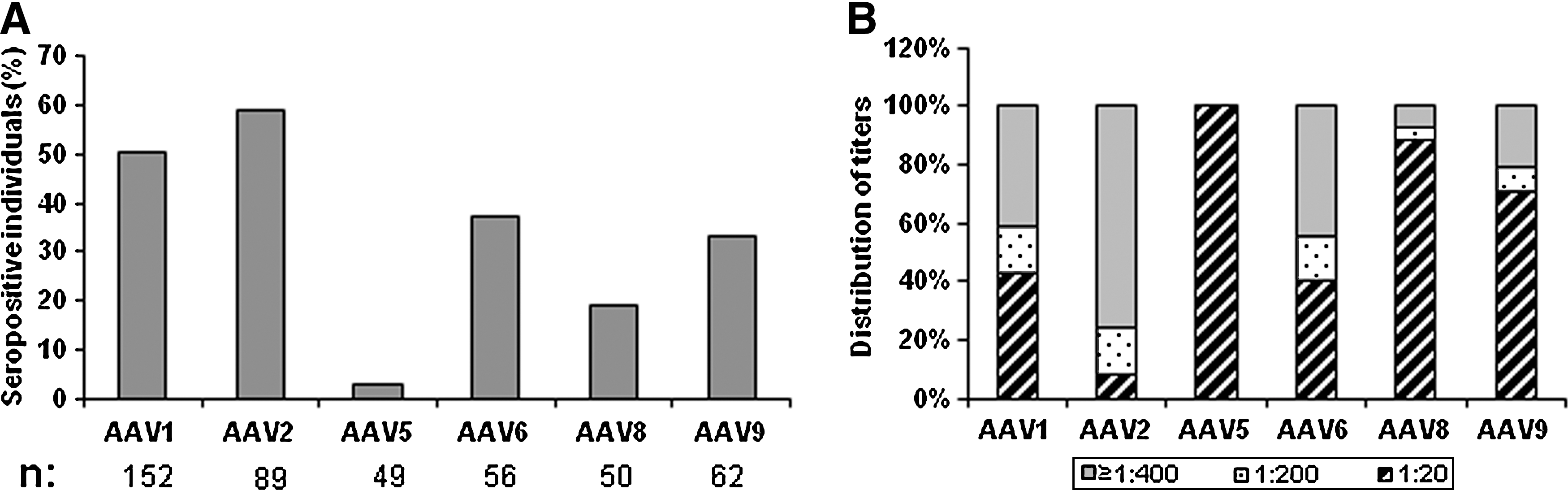
Prevalence of neutralizing factors in serum against AAV types 1, 2, 5, 6, 8, and 9. (
The magnitude of the neutralizing activity to AAV types 1, 2, 5, 6, 8, and 9 was measured by determining the neutralizing titer for each sample. Titer was defined as the highest dilution that still inhibited vector transduction by 50% or more. Then, for each serotype, percentages of seropositive individuals with titers of 1:20, 1:200, or ≥1:400 were determined (Fig. 3B). Interestingly, the distribution of donors with high titers (1:200 and ≥1:400) was highest for AAV1, AAV2, and AAV6, which were also the serotypes for which the highest seroprevalences were observed. Indeed, the majority of individuals who were seropositive for AAV8 or AAV9 presented low titers (1:20) and all individuals seropositive for AAV5 presented low titers (1:20). Of note, 41% of the tested population was seronegative to any of these serotypes (data not shown).
Discussion
The presence of humoral responses to the wild-type AAV common among humans is one of the limitations of in vivo transduction efficacy in humans when using cognate recombinant vector. Despite this predisposition in humans, AAV remains one of the most promising candidates for therapeutic gene transfer to treat many genetic and acquired diseases (Zaiss and Muruve, 2008). For this reason, characterization of the IgG subclass responses to AAV may lead to a better appreciation of the natural infection. Moreover, study of the prevalence of both IgG and neutralizing factors to AAV in human populations is of importance for the development of new strategies to overcome these immune responses. This study is the largest published survey of the seroprevalence of not only neutralizing factors, but also of IgG binding antibodies, to various AAV types in a large healthy adult population.
As previously described for AAV2 (Murphy et al., 2009), natural exposure to AAV types 1, 2, 5, 6, 8, and 9 can result in the production of antibodies of all four IgG subclasses, with a predominant IgG1 response. IgG1 was expected, as the subclass is commonly induced after viral infections such as measles, hepatitis B, human T-lymphotropic virus, rubella, and B19V, which like AAV is a member of the Parvoviridae (Morgan-Capner and Thomas, 1988; Lal et al., 1993; Franssila et al., 1996; Rey et al., 2000; Toptygina et al., 2005). IgG2 was also shown to be a constituent of the AAV-specific antibody response (Madsen et al., 2009; Murphy et al., 2009), but it is low as for B19V (Lal et al., 1993; Franssila et al., 1996; Toptygina et al., 2005). Of note, the IgG2 response in this study was highest with AAV8. The IgG3 response was low, whereas this subclass was previously described as a significant component of virus-induced IgG (Madsen et al., 2009; Murphy et al., 2009). The IgG4 response was low and variable. Variable levels of IgG4 were also observed in B19V infection (Franssila et al., 1996). Despite low levels of IgG3 and IgG4, some subjects studied presented significantly higher levels of IgG3 specific for AAV6 and of IgG4 specific for AAV types 1, 2, 5, and 6. Of note, the profile of IgG subclass responses, except for a few donors, was similar for all AAV types analyzed. Of note, IgG4 subclass synthesis has already been described as reflecting long-lasting or repeated exposure to antigen, either sequestered in immune complexes or expressed persistently or by repeated contact (Aalberse et al., 1983; Linde et al., 1988; Bird et al., 1990; Lundkvist et al., 1993; Maizels et al., 1995). This IgG subclass pattern, IgG1 > IgG3 ≈ IgG4, may reflect, as for other viruses, the fact that naturally infected patients were cured or eventually became chronic carriers of virus (L. Wang et al., 2005; Tsai et al., 2006). The specific role of IgG subclasses in the clearance of AAV remains unclear, as for other viruses, and depends on the context in which the immune system encounters AAV capsid antigens, the nature of helper virus infection generating different innate and adaptive immune responses, the local sites within the body where the vector encounters the immune system, and the genetic background of the population (Murphy et al., 2009).
Prevalences of anti-AAV1 and -AAV2 total IgG determined by ELISA were higher (67 and 72%) than those of other evaluated anti-AAV IgG. Surprisingly, the prevalence of AAV6 IgG was significantly lower (46%) than that of AAV1, whereas these types showed greater than 96% homology (Schmidt et al., 2006), indicating the importance of these few different amino acids in the immunogenicity of AAV in humans. Of note, we observed a lower prevalence of total IgG to AAV8 (38%), which has been shown to be a robust vector for achieving high levels of transgene expression in liver, muscle, and heart (Gao et al., 2002, 2004; Nakai et al., 2005; Z. Wang et al., 2005; Toromanoff et al., 2008). AAV9 has also been shown to efficiently target skeletal muscle, liver, and heart (Pacak et al., 2006; Vandendriessche et al., 2007). AAV5 has revealed higher transduction frequencies than AAV2 within the central nervous system (Burger et al., 2004) and arthritic joints (Khoury et al., 2007). In this regard, they were considered to be promising candidates for gene therapy.
As shown, all donors positive for IgG specific for AAV types 5, 6, 8, and 9 also had IgG specific for AAV2 and AAV1, indicating that cross-reactions are important, confirming the studies showing that AAV2 is the most frequent type present in humans (Tobiasch et al., 1998; Erles et al., 1999). The lowest coprevalences were observed between AAV5 and AAV1 or AAV2. Interestingly, that may be related to the capsid amino acid composition of AAV5, which differs from that of AAV1 and AAV2 by almost 40%, whereas the others differ from each other in the range of 15% (Gao et al., 2005).
It was possible to detect neutralizing properties to AAV in most sera found to be positive for IgG in the corresponding AAV ELISA, confirming the specificity of the results obtained by ELISA, but also indicating that neutralizing properties of sera were correlated with specific binding antibodies. Nevertheless, it cannot be excluded, as reported for other viruses (Levy et al., 1975; Nemerow et al., 1982; Harris et al., 1990; Ebenbichler et al., 1991), that in certain cases some other unidentified factors present in the serum can be responsible, at least in part, for the virus neutralization.
Of note, neutralizing antibodies may inhibit viral entry into nonimmune target cells, but immunoglobulin- or complement-opsonized virus may be targeted to and taken up by innate immune cells (including antigen-presenting cells [APCs], such as dendritic cells) through Fc receptors and complement receptors. Fc receptor cross-linking or targeting of opsonized virus particles to innate immune cells and the engagement of innate receptors may either induce signal transduction events or may facilitate virus entry into these APCs, which can lead to inflammatory responses. Thus, future investigations must define the role of antiviral antibodies in mediating AAV interactions with the immune system. Here we have shown that the two highest seroprevalences were for AAV2 (59%) and AAV1 (50.5%) neutralizing factors, as previously reported in Europe with about 55 and 46%, respectively (Calcedo et al., 2009). Interestingly, significantly lower seroprevalences of neutralizing factors were observed to AAV9 and AAV6 and especially AAV8 (19%), and low seroprevalences of neutralizing factors were observed to AAV5 (3.2%). Furthermore, a correlation was observed between the prevalence of neutralizing factors to an AAV type and the distribution of titers. Indeed, AAVs with the lowest prevalence in human serum samples were those for which low titers (≥1:20) were observed. Nevertheless, it was previously reported that even low levels of neutralizing antibodies (1:5–1:10) can completely abrogate transduction with high titers of vectors (Jiang et al., 2006; Manno et al., 2006; Scallan et al., 2006). Of note, we observed only one serum sample in which the titer of neutralizing factors against AAV1 exceeded that of AAV2 and no serum sample in which the titer of neutralizing factors against AAV types 5, 6, 8, and 9 exceeded that of AAV2. Regarding the T cell response, it might not always be possible to avoid memory AAV capsid T cell responses by switching serotypes. Indeed, AAV2 capsid-primed CD8+ memory T cells in humans cross-react with epitopes from AAV8 and AAV1 capsid and result in expansion of functional CD8+ cytotoxic lymphocytes that are indistinguishable from those elicited by AAV2 capsid epitopes (Mingozzi et al., 2007a).
In conclusion, vectors based on AAV5, AAV8, and AAV9 may have an advantage for gene therapy in humans, depending on the target tissue. Preexisting immunity should be more limited, because not only IgG but also the prevalence of neutralizing factors to AAV5 and AAV8 in sera in the healthy population are lower than for AAV types 1, 2, and 6. Furthermore, among these AAV5- and AAV8-seropositive individuals, 85–100% present low titers (1:20), which is also true for AAV9. Nevertheless, cross-reactions between AAV serotypes will probably limit the use of one of these serotypes as an alternative to another to allow readministration or to circumvent preexisting naturally acquired immunity to AAV in humans.
Footnotes
Acknowledgments
This work was supported by the Association Française contre les Myopathies (AFM). The authors thank K. Poulard for technical help. The authors also thank C. Rivière and Fréderic Barnay-Toutain for providing viral vectors. The authors thank Philippe Moullier and Susan Cure for critical reading of the manuscript.
Author Disclosure Statement
No competing financial interests exist.