
Abstract
Crigler–Najjar syndrome type I is a severe inborn error of bilirubin metabolism caused by a complete deficiency of uridine diphospho-glucuronosyl transferase 1A1 (UGT1A1) and results in life-threatening unconjugated hyperbilirubinemia. Lifelong correction of hyperbilirubinemia by liver-directed gene therapy using a helper-dependent adenoviral (HDAd) vector has been previously reported in the Gunn rat, a model of Crigler–Najjar syndrome, but was only achieved using high doses (≥3 × 10¹² viral particles [vp]/kg), which are likely to elicit a severe toxic response in humans. Therefore, in this study, we investigate strategies to achieve correction of hyperbilirubinemia in the Gunn rat using clinically relevant low HDAd doses. We have found that correction of hyperbilirubinemia in the Gunn rat can be achieved with a low dose of 5 × 10¹¹ vp/kg by using an HDAd vector bearing a more potent UGT1A1 expression cassette. Furthermore, by using hydrodynamic injection of the improved HDAd vector, correction of hyperbilirubinemia in the Gunn rat can be achieved using an even lower dose of 5 × 10¹⁰ vp/kg. Although hydrodynamic injection as performed in rats is not acceptable in humans, clinically attractive, minimally invasive methods have been successfully developed to mimic hydrodynamic injection of HDAd vector in non-human primates. Therefore, using an improved expression cassette combined with a more efficient method of vector delivery permits correction of hyperbilirubinemia in the Gunn rat using clinically relevant low HDAd doses and may thus pave the way to clinical application of HDAd vectors for Crigler–Najjar syndrome gene therapy.
Introduction
Lifelong correction of hyperbilirubinemia using helper-dependent adenoviral (HDAd) vectors expressing UTG1A1 has been reported in the Gunn rat, a model of Crigler–Najjar syndrome (Toietta et al., 2005). In that study, complete and lifelong phenotypic correction was achieved using 1 × 10¹³ viral particles (vp)/kg and 3 × 10¹² vp/kg, and lifelong partial correction was achieved with a dose of 6 × 10¹¹ vp/kg. These encouraging results suggest that Crigler–Najjar patients may benefit from HDAd-mediated gene therapy. Unfortunately, these doses are too high for human patients because they will likely elicit an acute toxic and potentially lethal response considering that a partial ornithine transcarbamylase (OTC)-deficient patient died following administration of 6 × 10¹¹ vp/kg of a recombinant adenovirus vector (Raper et al., 2003). Because adenovirus-mediated acute toxicity is dose-dependent (Nunes et al., 1999; Morral et al., 2002; Brunetti-Pierri et al., 2004), it is imperative that strategies be developed to permit efficient high hepatocyte transduction at much lower dose. In this regard, we have previously shown that HDAd vector delivered by hydrodynamic injection in mice significantly increases hepatic transduction efficiency and reduces vector systemic dissemination and activation of the acute inflammatory response (Brunetti-Pierri et al., 2005). Unfortunately, hydrodynamic injections, as performed in rodents, cannot be performed in humans because of the rapid injection of a large volume. However, we have successfully developed minimally invasive and clinically attractive methods to mimic hydrodynamic injection of HDAd vector in non-human primates without the need for rapid, large volume injection. It is important that these methods result in high-efficiency hepatocyte transduction using clinically relevant low vector doses (Brunetti-Pierri et al., 2007, 2009). In addition to higher-efficiency hepatocyte transduction, strategies to improve the potency of the expression cassette by inclusion of cis-acting elements to enhance transgene expression may also improve the therapeutic index of the vector to permit dose reduction. Therefore, in the current study, we investigate the efficacy of hydrodynamic injection of an HDAd vector bearing an improved UGT1A1 expression cassette in Gunn rats.
Materials and Methods
HDAd vectors
HDΔ28E4LacZ contains a MCMV-LacZ expression cassette and has been described in detail previously (Palmer and Ng, 2003). The expression cassette of the HDAd-rat phosphoenolpyruvate carboxykinase promoter (PEPCK)-UGT1A1-HCR is identical to that of the HDAd-PEPCK-UGT1A1 (Toietta et al., 2005) except for the insertion of a ∼1.7-kb XhoI–SalI fragment containing the human apoE
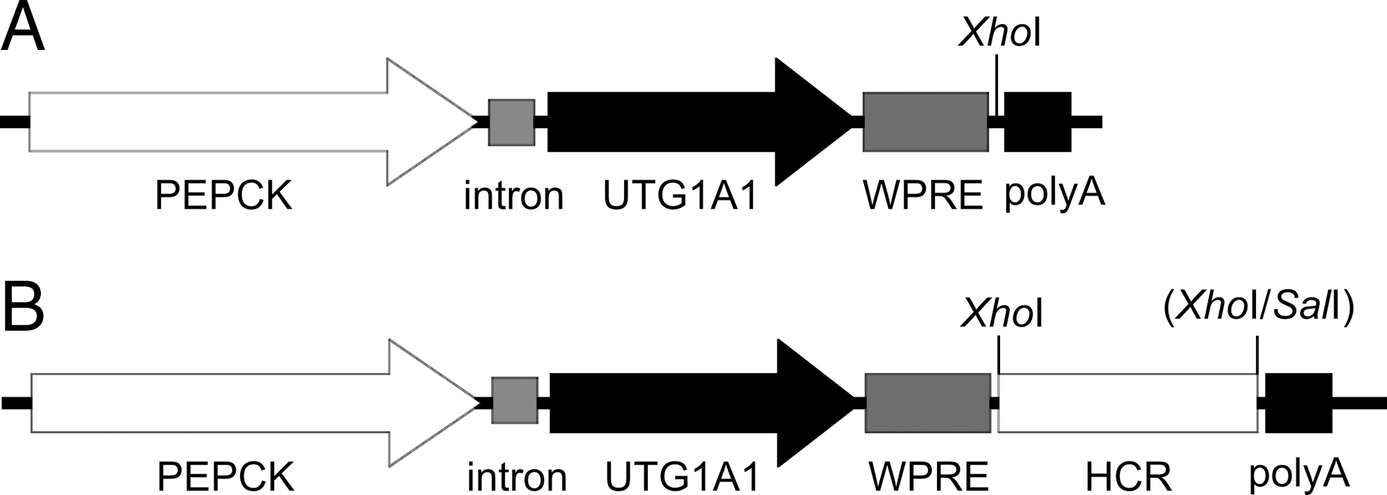
Expression cassettes in (
Animal studies
Female homozygous Gunn rats (HsdBlu:Gunn J/J) were purchased from Harlan (Indianapolis, IN) and were maintained on Teklad rodent feed (Harlan, Indianapolis, IN) under 12-hr light/dark cycles. Eight-week-old female rats (weighing 150–180 g) were used for all vector experiments. Wistar rats used as controls of the analysis of bilirubin conjugation were purchased from Harlan (Indianapolis, IN). Animal experiments were performed according to a protocol approved by the Baylor College of Medicine (Houston, TX) Institutional Animal Care and Utilization Committee. Conventional injections were performed injecting 0.5 ml of vector diluted in lactated Ringer's solution into the tail vein. The hydrodynamic injections were performed by injecting the vector diluted in lactated Ringer's solution (100 ml/kg) prewarmed to 37°C via the tail vein in less than 15 sec using a Thermo winged infusion set and a 25-ml syringe, as previously reported (Maruyama et al., 2002). The animals injected with HDΔ28E4LacZ vector were sacrificed 72 hr post-injection for bromochloroindolyl galactopyranoside (X-Gal) histochemistry and β-galactosidase activity, which were performed as previously described (Brunetti-Pierri et al., 2005). Blood samples from rats injected with HDAd-PEPCK-UGT1A1-HCR were collected by lateral saphenous vein puncture at baseline and at the various times after vector injection. Total serum bilirubin was assayed using the diazo dye method on a Roche COBAS Integra 400 Plus (Roche Diagnostics, North America, Indianapolis, IN), according to the manufacturer's instructions. Bile was collected by catheterization of the bile duct at 60 weeks post-vector and analyzed using high-performance liquid chromatography, as previously described (Toietta et al., 2005). Statistical analyses were performed with the t test.
Results and Discussion
We first sought to determine whether hydrodynamic injection of HDAd vector would result in an increase of hepatic transduction in rats. To accomplish this, HDΔ28E4LacZ at 1 × 10¹² vp/kg was conventionally or hydrodynamically injected into wild-type rats. At 72 hr post-injection, the livers were harvested for analyses. Consistent with our previous results in mice (Brunetti-Pierri et al., 2005), conventional injection of 1 × 10¹² vp/kg (Fig. 2A) yielded qualitatively fewer β-galactosidase-positive cells than hydrodynamic injection (Fig. 2B) in wild-type rats as determined by X-gal histochemistry. Splenic transduction was also investigated. We found little to no splenic transduction following either conventional (Fig. 2C) or hydrodynamic injection (Fig. 2D) of HDΔ28E4LacZ at 1 × 10¹² vp/kg as determined by X-gal staining. To quantitate the amount of hepatic transgene expression, total proteins were extracted from the livers, and the amount of β-galactosidase activity was determined. The results revealed that hydrodynamic injection resulted in fourfold higher activity than conventional injection (p = 0.035) (Fig. 2E).
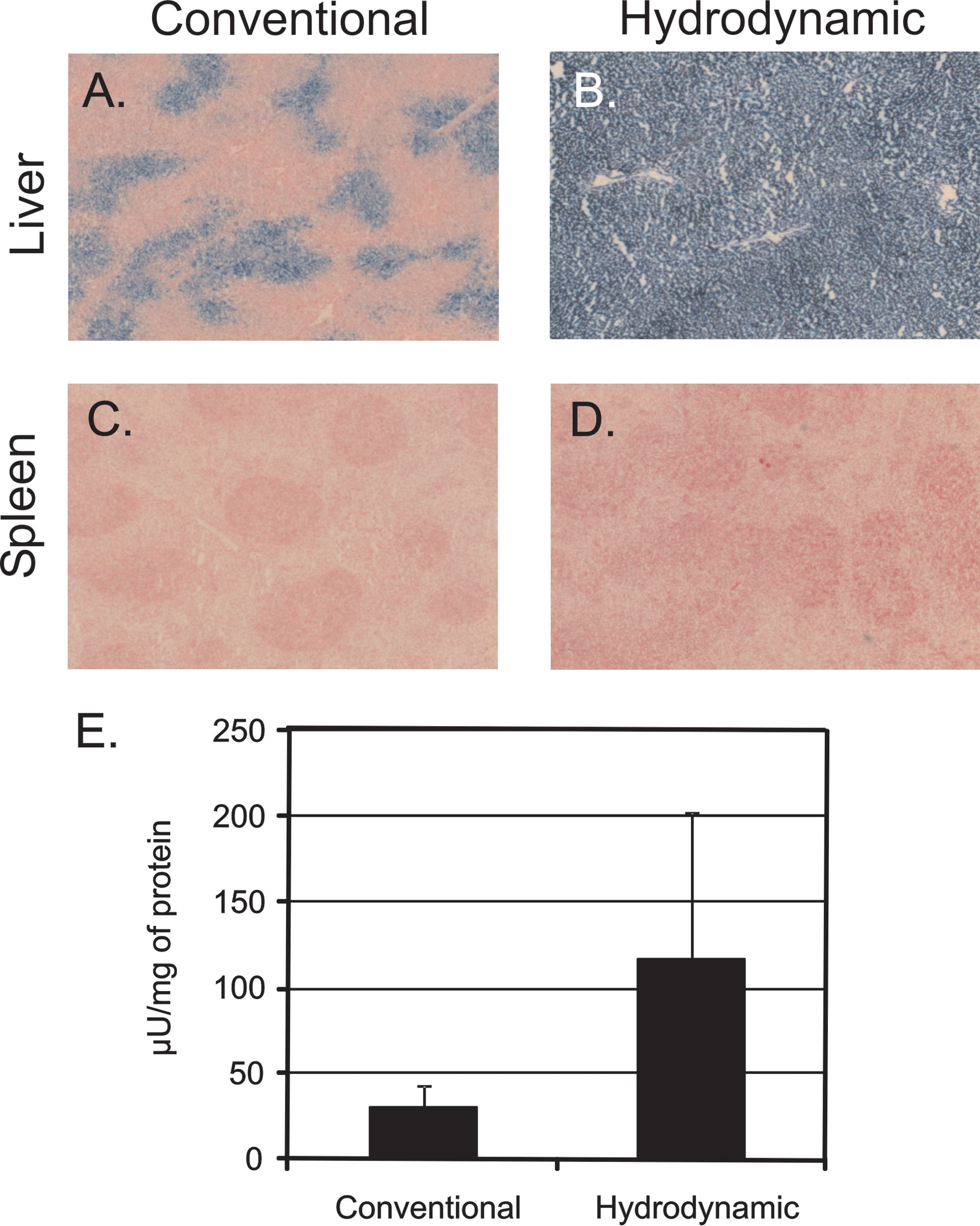
X-gal histochemistry of representative rat (
Toietta et al. (2005) showed that conventional tail vein injection of HDAd-PEPCK-UGT1A1 into Gunn rats resulted in lifelong phenotypic correction of hyperbilirubinemia, albeit using high vector doses (≥3 × 10¹² vp/kg). As a first step towards improving the therapeutic index of the vector, we modified HDAd-PEPCK-UGT1A1, the vector used by Toietta et al. (2005), to create HDAd-PEPCK-UGT1A1-HCR by inserting into the UGT1A1 expression cassette the human apolipoprotein E HCR (Fig. 1). The HCR is a powerful cis-acting liver-specific enhancer element shown to greatly increase liver-specific expression of associated genes (Simonet et al., 1993). We then administered, by conventional injection, four doses (5 × 10¹¹ vp/kg, 1 × 10¹¹ vp/kg, 5 × 10¹⁰ vp/kg, and 1 × 10¹⁰ vp/kg) of HDAd-PEPCK-UGT1A1-HCR into Gunn rats (Fig. 3A). With HDAd-PEPCK-UGT1A1-HCR at 5 × 10¹¹ vp/kg, total serum bilirubin was normalized for at least 60 weeks. At 1 × 10¹¹ vp/kg, total serum bilirubin did not normalize but did result in an average 60% reduction compared with pretreatment levels (p = 0.000296). The dose of 5 × 10¹⁰ vp/kg by conventional injection also resulted in a partial correction with an average reduction of 44% compared with pretreatment levels (p = 0.005). Finally, a dose of 1 × 10¹⁰ vp/kg by conventional injection resulted in a 16% reduction of serum bilirubin compared with pretreatment levels (p = 0.0019). These results demonstrate that inclusion of the HCR decreases the therapeutic dose of HDAd vector because we could achieve normalization of serum bilirubin at a dose of 5 × 10¹¹ vp/kg with HDAd-PEPCK-UGT1A1-HCR, whereas Toietta et al. (2005) could only achieve normalization of serum bilirubin at doses of at least 3 × 10¹² vp/kg with HDAd-PEPCK-UGT1A1. Furthermore, we achieved a 60% reduction in serum bilirubin with HDAd-PEPCK-UGT1A1-HCR at 1 × 10¹¹ vp/kg, whereas an HDAd-PEPCK-UGT1A1 dose of 6 × 10¹¹ vp/kg was required to achieve a comparable level of reduction by Toietta et al. (2005). Our conclusion that inclusion of the HCR can significantly improve the therapeutic index of HDAd is consistent with the results reported by Brunetti-Pierri et al. (2008). In that study, an HCR-bearing HDAd vector expressing OTC was able to achieve phenotypic correction in OTC-deficient mice with at least a fivefold lower dose than an HDAd vector lacking the HCR.
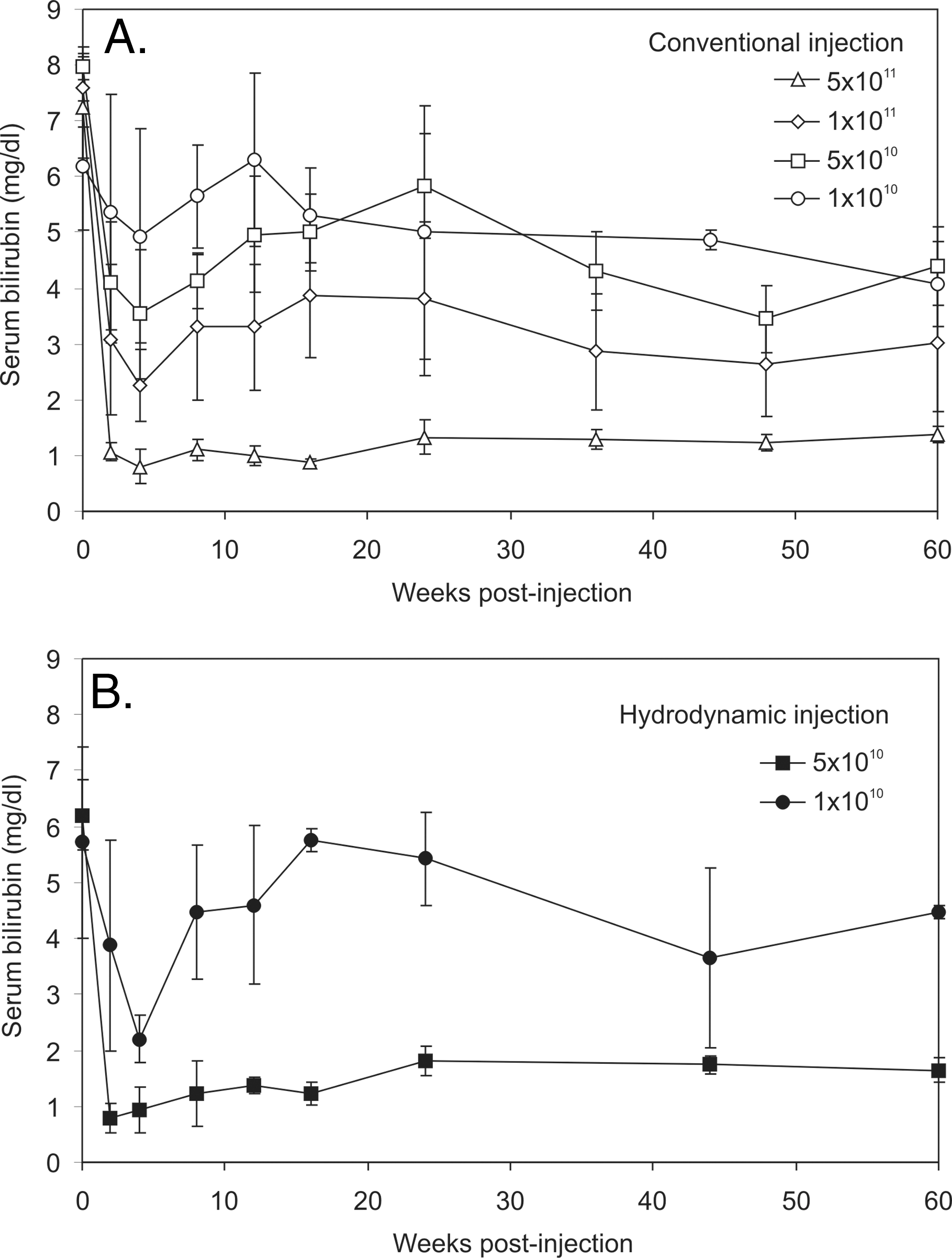
Serum bilirubin levels in Gunn rats injected at various doses by (
We next determined whether hydrodynamic injection of the vector could further reduce the therapeutic dose. To accomplish this, HDAd-PEPCK-UGT1A1-HCR at 5 × 10¹⁰ vp/kg and 1 × 10¹⁰ vp/kg was hydrodynamically injected into Gunn rats, and serum bilirubin levels were determined at various times post-injection (Fig. 3B). The results revealed that complete normalization of the serum bilirubin levels was achieved with 5 × 10¹⁰ vp/kg, whereas a 25% reduction of serum bilirubin levels compared with baseline (p = 0.0007) was achieved with a dose of 1 × 10¹⁰ vp/kg (Fig. 3B). These results demonstrate that hydrodynamic injection can further improve the therapeutic index of HDAd-PEPCK-UGT1A1-HCR such that clinically relevant lower vector doses can be used to achieve a therapeutic effect, at least in the Gunn rat model.
To confirm that the decline in serum bilirubin in the vector-injected animals was due to glucuoronidation of unconjuated bilirubin resulting in excretion of bilirubin monglucuronide (BMG) and diglucuronide (BDG) in the bile, bile was collected from one animal at each dose and injection condition at 60 weeks and analyzed by high-performance liquid chromatography (Fig. 4). As expected, BMG and BDG were absent in bile from untreated Gunn rats (Fig. 4A) but present in nonjaundiced wild-type control rats (Fig. 4B). Consistent with lowering of serum bilirubin, BDG and BMG were observed in the bile of Gunn rats treated by conventional injection of 5 × 10¹¹ vp/kg (Fig. 4C) and 1 × 10¹¹ vp/kg (Fig. 4E) as well as by hydrodynamic injection of 5 × 10¹⁰ vp/kg (Fig. 4D). Little to no BDG and BMG were observed in animals injected hydrodynamically with 1 × 10¹⁰ vp/kg (Fig. 4F) or conventionally with 5 × 10¹⁰ vp/kg (Fig. 4G). Taken together, these results demonstrate that conversion of unconjugated serum bilirubin to BMG and BDG for excretion is mediated by HDAd-PEPCK-UGT1A1-HCR in a dose-dependent manner and that lower vector doses can be effective if delivered by hydrodynamic injection.

Excretion of bilirubin glucuronides in the bile 60 weeks post-vector injection shown in high-performance liquid chromatograms of bile collected by cannulation of the bile duct in (
Acute serum bilirubin elevations are concerning in patients with Crigler–Najjar syndrome. Therefore, we investigated whether administration of HDAd vector was associated with acute bilirubin elevations. We did not observed elevations in serum bilirubin at 6 hr after either hydrodynamic or conventional injection of HDAd vector (data not shown). The lack of acute elevation of serum bilirubin following the hydrodynamic injections suggests that such acute stress may not be associated with significant decompensation in the Gunn rat.
Although hydrodynamic injection as performed in the rat is not possible in humans because of the rapid injection of a large volume, we have previously developed clinically attractive methods that successfully mimic hydrodynamic injection of HDAd vector in nonhuman primates (Brunetti-Pierri et al., 2007, 2009). In these minimally invasive methods, HDAd vector is injected into the animals following inflation of balloon occlusion catheters percutaneously positioned in the inferior vena cava to occlude hepatic venous outflow that results in increased intrahepatic pressure to mimic the hydrodynamic effect but without the need to rapidly inject large volumes (Brunetti-Pierri et al., 2007, 2009). These balloon occlusion catheter-based methods yielded up to an 80-fold improvement in hepatic transduction in non-human primates compared with systemic peripheral intravenous injection, and these improvements are greater than the improvement afforded by hydrodynamic versus conventional injection of HDAd vector into mice or rats.
In summary, we have demonstrated that long-term normalization of serum bilirubin in the Gunn rat can be achieved using clinically relevant low vector doses (<6 × 10¹¹ vp/kg) of an HDAd vector bearing a higher-potency hUGT1A1 expression cassette and delivered by hydrodynamic injection. Given that clinically attractive methods to mimic hydrodynamic injection in non-human primates have been successfully developed, the strategy describing here may be beneficial to Crigler–Najjar syndrome type I patients.
Footnotes
Acknowledgments
The authors wish to acknowledge Dr. Lenard Lichtenberger and Jim Romero for assistance with bile duct cannulation, Dr. Viraj Mane for assistance with the hydrodynamic injections, and Dr. Antony F. McDonagh at UCSF for performing the bile high-performance liquid chromatography analysis. We thank the Morphology Core Laboratory of the Gulf Coast Digestive Disease Center and Angela Major and Dorene M. Rudman for the enzyme histochemistry. This work was supported by the National Institutes of Health (grant K12 RR17665 to D.D., grant R01DK067324 to P.N., and grant R00 HL088692 to N.B.-P.).
Author Disclosure Statement
The authors have nothing to disclose.