
Abstract
Lentiviral vectors are beginning to emerge as a viable choice for human gene therapy. Here, we describe a method that combines the convenience of a suspension cell line with a scalable, nonchemically based, and GMP-compliant transfection technique known as flow electroporation (EP). Flow EP parameters for serum-free adapted HEK293FT cells were optimized to limit toxicity and maximize titers. Using a third generation, HIV-based, lentiviral vector system pseudotyped with the vesicular stomatitis glycoprotein envelope, both small- and large-volume transfections produced titers over 1×10⁸ infectious units/mL. Therefore, an excellent option for implementing large-scale, clinical lentiviral productions is flow EP of suspension cell lines.
Introduction
Expanded use of lentiviral vectors in early Phase I/II clinical trials necessitates efficient, large-scale production under current Good Manufacturing Practice (cGMP) guidelines. To meet this demand, several investigators have created packaging cell lines (PCLs) to produce lentiviruses (Kafri et al., 1999; Klages et al., 2000; Farson et al., 2001; Pacchia et al., 2001; Sparacio et al., 2001; Xu et al., 2001; Kuate et al., 2002; Ikeda et al., 2003; Kumar et al., 2003; Ni et al., 2005; Cockrell et al., 2006; Broussau et al., 2008). The more successful PCLs utilized inducible expression systems in an attempt to circumvent cellular toxicity from HIV proteins and the vesicular stomatitis virus glycoprotein (VSV-G) envelope (Kafri et al., 1999; Klages et al., 2000; Pacchia et al., 2001; Sparacio et al., 2001; Cockrell et al., 2006; Ni et al., 2005; Broussau et al., 2008). Unfortunately, using PCLs in large productions is problematic due to technical constraints. For example, the generation of PCLs takes several months of clone isolation and characterization. Furthermore, due to vector component gene silencing, reduced titers are observed in PCLs over time (Ni et al., 2005).
As an alternative to PCLs, transient transfection methods allow rapid generation of clinical product and can be adapted quickly to new transgenes and pseudotypes. Such a strategy offers better flexibility in productions for early Phase I/II clinical trials. The most common transient transfection technique used in lentivirus production is calcium phosphate precipitation of DNA (Graham and van der Eb, 1973; Bajgelman et al., 2003; Karolewski et al., 2003; Sena-Esteves et al., 2004; Kutner et al., 2009). Disadvantages of the calcium phosphate method include reagent pH sensitivity (Karolewski et al., 2003) and effectiveness only in adherent cell lines. Other transient transfection methods using lipofection (Felgner et al., 1987) or polyethylenimine have been reported (Kuroda et al., 2009; Segura et al., 2010). However, their use may add significant cost to large-scale, clinical productions when cGMP-grade transfection reagents must be used.
PCLs and current transfection techniques have advantages, but they also have important disadvantages that may compromise or complicate the generation of lentiviral vector in large production batches (>50 L). Therefore, other methods should be examined. Electroporation (EP) is an efficient means to load foreign DNA inside the cell (Tsong, 1991; Rols, 2006). The mechanism is thought to involve the electrophoretic association of DNA with the plasma membrane with subsequent entry into the cell (Phez et al., 2005). This technology has been developed extensively over the past decade and several manufacturers offer electroporation units. However, most EP systems are designed to transfect relatively small volumes (typically in a cuvette). Flow EP addresses this limitation by continuously passing the desired volume of a cell and DNA suspension between two electrodes (Rols et al., 1992). The procedure can be effectively scaled up for large bioprocessing applications while maintaining regulatory compliance (Li et al., 2002, 2010). Flow EP also eliminates the need for chemical transfection reagents. These factors are highly relevant to a successful clinical lentiviral production.
In the current study, we report the suitability of using flow EP for large-scale productions of lentivirus. The productions consist of a “standard” third generation lentiviral vector expressing enhanced green fluorescent protein (eGFP) pseudotyped with the VSV-G envelope. We evaluated the lentiviral production efficiency of flow EP in different volumes benchmarked against cuvette or “static” EP. Next, our optimized flow EP method was compared to a standard calcium phosphate transfection in terms of titer, producer cell toxicity, and residual plasmid DNA. For each aspect, flow EP was equal to or outperformed calcium phosphate transfection. Therefore, this method is highly suited to lentiviral productions for early phase clinical gene therapy trials with potential for further scale-up.
Materials and Methods
Cell culture
HEK293 FT cells were obtained from Invitrogen. These cells stably express the SV40 T antigen from the
Plasmids
We used a third generation lentivirus with components split over four plasmids. A plasmid containing an HIV-based, self-inactivating lentiviral vector, pCSCGW (pCSC-SP-PW-GFP; Marr et al., 2004) expressing eGFP from a CMV promoter was used for transgene expression. The remaining three plasmids included the following: pMDL, containing the HIV GAG-POL gene; pMD2.g, containing VSV-G; and pRSV-REV, containing the HIV rev gene (Zufferey et al., 1998).
Growth of 293FT(s) cells in 10-L Cellbags
293FT(s) cells grown to maximum cell density (typically 2×10⁶ to 3×10⁶ cells/mL) in several T175 flasks were harvested by centrifugation and used to inoculate 2 L of SFG media in a 10L Cellbag (GE Healthcare) to a final concentration of 5×10⁵ cells/mL. The 10-L Cellbag was mounted on a Wave bioreactor System 20/50 (GE Healthcare) and airflow was controlled with a CO2MIX20 (GE Healthcare). A CCA T/S blood gas analyzer with E-Glu cassettes (OPTI Medical) was used to monitor glucose, pH, pO2, and pCO2. The cells were further expanded to 5 L over 5 days with a complete media change-out occurring on day 3. Airflow was absent overnight on day 1 to prevent pH increase by excessive CO2 removal before cells were able to condition the 2 L of media. This was found not to be an issue when 5 L of media was used as on day 3. Media was added and removed from the Cellbag by using a peristaltic pump and tubing attached to the inoculation port.
Lentivirus production using EP
293FT(s) cells were harvested by centrifugation at 500×g for 5 min. After aspiration of the medium, cells were resuspended with 60% MaxCyte EP buffer (in sterile distilled water) (Li et al., 2002). The cells were again pelleted by centrifugation, the liquid aspirated, and resuspended in 60% EP Buffer. A viable cell count was obtained using a hemocytometer and trypan blue staining and the cell suspension adjusted to a concentration of 1×10⁸ cells/mL. Plasmid molar ratios of 2:1:1:0.1 (CSCGW:MDL:REV:MDG) were used in the EP transfection cocktail. Cells were electroporated using a GMP-compliant, MXCT GT Flow Electroporation unit (MaxCyte) (Li et al., 2002, 2010) by using one of the following described methods.
For static EP, 1.5×10⁷ cells and 5.9 μg of DNA (154.2 μL total volume) were added to an OC-400 EP cuvette (MaxCyte). After EP, cells were transferred to a T25 flask and incubated for 20 min in a 37°C cell culture incubator with 7.5 μg of DNase (Benzonase, EMD Biosciences or Pulmozyme, Genentech). LP media was added to the cells to a final volume of 4 mL. Viable cell count was obtained using a hemocytometer and trypan blue staining. Aliquots of virus-containing media were harvested 48 hr post-EP, filtered with a 0.45-μm syringe filter, and stored at −80°C.
For flow EP, 10–100 mL of cells at 1×10⁸ cells/mL and 39.2 μg of DNA/mL of cells were added to a CL-2 EP assembly (MaxCyte). After EP, cells were transferred to multiple T175 flasks in volumes of 10 or 20 mL and incubated for 20 min in a 37°C cell culture incubator with 50 μg/mL of DNase (Benzonase or Pulmozyme). After incubation, cell suspensions were transferred to a 10-L Cellbag and cultured in a Wave bioreactor using the following settings: 25 rocks/min, 7.5° rocking angle, and 5% CO2 airflow of 0.3 L/min. T175 flasks were washed with LP media one to three times, pooled, and then added to the Cellbag to maximize cell recovery. Viable cell count was obtained using a hemocytometer and trypan blue staining. LP media was added to the Cellbag to obtain a final volume of 2.1 to 2.3 L. In some experiments after cells underwent flow EP they were transferred to T25 flasks for DNase treatment and viral production as described for static EP. Aliquots of virus-containing media were harvested 48 hr post-EP, filtered with a 0.45-μm syringe filter, and stored at −80°C.
Lentivirus production using calcium phosphate transfection.
HEK293T cells were seeded at 1.7×10⁶ cells per T25 flasks or 5×10⁶ cells per T75 flask. The following day, the cells were transfected by the calcium phosphate method as previously described (Kahl et al., 2004). Cells were re-fed D10 media supplemented with 10 mM sodium butyrate (4 mL in T25 and 12 mL in T75 flasks) 16–18 hrs posttransfection. Lentivirus was harvested 48 hr posttransfection. Aliquots were filtered with a 0.45-μm syringe filter and stored at −80°C.
Infectious titer determination
HEK293T cells were plated in 12-well plates at a concentration of 1×10⁵ cells/well. Serial dilutions of lentivirus-containing media with HEK293T growth media with 8 μg/mL polybrene was added to the cells 2 hr postplating in a final volume of 1 mL. Each dilution had at least two corresponding wells as replicates. Cells were harvested 3 days later for flow cytometric analysis using a FACSCalibur and Cellquest software (BD Biosciences). Infectious titers were determined using the dilution yielding 2%–20% GFP-positive cells with the following equation: (dilution×%GFP positive×100,000)/100. All titers were determined from aliquots that underwent one freeze–thaw cycle.
p24 assay
To determine p24 concentrations, serial dilutions of lentivirus-containing media were used with the HIV-1 p24ca Antigen Capture Assay Kit (AIDS Vaccine Program/SAIC-Frederick, Inc.). The manufacturer's protocol was followed.
Cell toxicity assay
Aliquots of lentiviral production supernatants were used in the Cell Death Detection ELISAplus kit (Roche Applied Science) according to manufacturer's instructions. This assay detects nucleosomes released into the media to give an overall estimate of cell necrosis. Absorbance values were converted to arbitrary units by multiplying by 1000.
Real-time PCR detection of plasmid DNA
DNA was extracted from a 0.5-mL aliquot of 48 hr lentivirus production supernatant using phenol:chloroform:isoamyl alcohol (25:24:1, pH 7.9). Precipitation of DNA was accomplished by adding 1 μL of 5 mg/mL linear acrylamide (Invitrogen), 33 μL of 3 M sodium acetate, and 735 μL of ethanol to 333 μL from the aqueous phase of the proceeding step. The mixtures were incubated overnight at −20°C. DNA precipitates were pelleted, washed with 70% ethanol, resuspended in equal volumes of Tris buffer (pH 8.0), and concentrations determined. Real-time PCR was performed with 1 ng of the isolated DNA using primers to the ampicillin resistance gene as previously described (Sastry et al., 2004). Values are reported as nanograms of plasmid per milliliter of supernatant. This was obtained by multiplying the PCR results by the DNA dilution factor (variable) and then by three (due to precipitating 0.33 mL of supernatant).
Statistics
Data were analyzed by two tailed t test or one-way ANOVA where applicable, and p values under 0.05 were considered significant.
Results and Discussion
Optimization of EP conditions for lentivirus production
We have previously reported that static EP of lentiviral production plasmids generates high titers in HEK293T cells (Li et al., 2008). Considering the high cost of cGMP grade DNA used in clinical productions, achieving lower DNA concentrations requires further optimization of the EP conditions. Transfection efficiency with decreased DNA concentrations can be compensated for with increased electroporation energy (Kubiniec et al., 1990; Rols and Teissie, 1998). To apply this principle, a series of static, cuvette-based electroporations was performed with reduced DNA amounts but increasing levels of applied energy. Figure 1 shows a representative experiment where gradually increasing the total EP energy and using approximately one tenth of the DNA, we can obtain titers equivalent to the initially reported results (designated E1). Raising EP energy beyond the E5 value did not further increase titers (data not shown). Therefore, the E5 program settings were used in static EP for the remainder of this study.
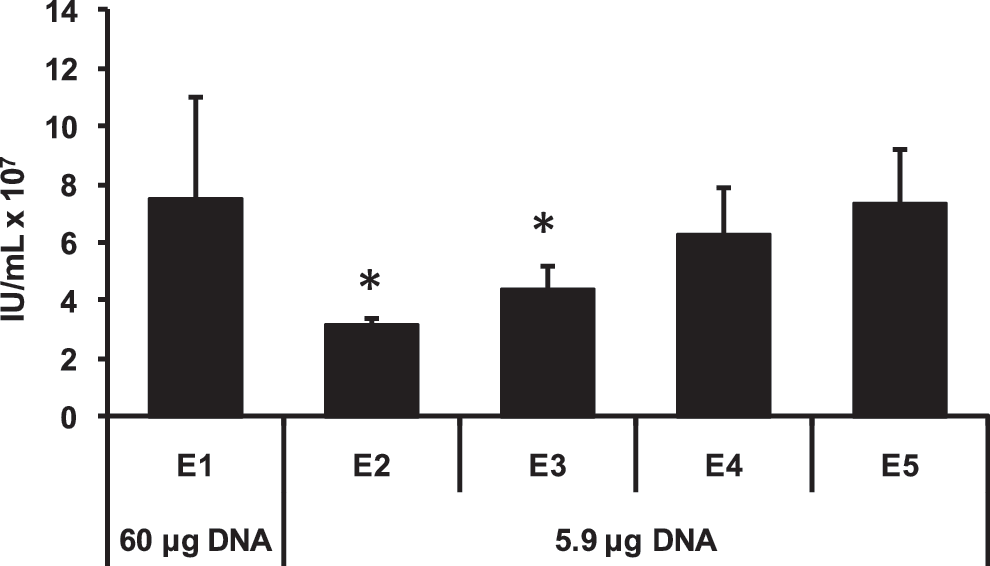
Optimization of EP conditions at reduced DNA concentrations. Static EP with lentiviral plasmids was performed as described in the Methods section using either 60 μg or 5.9 μg of plasmid DNA. E values are preloaded computer-controlled protocols with increasing energy delivered to the cells (i.e., E1<E2<E3<E4<E5). Viral titers were determined by flow cytometric assay of 48-hr supernatants. Results are an average of three replicates. IU, infectious unit. *Signifies statistically significant difference with p<0.01 compared to E5 as determined by Student's t test.
Application of static EP settings to a flow EP program for scale-up
The MaxCyte GT flow EP system was designed to use static EP energy settings in a flow EP protocol without further optimization; this effectively allows for direct scale-up (Li et al., 2002). To confirm this ability in regards to lentivirus production in 293FT(s) cells, flow EP was executed with 10 or 50 mL of cell suspension. Static EP was done with an aliquot of the cells as a control. An equivalent amount of cells from each EP method were then transferred to T25 flasks for lentiviral production. Additionally, the calcium phosphate transfection method routinely used in our lab for lentivirus production in HEK293T cells (Kahl et al., 2004) was performed as a comparison. As demonstrated in Fig. 2, the average titers for flow EP, static EP, and the calcium phosphate method were not statistically significant. Our titers were also comparable to previously reported values for adherent HEK293T cells (Zufferey et al., 1998; Karolewski et al., 2003; Kutner et al., 2009). Interestingly, flow EP requires one third less DNA than the calcium phosphate method in a T25 flask lentivirus production (5.9 μg versus 9.2 μg of DNA). These results establish that flow EP can be successfully used for lentivirus production and titers achieved are at least equivalent to the calcium phosphate method.
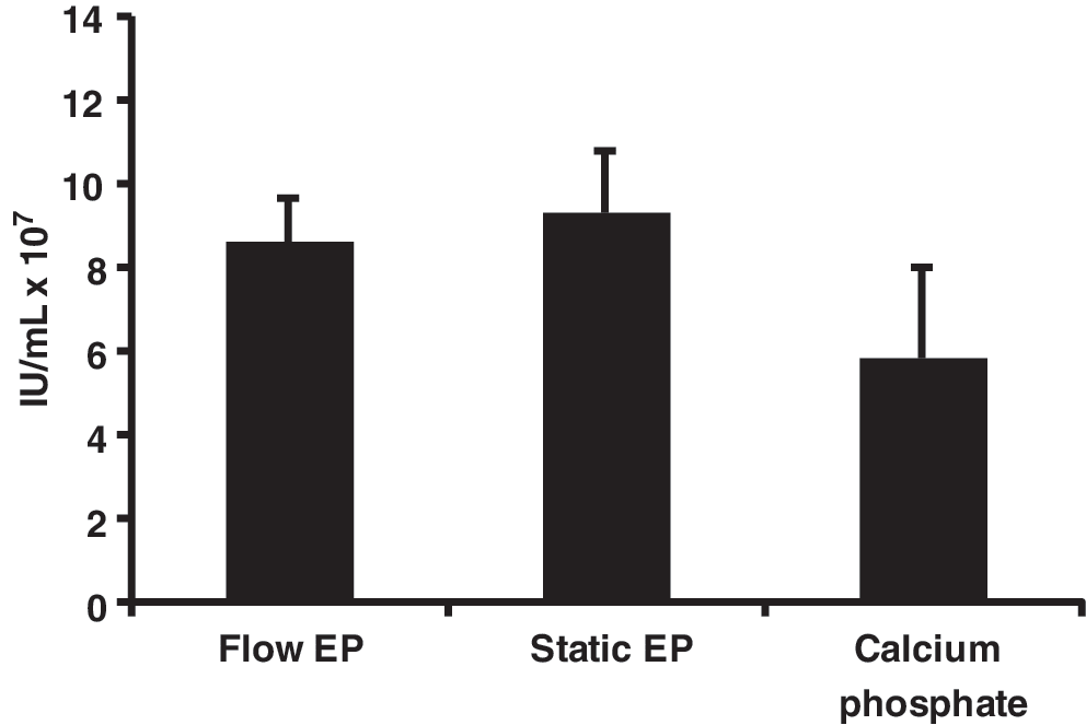
Testing optimized static EP conditions in a flow EP protocol. Flow EP (two replicates with 10 mL and two replicates with 50 mL), static EP, or calcium phosphate transfection was performed as described in the Methods section. Equal counts of cells from each EP method were added to 4 mL of media for viral production. Viral titers were determined by flow cytometric assay of 48-hr supernatants. The group means were not statistically different as determined by one-way ANOVA (F 2,8=3.15, p=0.098).
Pulmozyme DNase usage during recovery incubation increases titers
The addition of a bacterial sourced DNase (e.g., Benzonase, EMD Biosciences) to the cells immediately following EP improves viability which, consequently, improves lentivirus titers (Li et al., 2008). The mechanism of improved cell viability after EP is hypothesized to involve an increase in the plasma membrane sealing rate due to digestion of DNA on the plasma membrane (Li et al., 2008). DNase has previously been used in clinical lentiviral production protocols to eliminate plasmid contamination in the final product (Slepushkin et al., 2003; Levine et al., 2006). Pulmozyme, a human DNase approved by the U.S. Food and Drug Administration for use in cystic fibrosis therapy, was examined as a potential alternative. 293FT(s) cells in a volume of 10 mL were transfected by flow EP. Aliquots of transfected cells were incubated with or without 50 μg/mL of Pulmozyme in T25 flasks. An equivalent number of cells transfected by static EP were similarly treated with 50 μg/mL of Pulmozyme as a control. As seen in Fig. 3, the addition of Pulmozyme increased the 48-hr titer almost twofold. Importantly, this step must be performed in cell culture vessels to allow for gas exchange. We observed that using capped tubes, even with a low volume of liquid, would kill most of the cells when incubated at 37°C for 20 min (data not shown). Pulmozyme was included in the flow EP protocol for the remainder of the study.
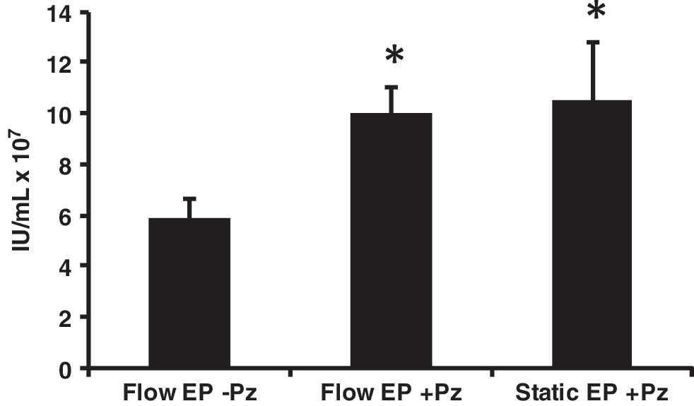
Increased titer with Pulmozyme treatment during EP recovery. Ten-milliliter flow or static EP was performed as described in the Methods section. Recovery incubation immediately following EP was performed with or without 50 μg/mL Pulmozyme in T25 flasks containing equivalent numbers of cells. Viral titers were determined by flow cytometric assay of 48-hr supernatants. Results are an average of two replicates. IU, infectious unit; Pz=Pulmozyme. *Signifies statistically significant difference with p<0.01 compared to Flow EP without Pulmozyme as determined by Student's t test.
Lentivirus productions in a Wave bioreactor using flow EP transfected cells
In contrast to using multiple T-flasks, roller bottles, or cell factories, Wave bioreactors can be adapted to varying batch sizes in a single, closed system. We performed three independent, large-scale lentivirus productions in 10-L Cellbags (maximum liquid capacity of 5 L). The overall workflow for cell expansion and production is shown in Fig. 4. As detailed in Table 1, titers were consistent between productions and comparable to the small-scale titers (see Fig. 2). The specific productivity of each experiment was also consistent with approximately 40 infectious particles harvested per viable transfected cell (Table 1). Expressing the infectious titer data as a ratio with p24 assay data, the flow EP productions yielded values of greater than 1×10⁵ IU/ng of p24 (Table 1). The ratios were slightly higher than previously reported values for calcium phosphate transfections (Mochizuki et al., 1998; Follenzi and Naldini, 2002). Elevated ratios indicate a higher percentage of infectious particles from total particles produced. Overall, flow EP is a viable method for large-scale lentivirus production.
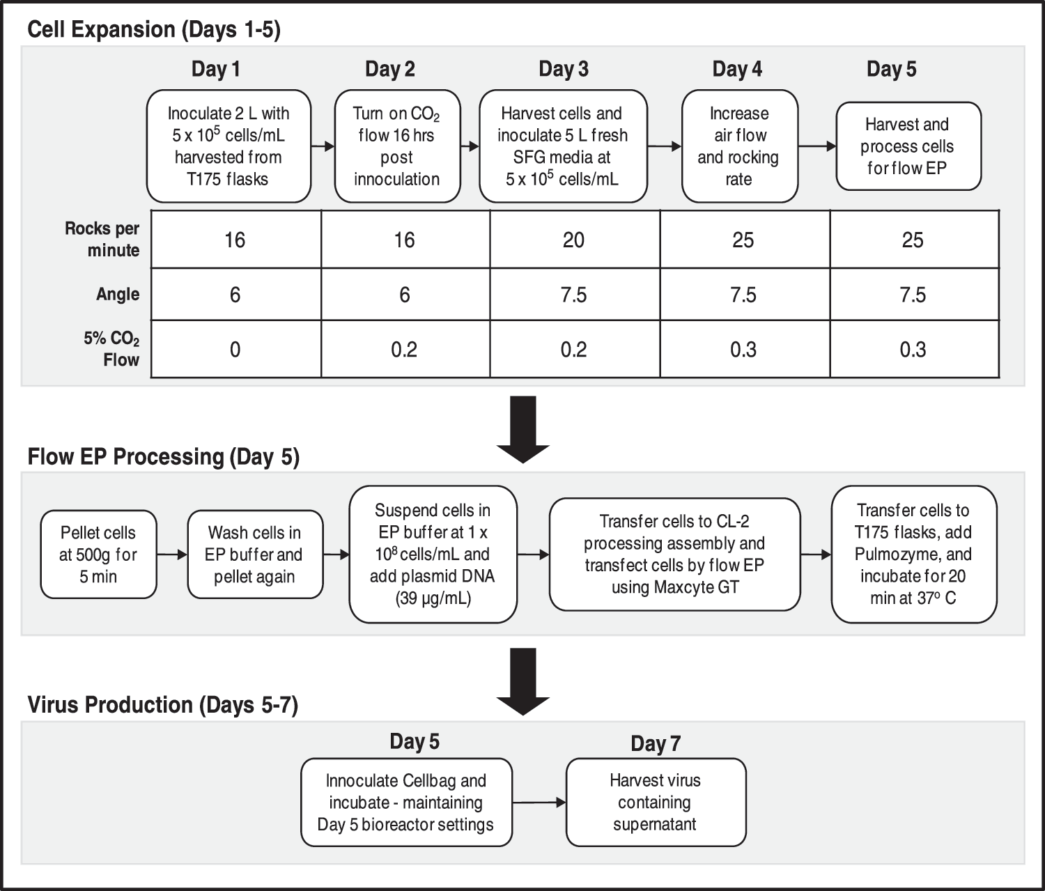
Workflow of cell expansion and lentivirus production in Wave bioreactor using 10-L Cellbags. Cell expansion is initialized in 2 L of media and then scaled up to 5 L over a period of 5 days. On the fifth day, cells are harvested and subjected to flow EP. The transfected cells are added to fresh media and viral production occurred over days 6 and 7. Wave bioreactor settings on days 6 and 7 were maintained at day-5 values.
The total cell count is based on viable cells counted immediately following the addition of electroporated cells to the Cellbag. A reduction in the number of T175 flask washes (used in the post-EP Pulmozyme treatments) from three to one led to the slightly lower final volume in experiment 3. Viral titers and p24 assays were performed using aliquots of 48-hr posttransfection harvests.
Harvested supernatant from lentivirus productions may include contaminants in the forms of plasmid DNA from transfection. DNase treatment postproduction, as reported previously (Slepushkin et al., 2003; Transfiguracion et al., 2003; Sastry et al., 2004), is a highly effective way to eliminate residual plasmid. Since a DNase digestion step is performed post-EP, the elimination of residual plasmid was quantified by using a real-time PCR assay (Sastry et al., 2004). Plasmid DNA was detected in each production (Table 2). Except for production 2, plasmid contamination was slightly less than a T75 flask calcium phosphate transfection not treated with DNase. All values were within a range previously reported for unconcentrated lentiviral supernatants (Sastry et al., 2004). Consequently, the flow EP transfection method may still benefit from postharvest DNase treatment similar to calcium phosphate transfections in clinical productions.
Released cellular histone–DNA complexes and residual plasmid DNA assays were performed using aliquots of 48-hr posttransfection harvests. The CaPO4 transfection sample was not treated with DNase. Assay data are representative of at least two separate experiments.
Statistically significantly different at p<0.01 compared to CaPO4 transfection as determined by Student's t test.
To study contaminants specifically from producer cells, a commercial ELISA kit was employed to detect histone–DNA complexes released from necrotic cells. Data from a representative ELISA assay in Table 2 demonstrate that flow EP productions consistently resulted in significantly less necrotic marker. The differences cannot be accounted for by cell density alone. Calcium phosphate transfections average 2×10⁶ to 3×10⁶ cells/mL posttransfection; this count is comparable to the productions reported here with 293FT(s) cells (2.1×10⁶ to 3.5×10⁶ cells/mL). Taken as a whole, the data suggest the flow EP method yields similar, and possibly lower, producer cell contaminants when weighed against calcium phosphate transfection. Since serum-free media is utilized, further downstream purification can be carried out to effectively reduce contaminants (Slepushkin et al., 2003; Segura et al., 2007; Lesch et al., 2011).
The purpose was to develop a scalable GMP-compliant flow electroporation system for lentivirus production. The results demonstrated that flow electroporation development was successful overall (Table 1). However, the original strategy of performing 5-L productions was unfortunately limited to 2 L due to modest cell densities obtained during expansion. Independent of Cellbag size, we observed that 293FT(s) cells reach a maximum density of approximately 2×10⁶ cells/mL (data not shown). Metabolic data provide clues to potential factors contributing to the modest cell densities. Metabolic readings at the time of lentivirus harvest were similar to those observed on Day 5 of cell expansion (Supplementary Table S1; Supplementary Data are available online at
Another limitation of the flow EP protocol reported here is liquid handling. A benchtop centrifuge was used to pellet cells after removal from the cellbags by peristaltic pump. Even with 0.5-L bottles, this proved time consuming and led to some cell loss. More importantly, for a clinical production in which sterility is paramount, a more streamlined cell-harvesting method with limited use of transfer vessels is necessary. Such a method could utilize tangential flow filtration for cell harvesting and buffer exchanges. A further reduction in transfer vessels could be realized by using a single Pulmozyme treatment vessel instead of multiple T175 flasks (e.g., 2-L Cellbag). While the liquid handling in our study was not optimal, it does not diminish flow EP being an efficient transient transfection method for lentivirus production when processing large numbers of cells.
In summary, we have successfully demonstrated the ability to produce lentivirus on a significantly large scale using flow EP. To our knowledge, this is the first report of a flow EP method used to produce lentivirus in a bioreactor. As a transient transfection method, flow EP has the advantage of providing flexibility in early stage clinical trial productions while avoiding the caveats associated with PCLs. Additionally, flow EP lowers production costs by eliminating the need for cGMP-grade chemical transfection reagents. Although 293FT(s) cells are used in the present study, we stress the fact that flow EP can be successfully used with any HEK293-based suspension cell line in lentivirus productions. Future studies should explore further refinements and scale-up of this method.
Footnotes
Acknowledgments
This work was supported by a grant from the National Heart, Lung, and Blood Institute (5R44HL088996-03 to Madhusudan Peshwa). Scott Witting was also supported by a grant from the National Heart, Lung, and Blood Institute (P30 HL101337 to Kenneth Cornetta). The authors thank Dr. Philip Zoltick for the lentiviral transgene plasmid (pCSCGW) and Sarah Dolan and Priya Vallanda for excellent technical assistance.
Author Disclosure Statement
Lin-Hong Li, Cornell Allen, James Brady, Rama Shivakumar, and Madhusudan Peshwa are employed by MaxCyte.
References
Supplementary Material
Please find the following supplemental material available below.
For Open Access articles published under a Creative Commons License, all supplemental material carries the same license as the article it is associated with.
For non-Open Access articles published, all supplemental material carries a non-exclusive license, and permission requests for re-use of supplemental material or any part of supplemental material shall be sent directly to the copyright owner as specified in the copyright notice associated with the article.