
Abstract
The goals of the National Heart, Lung, and Blood Institute (NHLBI) Center for Fetal Monkey Gene Transfer for Heart, Lung, and Blood Diseases are to conduct gene transfer studies in monkeys to evaluate safety and efficiency; and to provide NHLBI-supported investigators with expertise, resources, and services to actively pursue gene transfer approaches in monkeys in their research programs. NHLBI-supported projects span investigators throughout the United States and have addressed novel approaches to gene delivery; “proof-of-principle”; assessed whether findings in small-animal models could be demonstrated in a primate species; or were conducted to enable new grant or IND submissions. The Center for Fetal Monkey Gene Transfer for Heart, Lung, and Blood Diseases successfully aids the gene therapy community in addressing regulatory barriers, and serves as an effective vehicle for advancing the field.
Introduction
Although all age groups can benefit from new molecular and cell-based therapies, the youngest patients may provide both unique advantages and additional challenges (“children are not small adults”). Because many inherited disorders can be identified prenatally, fetal gene transfer may provide the best time for treatment because early intervention may eliminate the pathology associated with disease. In addition, the functional immaturity of the fetal immune system can overcome the barrier of immune responses that complicates gene transfer postnatally and in adults (Brantly et al., 2009; Mays and Wilson, 2011).
Animal models are essential to understand biological functions, study complex human diseases, and address safety of new therapies proposed for human use. Monkeys and humans share many features because of their close phylogenetic relationship; similarities in development, physiology, immunology, and anatomy aid in overcoming the roadblocks to clinical translation (Tanimura and Tanioka, 1975; Bontrop, 2001; Roth et al., 2004; Tarantal, 2005; Shively and Clarkson, 2009; Sykes, 2009). For example, monkeys have been widely used as preclinical models for human bone marrow transplantation and stem cell gene therapy (Trobridge and Kiem, 2010), and have shown substantial advantages when compared with other species. Long-term engraftment is challenging to assess in a short-lived mouse model, and differences between mouse and human host cell receptors may limit translation to the human clinical setting (Horn et al., 2003; Mezquita et al., 2008; Sykes, 2009; Beard et al., 2010; Trobridge and Kiem, 2010; Gibbons and Spencer, 2011). Studies also indicate that the histology and time course for allograft rejection in monkeys parallel those in humans because of similarities in MHC genes and immune ontogeny (Bontrop, 2001; Sykes, 2009), and tolerance is much easier to achieve in mice (Mestas and Hughes, 2004). Thus, nonhuman primates can accelerate the development of promising new gene- and cell-based therapies for humans in all age groups.
Studies supported by the National Heart, Lung, and Blood Institute (NHLBI) Center for Fetal Monkey Gene Transfer for Heart, Lung, and Blood Diseases (California National Primate Research Center at the University of California, Davis, Davis, CA) have addressed gaps and roadblocks through a proven and highly effective program. The established infrastructure can rapidly test new paradigms, move new hypotheses and emerging gene transfer vectors into a preclinical setting, and provide critical preliminary data for new grants and investigational new drug (IND) submissions. Novel aspects of the program include the following: a strong track record in a collaborative team approach, strengths in translational research, unique capabilities in the study of fetal/pediatric nonhuman primate models of human development and disease, and the use of novel in vivo imaging modalities including positron emission tomography (PET) and bioluminescence imaging (BLI). The development and application of new in vivo imaging technologies have provided new ways to monitor long-term gene expression and the trafficking of transplanted cells in real time (Tarantal et al., 2006, 2009, 2012c). The Center also highlights a state-of-the-art fetal therapy program with many ultrasound-related techniques and procedures that have been applied comparable to those used in humans (Tarantal, 2005).
Materials and Methods
Animals
All animal procedures conform to the requirements of the Animal Welfare Act, and protocols are approved before implementation by the Institutional Animal Care and Use Committee (IACUC) at the University of California, Davis. Normally cycling, adult female rhesus monkeys (Macaca mulatta) with a history of prior pregnancy are bred and identified as pregnant according to established methods (Tarantal, 2005). Pregnancy in the rhesus monkey is divided into trimesters by 55-day increments, with 0–55 days of gestation representing the first trimester, 56–110 days of gestation representing the second trimester, and 111–165 days of gestation the third trimester (term, 165±10 days). Activities related to animal care (diet, housing) are performed according to California National Primate Research Center standard operating procedures (SOPs). Newborns are typically delivered by cesarean section at term according to established protocols and raised in the gene therapy nursery for postnatal studies (Tarantal et al., 2005). Animals in older age groups (juvenile to adult) are maintained under established housing conditions with environmental enrichment based on SOPs and by study assignment.
Process
A call for proposals (Fig. 1) is circulated annually to NHLBI-supported investigators, posted on the Center for Fetal Monkey Gene Transfer for Heart, Lung, and Blood Diseases website (

Annual call for Letters of Intent.
Results and Discussion
A series of basic questions in gene delivery have been addressed including the efficiency of gene transfer using lentivirus (HIV or SIV) and adeno-associated virus (AAV) vectors to target specific cells and tissues; the potential effects of gene transfer on prenatal and postnatal development; the potential risk(s) to the fetus, infant, and dam; and the assessment of inadvertent germ-line transmission. Studies have also compared systemic and organ-targeted gene transfer approaches, and consistently shown high levels of transduction and transgene expression for extended periods of time (up to 10 years) without evidence of adverse effects. Examples of accomplishments include the following:
• Demonstrated systemic fetal administration of HIV-1-derived lentiviral and AAV vectors results in prolonged transgene expression in the muscular component of the diaphragm, with no evidence of adverse effects (Jimenez et al., 2005a). These findings suggest the potential for fetal gene transfer to be developed into a durable therapeutic modality for the treatment of diseases for which only a small amount of protein is needed for correction (e.g., hemophilia)
• Showed that direct intrapulmonary and intramyocardial gene delivery results in persistent transgene expression without evidence of adverse effects (Tarantal et al., 2005). These studies support that the route and timing of vector administration are crucial for limited biodistribution to other anatomical sites
• Conducted extensive analysis of gonads after various routes of lentiviral vector administration prenatally and confirmed no evidence of germ cell gene transfer in males or with organ-targeted approaches (Lee et al., 2005b; Tarantal et al., 2005). These studies further support the importance of organ-targeting and vector design (Pacak et al., 2006) to limit biodistribution
• Used SIV-based lentiviral vectors (Kahl et al., 2008) to transduce young monkey CD34+ hematopoietic stem/progenitor cells for autologous transplantation, and used nonmyeloablative conditioning regimens such as busulfan and fludarabine (Kahl et al., 2006; Tarantal et al., 2012a)
• Addressed the role of overexpression of transforming growth factor-β1 during fetal lung development, using an intrapulmonary gene transfer approach (Tarantal et al., 2010), providing a new model to explore lung disease that may allow greater insight into human lung development
• Reported that AAV8-mediated hepatic gene transfer in infant monkeys is safe and efficient but less stable when compared with adolescent animals (Bell et al., 2011; Wang et al., 2011)
• Showed that in utero delivery of an AAV2/5-human FVII vector in the late third trimester confers therapeutic expression of human FVII at birth, which was maintained above baseline levels for at least 2 months, and that readministration with capsid proteins of another serotype (AAV2/8) ∼1 year after fetal gene delivery resulted in a further increase in plasma levels (Binny et al., 2012)
• Evaluated stem and progenitor cell age-related differences (fetal through aged) and showed significant differences across the lifespan (e.g., endothelial, hematopoietic, mesenchymal) (Lee et al., 2004, 2005a, 2006; Hacia et al., 2008; Kim et al., 2008; Shelley et al., 2012)
• Developed in vivo imaging techniques (PET, BLI) (Tarantal et al., 2006, 2009, 2012c; Tarantal and Lee, 2010) that demonstrated long-term gene expression, and showed engrafted foci of human cells at sites not appreciated by the collection of blood or bone marrow
• Showed high levels of firefly luciferase expression with no adverse effects up through ∼8 years postnatal age when organ-targeted fetal gene transfer approaches were used (e.g., intrathoracic, intrapulmonary, intramyocardial, intraportal, intrahepatic; lentiviral vectors or AAV serotypes) (Fig. 2). Imaging findings also showed good correlations with outcomes at the tissue level (Tarantal et al., 2009; Tarantal and Lee, 2010; Tarantal, unpublished data)
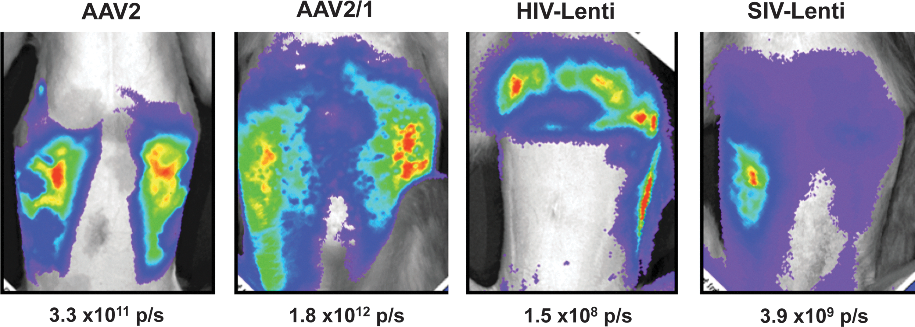
Examples of a subset of animals transferred prenatally during the late first trimester with AAV2, AAV2/1, HIV, or SIV-based vectors that indicate long-term transgene expression (up to 6 years) without evidence of adverse effects. These animals have exhibited stable expression in the muscular component of the diaphragm and peritoneum (1×10⁷–4×10⁹ photons/sec [p/s]) when using the prenatal systemic (intraperitoneal) ultrasound-guided approach for fetal gene transfer.
The Center for Fetal Monkey Gene Transfer has supported projects that provided data for new IND applications. Each aspect of the clinical development plan was facilitated by interactions with the Center and showed that this program aids the gene therapy community in addressing regulatory barriers. The leap from preclinical discovery to human subject research is challenging, and the support and resources provided by the Center are a crucial component in this process. Examples include the following:
• Studies that assessed the use of busulfan in young monkeys to model nonmyeloablative conditioning have been used to translate this approach into an ADA-SCID clinical trial. These and related preclinical, translational studies have also involved the production of high volumes of lentiviral vectors, which has been instrumental in moving vector production methods to the levels necessary for human clinical trials.
• Studies have been instrumental in achieving the overall goal of using AAV expression of human acid α-glucosidase (GAA) in patients with Pompe disease. Initial studies established the usefulness of AAV serotype 1 in rhesus monkey muscle, and the results of this study were included in an IND submission for the use of AAV in 3- to 14-year-old patients who have developed ventilator dependence. Studies in the Center also identified novel properties of AAV2/9 in skeletal and cardiac muscle, important for enabling an IND application. Each aspect of the clinical development plan for Pompe disease was facilitated by studies in monkeys (Smith et al., 2012).
• A current ongoing study in young monkeys is focused on identifying a safe and robust AAV delivery method for regional limb gene transfer for patients with Duchenne muscular dystrophy and involves a side-by-side comparison of AAV9 with an engineered capsid that shows strong detargeting of liver and preferred transduction of heart and skeletal muscle (Mitchell et al., 2010). BLI is used to investigate biodistribution and tropism; outcomes have been incorporated in a pre-IND submission.
• Through funds from another NIH institute, studies in monkeys assessed the safety and gene transfer efficiency of a lentiviral vector, which showed a lack of toxicity and no adverse events in fetal and juvenile monkeys. These studies were critical in gaining approval for an IND application and conducting the first-in-human trial of an expressed small interfering RNA (siRNA) in a lentiviral vector (DiGiusto et al., 2010).
The Center has developed many techniques that are routinely used to aid investigators in the conduct of their studies. Examples include the following:
• Primers and probes for quantitative PCR (qPCR) to assess biodistribution (DNA and RNA) (Jimenez et al., 2005a; Tarantal et al., 2005, 2012a). Because of genomic homology, primer and probe sets based on human sequences may also amplify rhesus monkey genes, and thus the Center has developed a catalog of multiple sets of primers and probes that can distinguish between the species to meet the needs of various investigators and projects.
• Primer and probe sets to identify male DNA sequences (human or rhesus) in serum and tissues for the multicopy testis-specific Y-encoded (TSPY) gene (high sensitivity), and to quantify the single-copy sex-determining region Y (SRY) gene by PCR (Jimenez and Tarantal, 2003a, b). The rhesus Y chromosome assays are used routinely to identify male sequences in maternal blood samples in early gestation (before sexual differentiation), to select dams for studies that are focused on diseases that are male-specific, or to balance sex ratios (Wang et al., 2011).
• Human TSPY and SRY assays are also used to identify human cells in the rhesus host posttransplant and in parallel with techniques that detect human cells in monkey tissues using a human-specific centromere probe. These methods provide an efficient means to further analyze specimens by immunohistochemistry and antibodies that cross-react only with human specimens (Tarantal et al., 2000, 2012b).
• Streamlined the process for rapid and accurate assessment of tissues that express transgenes such as the enhanced green fluorescent protein (eGFP) by performing whole tissue fluorescence imaging to identify regions for further assessment by qPCR or immunohistochemistry (Tarantal et al., 2005).
• Adapted reporter systems, such as the gene for firefly luciferase and its substrate luciferin, for BLI and those that encode a phosphorylating enzyme (e.g., HSV-sr39TK) such that a radiolabeled substrate for the enzyme is trapped only in cells that express a reporter for PET imaging (Cherry, 2004; Tarantal et al., 2006). The advantages of in vivo imaging include that each individual can be followed longitudinally in real time, conferring a large statistical advantage, and animal-to-animal variability is reduced as a confounding variable. Studies have also shown that in vivo BLI is highly effective for monitoring long-term gene expression, identifying foci of engrafted cells, and assessing cell trafficking when combined with PET (Tarantal et al., 2012c).
In summary, the Center for Fetal Monkey Gene Transfer for Heart, Lung, and Blood Diseases is a unique resource that has served an important role in the gene therapy community by addressing essential questions in gene delivery and consistently providing investigators with opportunities to test new vector constructs and approaches that advance the field. The Center provides collaborative opportunities to aid investigators in pursuing studies in rhesus monkeys of all age groups to address crucial gaps and roadblocks, and to provide the data necessary for NIH grant submissions and IND applications. Monkeys are a challenging species and specialized expertise and resources are needed to effectively and safely conduct these studies. Any investigator funded by the NHLBI is eligible to submit a Letter of Intent requesting to conduct a study with monkeys or to obtain specimens (e.g., serum, cells, tissues) for in vitro investigations. The potential ramifications of gene delivery in humans underscores the importance of rigorous assessments of long-term gene expression and safety, and the Center for Fetal Monkey Gene Transfer for Heart, Lung, and Blood Diseases provides these opportunities to investigators nationally.
Footnotes
Acknowledgments
The Center for Fetal Monkey Gene Transfer for Heart, Lung, and Blood Diseases is supported by NIH grants (HL069748 and HL085794) and by the California National Primate Research Center base operating grant (NIH grant RR00169 and OD011107).
Author Disclosure Statement
The authors have nothing to disclose.