
Abstract
Duchenne muscular dystrophy (DMD) is a severe hereditary neuromuscular disorder caused by mutations in the dystrophin gene. Antisense-mediated targeted exon skipping has been shown to restore dystrophin expression both in DMD patients and in the mdx mouse, the murine model of DMD, but the ineffective delivery of these molecules limits their therapeutic use. We demonstrated that PMMA/N-isopropil-acrylamide (ZM2) nanoparticles (NPs), administered both intraperitoneally and orally, were able to deliver 2′OMePS antisense inducing various extents of dystrophin restoration in the mdx mice. Defining NP biodistribution is crucial to improve effects on target and dose regimens; thus, we performed in vivo studies of novel ZM4 NPs. ZM4 are conjugated with NIR fluorophores as optical probes suitable for studies on the Odyssey Imaging System. Our results indicate that NPs are widely distributed in all body muscles, including skeletal muscles and heart, suggesting that these vehicles are appropriate to deliver antisense oligonucleotides for targeting striated muscles in the DMD animal model, thus opening new horizons for Duchenne therapy.
A
We were pioneering designing novel cationic core-shell nanospheres (T1 and ZM2), made up of a core of poly(methyl methacrylate) or NIPAM-PMMA, surrounded by a shell bearing cationic groups. Our NPs bind 2′OMePS oligoribonucleotides and have a functional effect, inducing body-wide dystrophin rescue both in skeletal and in cardiac muscles (Rimessi et al., 2009; Ferlini et al., 2010; Bassi et al., 2012). The main added value of using our NPs was related to the very-low-dose regimen of AONs able to restore dystrophin synthesis.
In order to study the biodistribution in mdx mice after intraperitoneal (IP) administration, we designed novel NP-marked ZM4. ZM4 exposes at the surface, or within the outer shell, the binding groups. The quaternary ammonium groups reversibly adsorb AONs, while reactive primary amino groups are able to covalently conjugate NIR-797 fluorophore as optical probe, thus leading to ZM4-IR (Fig. 1a). ZM4 presents the same physical/chemical characteristics of ZM2 (Ferlini et al., 2010).
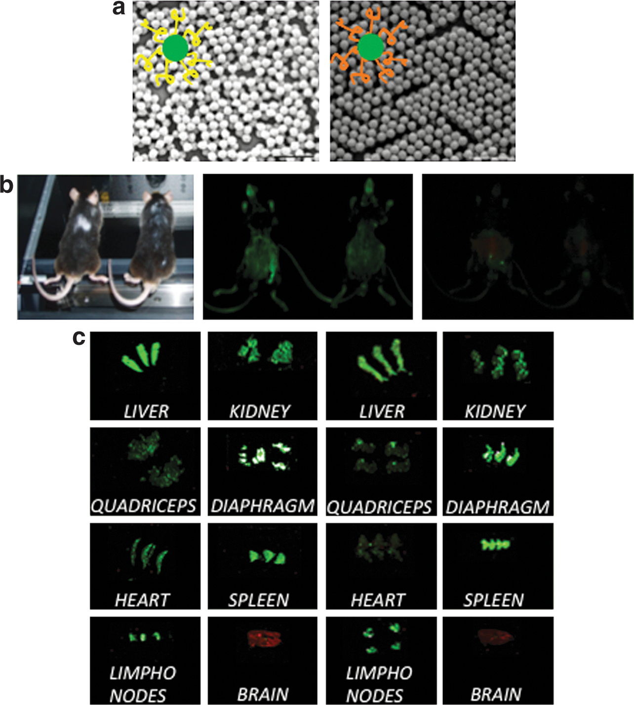
The in vivo ZM4 biodistribution, after single or multiple IP administrations, was evaluated by the Odyssey Imaging System (Li-Cor Biosciences), which is able to track over time the specific distribution of infrared dye-labeled NPs in live animals. After a single IP administration, ZM4-IR remained mainly in the peritoneal cavity for about 24 hr and then diffused widely throughout the body. Fluorescence persisted for up to 22 days (Fig. 1b).
We also treated mdx mice with multiple ZM4-IR IP administrations for 2 months and analyzed biodistribution. Mice were sacrificed at 7, 30, and 60 days after the last injection, and 10 μm cryosections were analyzed with Odyssey. All muscles, including the heart, showed fluorescence until 60 days. The fluorescence, absent in the liver, persisted in kidneys, spleen, and lymph nodes (Fig. 1c). We also analyzed sagittal cryosections from the brain, and, at all the stages analyzed, the fluorescence was exclusively localized in blood vessels and no signal was detectable in the nervous tissue and in the brain ventricles, demonstrating that ZM4 does not cross the blood–brain barrier.
These results, together with the previous ones we reported on dystrophin restoration (Ferlini et al., 2010; Bassi et al., 2012), support the rationale of using NPs for AON delivery, even if improvements in biodegradability are needed. Moreover, our approach to profile biodistribution “in vivo” might be adopted also for other similar compounds.
Footnotes
Acknowledgments
The Telethon Italy Grant GGP09093 (to A.F.) is acknowledged. We are grateful to the Industria Chimica Emiliana (ICE Reggio Emilia). A.F. is principal investigator of two antisense-based clinical trials (Prosensa, The Netherland, and GSK, UK) based on PRO044 and drisapersen.