
Abstract

Ancient History
I
In considering graduate studies, I was very interested in exploring the possibility of using viral vectors to deliver genes to cells, and Alex Rich and virtually everyone else I sought advice from about possible PhD programs suggested I try to work for Paul Berg, a faculty member of the Department of Biochemistry at Stanford University School of Medicine. At the time, Paul was widely recognized as a pioneer in the development of recombinant DNA technology. While trained as a classical biochemist, Paul had by the mid-1970s become an expert in virology as well, and members of his laboratory, particularly Tom Shenk, Janet Mertz, and Steve Goff, were actively involved in efforts to genetically characterize the life cycle of SV40 through the generation of engineered mutants of the virus (Mertz et al., 1975). I decided to apply to the Department of Biochemistry with the express intention of working for Paul, despite the feedback from many, including Paul, that one should not apply to a PhD program with his/her heart set on working for a specific faculty member. I was very lucky to be accepted into the program, and even more lucky that, after much resistance, Paul accepted me into his lab.
At the time I joined the lab, Steve Goff, my new bay mate, was directly working on efforts to transmit foreign sequences to cells using engineered recombinant SV40 genomes and mutant SV40 helper viruses (Goff and Berg, 1976). I joined the lab's virus vector efforts with a specific interest in developing methods for introducing foreign protein coding sequences into the SV40 genome in such a way that the coding sequences could be expressed using viral signals normally used to express viral gene products. In parallel to studies aimed at transmitting recombinant viral genomes as infectious virus particles, my thesis work also focused on the development of plasmid DNA vectors useful for the transfection of cells, in which SV40 promoter, splicing signals, and polyadenylation sites were used to promote the expression of inserted protein coding sequences. Through both of these kinds of studies, we were able to develop a series of virus- and plasmid-based vectors generally useful for the expression of genes in mammalian cells. In studies with recombinant SV40 viral vectors, a notable achievement was our demonstration of the ability to generate recombinant viruses encoding human beta hemoglobin, which, upon infection of cells, led to the efficient expression of the authentic human protein (Mulligan et al., 1979). In studies with plasmid vector constructs, we developed some of the first DNA vectors that carried “selectable markers” that enabled the identification and isolation of cells stably transduced by the marker containing constructs and cotransfected expression constructs.
In one important series of studies in this area, we developed a new dominant acting selectable marker based on the engineered expression of Escherichia coli guanine-xanthine phosphoribosyl transferase (Eco gpt), a bacterial enzyme analogous to the human HGPRT enzyme, the enzyme deficient in patients with the inherited Lesch–Nyhan syndrome (Mulligan and Berg, 1981). In addition to the demonstration that the Eco gpt gene could be generally used to select stable integrants in a wide variety of cells, we were also able to show that the stable introduction of the Eco gpt gene into human fibroblastic cells derived from Lesch–Nyhan syndrome patients corrected the metabolic defect of the cells (Mulligan and Berg, 1980). Although a very primitive “gene therapy experiment,” I can well remember heated discussions within the department with my fellow students and postdocs about both the scientific and ethical implications of that simple study. My time at Stanford was the most exciting time of my scientific career. Paul was a truly magnificent mentor. He made us all feel that we were doing the most important and most exciting scientific work in the world. I have never forgotten how motivating a PI's enthusiasm for one's work can be for individuals in the lab.
Early Progress in Vector Development
As I neared the end of my thesis work, I had become convinced of the power of viral vectors for gene transfer and of their likely utility in gene therapy, and decided to continue to conduct research in this area as a postdoc. Given that during its normal viral life cycle SV40 replicates efficiently and ultimately kills the infected cells, I decided to consider applying the principles of vector development gleaned from work with SV40 to the development of vectors derived from other viruses possessing life cycles more conducive to applications in gene therapy. For a variety of reasons, I believed that the retrovirus represented an ideal virus to adapt as a gene transfer vehicle. In contrast to SV40, part of the retroviral life cycle involves the highly efficient and stable integration of viral sequences into the genome of the infected cells. Additionally, early experimental work with retroviruses revealed the ability to generate viral pseudotypes possessing expanded or restricted host ranges (Coffin et al., 1997). Accordingly, vectors derived from retroviruses should offer the ability to stably transduce a variety of cell types at high efficiency. For these reasons, I decided to pursue the development of retroviral vectors as a postdoctoral fellow, and my instinct was to return to MIT, given my very positive previous research experiences as an undergraduate and the virological expertise of a number of MIT investigators.
With a lot of help and kind words from Paul and Alex, in 1980 David Baltimore and Phil Sharp offered me a unique postdoctoral arrangement at the Center for Cancer Research at MIT that I couldn't resist: I would have the ability to work as an independent postdoctoral fellow under the wing of David, be located in Phil's laboratory, and to interact closely with John Potts at the Massachusetts General Hospital, a terrific physician scientist I had gotten to know while in Alex's lab who was interested in applying recombinant DNA technology to medical problems. As a postdoc in David's and Phil's lab, my main focus was to develop a fundamental framework for the generation of recombinant retroviruses that would enable the efficient transfer and integration of foreign genetic sequences, yet prevent the subsequent transmission of either recombinant genomes or wild-type retrovirus to new cells. Early efforts at the development of retroviral vectors employed a strategy whereby plasmid DNA encoding wild-type retroviral DNA was introduced into cells along with recombinant proviral DNA constructs in which portions of the viral genome were replaced by foreign sequences (Wei et al., 1981; Tabin et al., 1982). This approach led to the production of a mixed stock of virus containing both wild-type retrovirus and viral particles carrying the desired recombinant retroviral genome. A major limitation of this and related approaches was that because of the presence of wild type in the virus prep, transmission of the desired recombinant genomes resulted in cells carrying both recombinant and wild-type proviral sequences and, consequently, the transduced cells themselves become virus-producing cells.
Since the viral products necessary to achieve reverse transcription and integration of the viral genome are carried into the infected cell via the infecting virus particle, I reasoned that it should in theory be possible to generate a recombinant virus carrying solely a recombinant genome that would be competent to be encapsidated into virus particles and transmitted to cells via virus infection, but incapable of being further transmitted to new cells. To generate such a virus, it would be critical to design a means of providing for the production of all of the viral proteins necessary for the encapsidation of the recombinant genome, yet not allow the encapsidation and transmission of viral “helper” genomes encoding the viral proteins necessary for virus production. To explore this idea, I recruited Richard Mann, a graduate student in David's lab, to work with me, and we set out to try to find a solution to the problem. At the time, little was known about the cis-acting sequences in murine retroviral genomic RNA necessary for its encapsidation into virus particles other than that, in contrast to the full-length genomic RNA, the subgenomic env encoding mRNA, which contain the same 5′ and 3′ termini as genomic RNA, was not encapsidated (Rothenberg et al., 1978).
Based on this finding and studies of avian retroviral mutants possessing a defect in packaging viral RNA (Linial et al., 1978), we generated a series of deletions of a wild-type murine leukemia virus proviral genome, focusing on noncoding regions upstream of the start of the gag coding sequences, yet downstream of the region common to full-length and subgenomic RNAs. Through the analysis of the infectivity of proviral DNA carrying such deletions, we were able to identify a small region of the viral RNA responsible for the encapsidation of full-length viral genomic RNA. Specifically, upon transfection of cells with proviral DNAs carrying deletion of this region, which we termed “psi,” both viral RNA and all of the viral proteins were produced, but no infectious virus could be recovered from the transfected cells. More importantly, after co-transfection of cells with psi-minus proviral DNA and a recombinant proviral DNA genome containing the cis-acting sequences necessary for reverse transcription and integration, psi sequences, and foreign nonviral sequences, but lacking sequences encoding the viral gag-pol and env sequences, the virus produced from the cells efficiently transmitted the recombinant viral genome to recipient cells, but not the wild-type “helper” genome. As expected, the transduced cells were not capable of further transmission of the recombinant genome, since they harbored no proviral sequences encoding the viral proteins. We termed the virus produced by such a system “helper-free defective retrovirus.”
The development of this first system for the production of helper-free defective retroviral vectors (Mann et al., 1983) provided an exciting conceptual framework for the further development of possible gene therapies. In contrast to available DNA transfection methodologies, which resulted in poor efficiencies of stable transduction of cells and were not applicable to primary cell types, helper-free defective retroviral vectors offered the technical means to efficiently and stably transduce both cell lines and primary cells without the need for any biochemical selection. Subsequent to our report describing psi-2 cells, Howard Temin's lab described a similar system for the production of helper-free avian retroviral vectors (Watanabe and Temin, 1983).
Exploration of Therapeutic Applications of Gene Transfer Technology
In 1981, David approached me about possibly joining him as a faculty member at the Whitehead Institute for Biomedical Research, a new institute that was to be established at MIT. I could not conceive of a better opportunity, or a better boss, and therefore I jumped at the opportunity and accepted immediately, never considering a job search elsewhere. Although the Whitehead was not to open for a while, Salvador Luria, the director of the Cancer Center at the time, graciously offered me lab space and a junior faculty position within the Cancer Center in advance of the Whitehead opening and I set up shop immediately. As a new independent investigator at MIT, I had the amazing good fortune to be able to recruit a number of truly outstanding individuals to join the lab as postdoctoral fellows, including Connie Cepko and Ihor Lemischka, two former graduate students in the Sharp lab, and several terrific MIT undergraduates, including Andy Chess and David Altshuler, and these hires enabled my new lab to get up and running very quickly.
Over the following years at MIT, our efforts in the area of vector development focused primarily on the design and characterization of both retroviral and lentiviral vectors that employed different strategies for achieving the efficient expression of either protein coding sequences or chromosomal genes carrying their own signals for expression, and on the further design of retroviral and lentiviral vector packaging cell lines useful for the production of recombinant viral vectors. A major early focus of the lab was to generate new retroviral packaging cell lines that would enable the production of recombinant virus possessing human host range, and possess the necessary safety features to enable the generation of recombinant virus suitable for use in clinical studies. In particular, while the original psi-2 packaging system we developed enabled us and others to begin to evaluate a variety of therapeutic gene therapy strategies in animal models, the design did not prevent the transmission of a low level of helper sequences to recipient cells, and therefore the production system was not suitable for human use.
Over the years, we and others developed several further generations of packaging cells capable of producing recombinant virus possessing human host range (Danos and Mulligan, 1988; Miller, 1990; Miller et al., 1991). In such lines, the separately engineered expression of the individual viral genes prevented the emergence of helper virus when used in conjunction with vectors possessing no sequence overlap with the constructs encoding the packaging gene products. One of the second-generation packaging cells developed in the lab by Olivier Danos, termed psi-CRIP cells (Danos and Mulligan, 1988), was employed with our MFG vector in the first demonstration of successful gene therapy in humans by Alain Fischer, Marina Cavazzana, and their coworkers (Cavazzana-Calvo et al., 2000).
With regard to vector design, in addition to the development of a variety of strategies to promote the expression of protein coding sequences, a multiyear interest in the lab was to develop vector designs that would enable the transfer of genomic DNA sequences in such a way that the expression of the transgene would be regulated in the same manner as they would in their normal chromosomal location. Much of our specific focus in this area involved efforts to obtain the proper regulated expression of human beta-hemoglobin. These efforts necessitated both the empirical identification of transcriptional control elements sufficient to properly control the levels and tissue specificity of globin gene expression and the design of vectors that would enable the efficient transmission of the genomic sequences. A number of postdocs on the lab, notably Roger Cone, Elaine Dzierzak, and Michel Sadelain, made critical contributions to the design of such vectors over the years (Cone et al., 1987; Dzierzak et al., 1988; Sadelain et al., 1995), providing the foundation for the recent clinical studies Michel and others are currently conducting that are aimed at the treatment of hemoglobinopathies (Cavazzana-Calvo et al., 2010; Boulad et al., 2014) (Fig. 1).
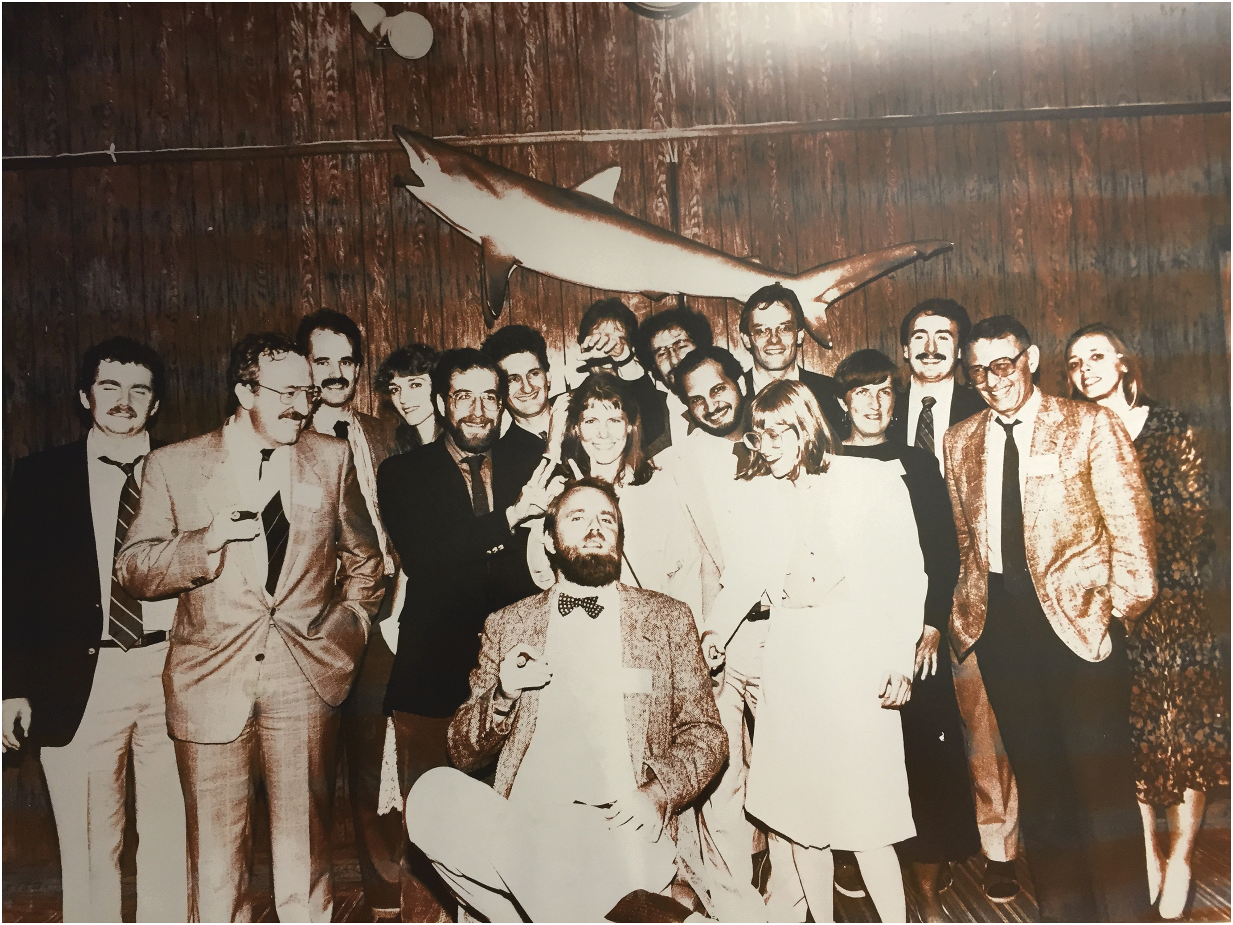
Mulligan lab (circa 1986–87) with Paul Berg at Whitehead Institute Retreat at the Chatham Bars Inn, Chatham, Massachusetts. Seated: Richard Mulligan. First row: Brad Guild, Pierre Lehn, Dan Silver, Heidi Stuhlmann, Jim Barsoum, Lisa Spain, Mary Collins, and Paul Berg. Second row: Jeff Morgan, Cori Bargmann (Weinberg lab), Olivier Danos, Paul Robbins, Mitch Finer, James Wilson, Doros Platika, and Elaine Dzierzak.
In parallel to the lab's efforts in the area of vector development, a major effort was made over the years to explore specific therapeutic strategies for treating both inherited and acquired diseases via gene transfer. Our studies involved efforts to transduce a variety of potentially important target cell types, including bone marrow–derived hematopoietic stem cells, fibroblasts and keratinocytes, hepatocytes, endothelial cells, and T lymphocytes. In studies with most of these cell types, a major goal of our efforts was to develop methods for manipulating the primary cells during viral transduction in such a way that the resulting transduced cells would function appropriately and properly express the transferred genes after their transplantation into animals. Jeff Morgan's early work with keratinocytes (Morgan et al., 1987) and Jim Wilson's studies with hepatocytes (Wilson et al., 1988) particularly exemplified our focus on maintaining the functionality of transduced cells.
Our most focused and sustained efforts in this area over the years involved our interest in developing a gene transfer methodology that would enable the treatment of diseases affecting hematopoietic cells, such as sickle cell anemia, severe combined immunodeficiencies, and beta-thalassemias. An obvious approach to the treatment of such diseases would be to transduce bone marrow–derived hematopoietic stem cells in vitro and subsequently to introduce the transduced cells back into a patient via bone marrow transplantation. At the time we began our studies in this area, very little was known about the general physical and functional characteristics of hematopoietic cells, and importantly, even less was known about how to manipulate stem cells in vitro in such a way that the cells would proliferate yet the reconstitution properties of the cells would be preserved. I reasoned that it was likely that in order to achieve the practical goal of development of a gene transfer/cell transplantation protocol for hematopoietic cells, we would need to become committed to obtaining a far more detailed and fundamental understanding of stem cell characteristics and function. For this reason, we embarked on what turned out to be over a decade of basic research efforts in this area.
Given the inability to physically isolate reconstituting stem cells in a pure form, and due to our view that there was no reliable in vitro assay that directly scored for the cells, we chose to focus the majority of our gene transfer efforts on murine bone marrow transplantation models involving the transplantation of transduced bone marrow into lethally irradiated recipients. Since retroviral transduction of cells genetically marks individual transduced cells in a unique way because of proviral integration, we reasoned that the most rigorous way to demonstrate the transduction of reconstituting cells would be to demonstrate that different hematopoietic cell types in animals engrafted with transduced cells carried identical proviral integration sites. With the help of David Nathan and others from Children's Hospital familiar with bone marrow transplantation models, two of my postdocs, David Williams and Ihor Lemischka, set out to define the optimal conditions for transduction of murine bone marrow, and to establish a robust proviral marking assay useful for characterizing transduced cells in engrafted animals. In 1984, we published the first report of the transduction of reconstituting hematopoietic stem cells using replication defective retroviral vectors (Williams et al., 1984). This result was huge for us, as it clearly indicated the feasibility of developing ex vivo gene therapies for diseases affecting hematopoietic cells. The study stimulated all of our subsequent studies over the next decade focused on the development of gene therapies for different inherited diseases of the blood, including SCID, beta-thalassemia, and Wiskott–Aldrich syndrome (Dzierzak et al., 1988; Wilson et al., 1990; Klein et al., 2003).
With regard to our basic studies of hematopoiesis, an interesting finding from David's and Ihor's studies was that despite the estimated number of stem cells present in the bone marrow samples that were transduced, only a small number of stem cell clones appeared to be transduced, as evidenced by proviral marking. Ihor subsequently explored this phenomenon in more detail, and found that the marking patterns were indicative of a “clonal succession” illustrative of dynamic changes in the contributions of different stem cell clones to the production of differentiated hematopoietic cells over time (Lemischka et al., 1986).
Another very productive series of basic studies, performed by Peggy Goodell, initially focused on efforts to identify and physically purify naturally proliferating hematopoietic stem cells through the use of fluorescent dye staining and cell-sorting techniques. In addition to succeeding in identifying and purifying a proliferating stem cell population, a very serendipitous finding from Peggy's work was that dual-wavelength analysis of Hoechst-stained whole bone marrow provided a novel means of purifying a highly enriched population of reconstituting stem cells we termed side-population or SP cells (Goodell et al., 1996). Subsequent studies by our group and others demonstrated the utility of this cell-sorting technique for the identification and purification of stem cell populations from other tissues (Goodell et al., 2005; Kotton et al., 2005). In other studies aimed at obtaining a more basic understanding of the developmental potential of hematopoietic stem cells, we collaborated with Lou Kunkel's group at Children's Hospital to determine whether blood stem cells might have the developmental potential to give rise to nonhematopoietic cells. In a mouse model of muscular dystrophy, we were able to show in bone marrow transplantation experiments that hematopoietic stem cells are, under certain experimental conditions, capable of giving to muscle, as evidenced by the detection of bone marrow-derived nuclei in the muscle fibers of transplanted recipients (Gussoni et al., 1999).
Another area of interest that the lab pursued for many years involved the notion that gene transfer technology might be used to enhance the capacity of the immune system to recognize and kill tumor cells. These studies, initiated by Glenn Dranoff, involved a long and fruitful collaboration with Drew Pardoll and his colleagues at Johns Hopkins School of Medicine. Drew's group had shown the ability of tumor cells transduced to express IL-2 to elicit antitumor immunity against nontransduced tumor cells (Fearon et al., 1990). Given our expertise with the generation and manipulation of retroviral vectors, we wished to make use of retrovirus vector technology to screen a much larger number of cytokines and other gene products to identify other gene products that could stimulate antitumor immunity.
Glenn generated a large number of recombinant viruses encoding different potentially interesting gene products, and developed what was then a new strategy for evaluating the “vaccination” potential of different transduced tumor cell lines that involved the use of irradiated rather than live cells to vaccinate animals. Through his studies, granulocyte-macrophage colony stimulating factor (GM-CSF) was identified as having the most potent immunostimulatory activity of the many gene products that were tested, when expressed in tumor cells (Dranoff et al., 1993). We immediately recognized the value of further understanding at a basic level the mechanism(s) underlying the effects of GM-CSF on tumor immunity and initiated what turned out to be a multiyear effort to understand the normal role of GM-CSF, and to clarify the manner in which GM-CSF interacted with other gene products to exert its function (Dranoff and Mulligan, 1994). Glenn has continued and extended the scope of such studies since leaving the lab, and over the past decade, his group's work has stimulated an enormous interest in the prospects for recruiting the immune system in the treatment of cancer, a subject now of obvious great interest.
Flirtations with Industry and Translational Studies
Early on in my career, David Baltimore counseled me on the issue of whether I should become actively involved in bringing our technologies to the clinic. He indicated that it would likely be many years before the optimal path from lab to clinic was understood, and that navigating complex translational and regulatory issues too early in the development of the field would adversely affect our productivity in the areas we were most capable of making contributions to. While I took his advice for years, as time passed and there was little effective movement of gene transfer technology into the clinic by others, I finally broke down and helped found Somatix, a gene therapy company established originally to bring our work with both transduced keratinocytes and tumor vaccines to the clinic.
In 1994, I chose to take a 2-year sabbatical from MIT to spend time at Somatix, which had recently relocated from Cambridge to Alameda, California. While my time in California was very interesting and productive, near the end of my time at the company, I began to question whether a small biotechnology company was the ideal vehicle to translate preclinical gene therapy advances into clinical studies, and therefore began to consider other possibly more fruitful avenues to advance translational studies. As I prepared to return to MIT, I was contacted by David Nathan about the possibility of moving my research effort from MIT to Harvard. David was a powerful champion of gene therapy and he appealed to my interest in being associated with translational gene therapy research by offering me the opportunity to direct a new Harvard-wide gene therapy initiative that he, Dan Tosteson, the dean of HMS, and Phil Leder, the chairman of the Department of Genetics, were establishing. After much discussion and soul-searching, I decided to accept the Harvard job offer, and upon my return to Boston in 1996, I moved my laboratory from MIT to Children's Hospital and HMS and became a Howard Hughes Medical Institute Investigator.
Over the next decade, in addition to continuing most of the research efforts initiated while at MIT in my new lab at Children's Hospital, I established a large laboratory within the HMS Department of Genetics, termed the Harvard Gene Therapy Initiative (HGTI), dedicated to translational gene therapy activities. In addition to operating a research-grade vector core to supply Harvard investigators with state-of-the-art vectors developed in our research laboratory, we established a good manufacturing practice vector production facility capable of production of viral vectors and other biological reagents suitable for clinical use. Through support from the Harvard hospitals, and the AFM, a French foundation dedicated to the treatment of inherited diseases via gene therapy, the HGTI was able to successfully produce a variety of clinical-grade AAV and adenovirus vectors and retroviral-transduced cell lines that were subsequently used in clinical trials evaluating gene therapies for muscular dystrophy and cancer at Harvard and elsewhere. As funding from the hospitals and AFM wound down and local interest in clinical gene therapy waned, we ultimately chose to shutter the HGTI manufacturing facility and return exclusively to our basic research activities.
Much of our efforts since that time have been focused on the development of two potentially novel technologies that we believe may significantly impact upon the future practice of gene therapy. In one series of studies initiated by Laising Yen almost a decade ago, we have been exploring a general approach to controlling the expression of therapeutic genes via small molecules that depends upon the regulation of RNA self-cleavage. The basic idea involves the notion that if self-cleaving ribozymes can be identified that can function in mammalian cells when embedded in a vector mRNA, it may be possible to control gene expression at the RNA level by inhibiting self-cleavage of the ribozyme-containing mRNA. While in the lab, Laising succeeded in generating specific RNA motifs capable of efficient self-cleavage in cells, and provided proof of principle for the gene regulation idea by demonstrating the ability of both antisense oligonucleotides and a nucleoside analog to inhibit self-cleavage of ribozyme-containing transcripts in cells in vitro and, in the case of the nucleoside analog, in cells in vivo (Yen et al., 2004).
Very recent efforts in our laboratory have been directed toward determining whether RNA aptamer sequences specific for a small molecule can be “grafted” to specific regions of the self-cleaving RNA motifs in such a way that engagement of the RNA with the small molecule leads to inhibition of self-cleavage. This in principle would enable the development of gene control systems “tailored” to any small molecule for which a specific aptamer can be developed. While the principle of “allosteric” ribozyme activity has been demonstrated in test tube experiments by Ron Breaker's laboratory (Soukup and Breaker, 1999), efforts to date to develop such a system that is functional in mammalian cells have not resulted in levels of regulation of any practical value (Bayer and Smolke, 2005). In a second series of studies, to date unpublished, we have begun to evaluate strategies to systemically deliver genes to cells via the transplantation of transduced cells that are engineered to fuse to cells in vivo that they come in contact with. These studies are based on the finding years ago that specific mutants of the murine leukemia virus envelope efficiently induce the fusion of cells expressing the mutant env and cells expressing the appropriate viral receptor (Januszeski et al., 1997). In preliminary studies, we have shown that the local transplantation of cells engineered to express the fusogenic env mutant into mouse muscle results in the efficient transfer of donor cell nuclei in muscle. Ongoing studies are addressing whether the systemic delivery of cells possessing fusogenic potential via the transplantation of transduced bone marrow-derived stem cells can lead to the systemic delivery of donor nuclei to recipient tissues. If successful, such an approach might offer a novel means to systemically deliver genes to tissues such as muscle, a technical capability that may be critical for the development of gene therapies for diseases such as muscular dystrophy that require gene correction in a large number of cells throughout the body.
Reflections
In this narrative, I have tried to illustrate some of the principles and themes that have characterized our research activities over the years. One of the most important of our guiding principles has been that practical studies of technology development and the development of specific gene therapy models must always be married to rigorous studies of the relevant underlying basic biology in order to maximize the chances for success. For example, our efforts to develop retroviral and lentiviral vectors necessitated that we study the biology of these viruses in detail, including an understanding of the cis-acting signals necessary for packaging, replication, and integration, and the mechanisms underlying expression of the different viral gene products. Our efforts to develop a transduction protocol for hematopoietic stem cells necessitated that we understand the basic biology of stem cells, including their physical characteristics, their response to growth factors, and the dynamics of their contribution to different differentiated blood cells after transplantation. Our efforts to develop effective tumor-based vaccines to enhance the recognition of tumor by the host necessitated that we understand the mechanism of action of gene products identified by our retroviral screens, and their network of interactions with other immune-related gene products.
An important corollary to the principle of marrying practical and basic experiments is that expertise in fundamental biological disciplines such as virology, cell biology, and biochemistry is often critical to tackle even practical problems in gene therapy research. In the scientific generation I grew up in, individuals involved in gene therapy research were all trained in one of these different basic disciplines. As time passed, however, an increasing number of investigators in the field began to be trained as “gene therapists,” often never receiving advanced training in the core basic science disciplines. Over the years, I have always tried to recruit trainees who could bring to the lab an expertise in a basic science discipline that could help us solve a particular problem or enable us to expand our efforts in an area we lacked expertise. I always advise young trainees interested in gene therapy to receive training in a fundamental discipline before entering the gene therapy arena. Hopefully, in the next chapter of gene therapy research, the field will again attract more individuals with advanced training in the core basic sciences.
A second theme of our studies of vector development over the years that may not be evident from the narrative is that we have always tried to develop technology that provides a specific capability that enables us to explore a possible therapeutic strategy or to address a specific biological problem that otherwise would not be approachable. It is very hard to develop useful technology without a specific context or desired design feature in mind, and indeed the “blind” development of technology usually results in technology that is of little practical value. When my postdocs or students embark on an effort in the area of vector development, they always have an application in mind and, not surprisingly, are highly motived to develop a specific piece of technology, knowing that they will appropriately get the first stab at applying the technology to a problem unapproachable by others.
In closing, it is worth commenting on the technical progress that has been made in the field over the years, and on the recent resurgence in interest in gene therapy by both investigators and industry. Previous to the recent era of clinical studies, I think it is fair to say that the results of most clinical trials in gene therapy were modest and equivocal, yet most often interpreted in the most generous and positive light by both investigators and the press. This clearly engendered the view among many that “hype” best characterized the field and that we were in fact far away from having the appropriate technology to succeed in the clinic. Clearly, a fundamental reason why people today are becoming much more interested in gene therapy is that over the past 5–10 years there has been unequivocal clinical success in several therapeutic areas. Gene therapy is working. While I have often heard investigators suggest to the press that the current success in the clinic is attributable to “dramatic and fundamental recent improvements in technology,” I don't think this is the best explanation for what has happened. In fact, progress in the development of gene therapy technology has been remarkably steady and incremental over the past two decades, and the technology that has been so successfully used in recent years is a product of years of development by different groups. Rather, I believe that much of the current success in gene therapy can be attributed to the meticulous translational efforts of a small number of clinical investigators who have uniquely been able to identify and successfully address the scientific and technical details that made the difference between clinical success and failure.
The skill set of a truly outstanding translational investigator is unique, as it comprises a complicated interdisciplinary mix of clinical and scientific expertise, and it is therefore the rare individual that possesses all these skills. In the case of the success of gene therapies involving diseases affecting hematopoietic cells, tremendous credit must be given to investigators such as Alain Fischer and Marina Cavazzana and their colleagues in Paris who personify the profile of an outstanding translational investigator (Cavazzana-Calvo et al., 2000; Cavazzana, 2014). In the case of cancer gene therapies, similar credit must be given to investigators like Carl June at the University of Pennsylvania (June 2014). These and other translational investigators deserve a special place in the history of the gene therapy field.
Not surprisingly, industry has been taking notice of the recent clinical successes in gene therapy, and this is very good news for both patients and investigators. I think a critical factor in the new-found interest in gene therapy among both the biotechnology and big pharma industries is their recognition, over the past decade, of the tremendous economic value of the development of treatments for orphan diseases. A number of biologicals and small molecule drugs developed by Genzyme and other companies for the treatment of different rare lethal inherited diseases are truly life-saving, and payers recognize both the societal and economic value of developing true cures for diseases. Gene therapy is obviously a most logical next step in the treatment of these diseases, and industry is now beginning to appreciate that opportunity, as well as understanding how the technology could be further extended to the treatment of more common acquired diseases such as cancer, heart disease, and so on.
Over the past several years, there has been an explosion of new venture-capital-backed companies focused on the development of gene therapies for different orphan diseases as well as for cancer, and several large cap biotechnology and pharmaceutical companies are developing programs in these areas as well. I believe this ground swell of interest among industry will usher in a new chapter in gene therapy research that will profoundly accelerate the development of truly life-saving therapies for patients. The amount of financial resources that will soon be devoted to such efforts is staggering. While these resources will certainly enable a dramatic expansion of critical preclinical efforts to explore new therapeutic approaches for different diseases in both academic centers and within companies, the greatest impact the involvement of industry will have on the further development of gene therapy will likely be in the development of methods for the manufacture of gene transfer vectors and other reagents suitable for use in patients, and in clinical development.
As many of us have learned the hard way, the academic arena is not ideally suited for the development of large-scale manufacturing processes and, not surprisingly, to date, little progress in the development of robust methods for the production of clinical-grade gene transfer reagents has been made. We can expect to soon see industry take up the mantle of manufacturing from the academic sector and this will greatly accelerate the delivery of gene-based therapies to patients. Similarly, the expertise of industry in the development of the types of clinical development activities necessary to obtain approval of a drug by the FDA, and the financial resources they can bring to bear on these activities, will dramatically reduce the time it will take to translate a gene therapy idea into a clinical treatment. The next chapter in the development of gene therapy will be a very exciting one for everyone involved.
Footnotes
Acknowledgments
I have been enormously fortunate to have worked with an extraordinary group of postdocs, students, and technicians over the past 40 years. I am so grateful to each and every one of those individuals for sharing a portion of their scientific lives with me. While extremely proud of all of the accomplishments of the lab over the years, it makes me most proud to see that so many members of the lab have gone on to become contemporary leaders of the gene therapy field as independent investigators. I have also been blessed to have had truly outstanding mentors at every stage of my professional life, who have guided virtually every aspect of my career, providing deep scientific wisdom, sage professional and personal advice, and incredible advocacy and support. I am so grateful to each of them. Lastly, I wish to acknowledge Devon Young, my trusted assistant for the past 33 years. Devon is an amazing individual who has played so many critical roles in the operation of our laboratory over the years. As the only individual in the lab who ever behaved like a real adult, Devon kept myself and others out of trouble on countless occasions, and served as a stable beacon for each generation of trainees passing through the lab. There couldn't have been a Mulligan lab without Devon.
Author Disclosure Statement
R.C.M. is a member of the board of directors of Biogen-Idec, a founding partner at Sarissa Capital Management, and a shareholder of Kadmon Corporation.