
Abstract
Identifying and tracking cells in the brain is a challenging task, requiring advanced molecular biology techniques and precise anatomical information. We have applied the principle of RGB marking to the long-term marking and tracking of progenitor and mature cells in the adult brain. Briefly, multicolor RGB marking is based on the simultaneous, lentiviral vector-mediated expression of three genes encoding fluorescent proteins in the three basic colors (red, green, and blue). Here, we show the application of RGB marking to the stable multicolor marking of adult granule cells in the hippocampus. This technique provides each individual cell with a characteristic hue, which facilitates their anatomical identification and spatial tracking. Multicolor RBG marking of mature neurons can provide an effective approach to dissect the function of individual cells, as it allows combination with functional techniques, facilitating the understanding of complex neuronal populations such as that of the dentate gyrus.
T
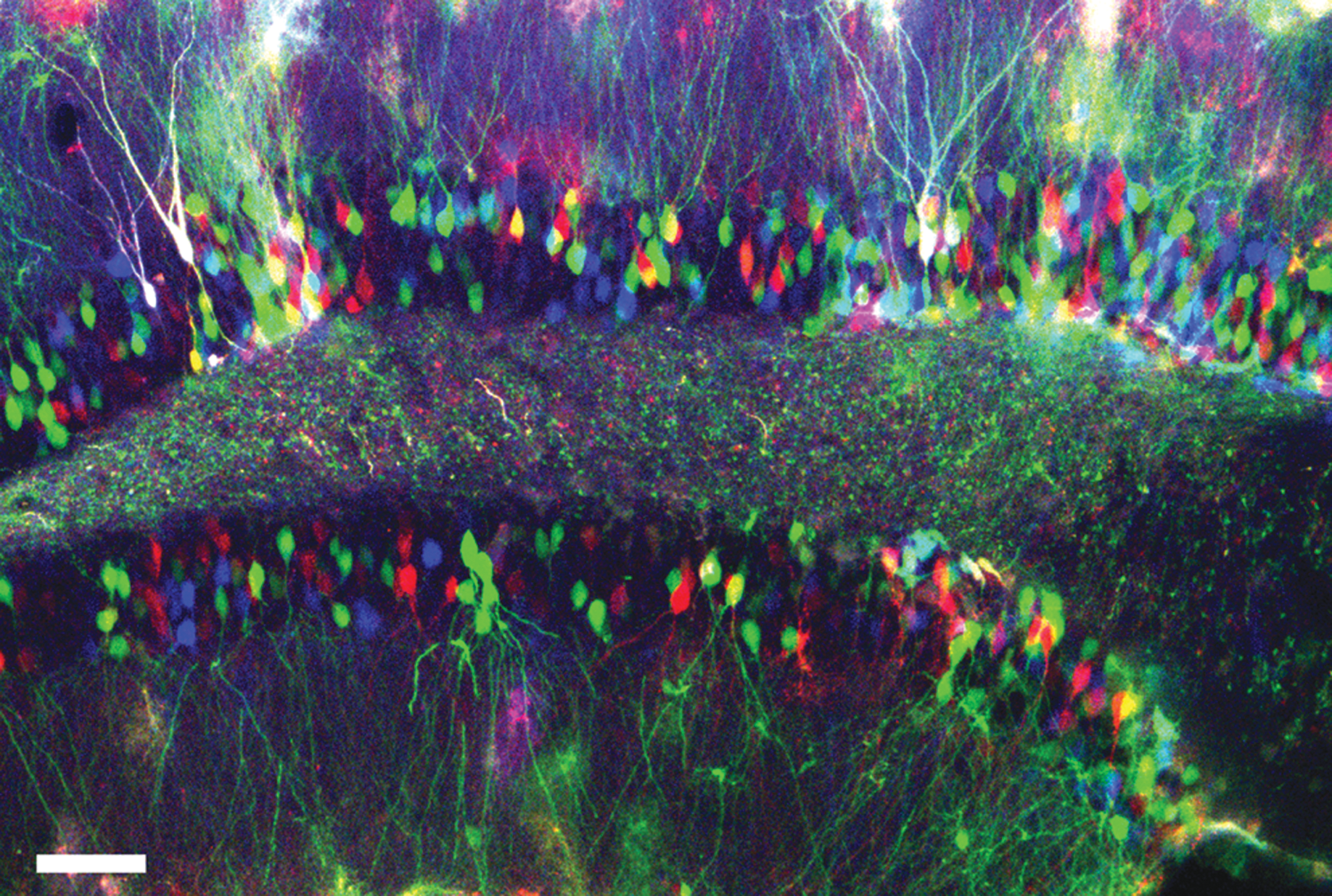
RGB marking of granular neurons after intraparenchymal administration of VSVG-CMV lentiviral vectors, analyzed 2 weeks after injection. Fluorescence is shown in red (mCherry), green (Venus), and blue (Cerulean). All images were analyzed by confocal microscopy. Scale bar, 25 μm.
This technique facilitates single-cell analysis and cell tracking over long periods of time and can be easily implemented in most neuroscience laboratories. Moreover, the use of lentiviral gene ontology (LeGO) vectors 8 allows the coexpression of fluorescent proteins and genes of interest and/or short-hairpin RNAs (shRNAs), providing a flexible and potent toolbox for multiple and simultaneous gene modification. We have made RGB marking in the brain available in different formats: using the spleen focus-forming virus or cytomegalovirus promoter, which enables targeting of different cell types, and using lentiviral or gammaretroviral vectors, which enables targeting broad or selected (proliferating) cell populations. 1 Multicolor RGB marking in the brain could be applied for time-lapse imaging, to develop gene therapy experiments and fate modification approaches, adapting well to the current needs of the research in neuroscience.
In the image (Fig. 1), fluorescence is shown in red (mCherry), green (Venus), and blue (Cerulean). The image was analyzed with an SP5 Leica confocal system. Scale bar, 25 μm.
Footnotes
Acknowledgments
The authors are indebted to many colleagues for kind support with various cells and constructs: R.Y. Tsien (Howard Hughes Medical Institute, USA) for mCherry cDNA, A. Miyawaki (RIKEN, Japan) and T. Schroeder (ETH Zürich, Switzerland) for Venus cDNA, D.W. Piston (Vanderbilt-Ingram Cancer Center, USA) for Cerulean cDNA, and Axel Schambach (MH Hannover, Germany) for the retroviral vector RSF91.GPF.pre*. This research was funded by the European Union Seventh Framework Programme under grant agreement IEF273243, by the Medical Research Council, Wessex Medical Research, and the DFG (SFB841, project SP2). K.R. was supported by a young investigator grant within the FFM Programme of the Univ. Med. Centre Hamburg-Eppendorf (NWF-12/09).
Author Disclosure Statement
The authors declare no competing financial interests exist.