
Abstract
Cardiovascular gene therapy aims to treat coronary and peripheral artery disease, heart failure, and arrhythmia. The chosen transgene, delivery method, gene therapy vector type, high-quality vector production, and dose are all determining factors of the therapeutic outcome. High-resolution vascular imaging and increased knowledge of vascular biology in physiological and pathological conditions enable the finding of novel molecular targets for cardiovascular gene therapy. Transgenic and knockout mouse models have provided researchers several powerful experimental tools for studying the effects of single genes on cardiovascular diseases. For preclinical efficacy, safety, and toxicology studies, large animal models are needed before entering into clinical testing. This review focuses on commonly used animal models in cardiovascular gene therapy and describes recent advancements in the field of vascular biology. Emphasis is also given on high-resolution imaging of microvasculature and its impact on our knowledge of vascular function.
Introduction
V
Emerging data on epigenetic regulation of vasculature, exosomes, noncoding RNAs, mechanotransduction, and cell signaling in vivo have transformed our views on vascular biology. Growing knowledge of vascular function at the cell and molecular level enables also finding of new therapeutic targets for cardiovascular diseases (CVDs). Availability of several transgenic and knockout mouse models greatly enhances possibilities to obtain physiologically significant results using gene transfer applications. Besides ApoE and LDL receptor knockout mice for hyperlipidemia and atherosclerosis research, 3 several mouse models with deletion of specific apolipoproteins, cytokines, growth factors, or extracellular matrix components have been generated. New mouse models, for example, for modeling vulnerable plaques, have also been recently introduced. 4 Final preclinical efficacy, safety, biodistribution, and toxicology studies of gene therapy vectors are usually performed in larger animals, such as rabbits or pigs, since they produce a more realistic outcome about the usefulness of CVD gene transfer applications regarding human clinical applications. Particularly, pigs have been increasingly used for myocardial infarction, cardiac ischemia-reperfusion, and in-stent restenosis studies. Pig heart is very similar to human heart and allows the use of similar gene vector dose, catheter interventions, and imaging techniques as for humans.
Detection of the therapeutic outcome in preclinical animal models by imaging of microvasculature in three dimension enables better understanding of vascular morphology and function, as well as biodistribution of the virus vectors after gene delivery. Fast development of new imaging modalities with higher spatial resolution is going to improve vascular imaging and analysis even further. 5 The challenge is now how to interpret the most useful data, for example, from bioinformatics and vascular imaging, for development of new therapeutic vectors for the treatment of CVDs.
Virus Vectors for Cardiovascular Gene Transfer
Ad vectors have been the most commonly used vector type in clinical trials aiming to treat CVDs because of their high gene transfer efficiency and ease of production in good manufacturing practice quality. Although systemic delivery of Ad vectors is limited because of induced immune response and virus interactions with circulating blood and cell components, localized intramuscular and intramyocardial delivery of Ad vectors has shown great promise in developing treatment for CVDs. Vascular gene transfer of endothelial cells, cardiomyocytes, or medial smooth muscle cells have shown to be obtained after Ad vector administration by intramyocardial injection, coronary perfusion, or intravascular delivery to transiently isolated vessel segments. 6 –11 To date, Ad vectors have been used in clinical trials for coronary and peripheral artery disease by expressing transgenes such as HGF, HIF-1α, and VEGF. 2,12 –14 A clinical trial for heart failure by Ad vector expressing adenylyl cyclase type VI is ongoing.
Besides Ad vectors, AAV and LV vectors have been used in cardiovascular gene transfer applications. With AAV vectors, high gene transfer efficiency has particularly been detected in cardiomyocytes after intramyocardial delivery. Persistent gene expression by AAV vectors has been shown in preclinical trials in cardiomyocytes and skeletal muscle. 7,15 AAV9 can efficiently target and transduce cardiomyocytes after systemic delivery, 16 whereas AAV2 has been shown to have the most promising efficiency after intracoronary delivery to heart transplants ex vivo. 17 AAV1 instead has shown to be the most efficient gene transfer vector after intramyocardial injection 18 and has been used in phase 2 clinical trials for heart failure by expressing Serca2a as a transgene. 19 Also, LV vectors have been developed for cardiovascular gene transfer because of their ability to induce long-term gene expression in cardiomyocytes in preclinical trials. 20,21 Instead, in rabbit skeletal muscle LV-mediated transduction was shown to be short-term (<3 weeks) and intraluminal administration of the vector to carotid artery led to low gene transfer efficiency. 7 Besides safety concerns with LV vector because of random integration, a limitation with both AAV and LV vectors has been their less efficient vector production in comparison to Ad vectors, which can limit their use in large animal models and clinical trials. Development of production and purification methods for AAV and LV vectors in large scale is therefore highly important in order to broaden their use in cardiovascular gene therapy.
To enhance cardiovascular gene transfer and reduce off-target effects of the virus vectors, transcriptional and transductional vascular targeting strategies have been utilized. In transductional vascular targeting, various polymers, magnetic particles, microbeads, lipids, bispecific molecules and endothelial or smooth muscle cell targeting peptides, single-chain antibodies, or ligands have been used. In transcriptional vascular targeting, cell-specific or disease-specific promoters (e.g., Tie1, Tie2, and antioxidant response element) or miRNA target sequences have been used in order to restrict transgene expression to the desired target tissues or cells. 22 –24 Although significant advancements have been obtained in vectorology, most of the surface-modified virus vectors cannot be produced at the moment in sufficient quantity or quality for studies in large animal models or clinical trials. Besides transductional and transcriptional targeting, vascular tropism can also been increased by vector pseudotyping and by using other vector serotypes or nonhuman virus vectors. Recently, Ad serotype 49 was shown to induce higher vascular gene transfer efficiency than Ad5 after ex vivo delivery to coronary artery bypass graft tissues. 25 Promising results have also been obtained after systemic administration of nonhuman Ad vectors in a murine model. 26
Animal Models for Cardiovascular Gene Transfer
Several transgenic and knockout mouse models have been developed for CVDs. 27 Since mice are cheap and easy to handle, most of the early animal studies are conducted in mice. ApoE and LDL receptor knockout mice are standard models for hyperlipidemia and atherosclerosis research. 3 Various knockout models in which specific apolipoproteins, cytokines, growth factors, or extracellular matrix components are deleted have also been developed. 27 –29 Albeit the small size of a mouse, many surgical operations can be performed including coronary ligation for myocardial infarction and reperfusion studies. 30 Carotid artery denudation and cuff models have been used for restenosis and atherosclerosis studies. 4 Peripheral skeletal muscle ischemia using ligation of femoral arteries is a common screening tool for proangiogenic factors. 31 Transaortic banding or angiotensin infusion is commonly used in heart failure studies. 32 Recently, the so-called vulnerable plaques have also been modeled in mice. 4 Mice allow adequate number of experimental animals per study group and interventions based on systemic tail vein delivery, intraperitoneal or periorbital delivery. In addition, intramuscular applications in peripheral skeletal muscles, direct intramyocardial injections, and intraretinal injections are feasible. However, due to the small size, significant damage can be caused by local injection techniques. The relevance of these models to human clinical situation remains unclear.
Rats are commonly used in cardiovascular gene therapy studies. They have been useful for carotid artery restenosis studies and acute myocardial infarction studies using coronary ligation. 33 In addition, transaortic banding and hypertensive models have been used for heart failure studies 34 and femoral artery ligation has been used for peripheral muscle ischemia studies. Due to their larger size, rats provide an easier model than mice for targeted gene transfer, for example, in heart with less local damage caused by needle injections. However, rats are very resistant to hyperlipidemia without extensive hormonal and dietary modifications. This limits the usefulness of rat models in vascular studies other than those focusing on restenosis and in-stent restenosis. Currently, some genetically modified rats have been made for cardiovascular studies, which can improve their value in cardiovascular gene therapy studies. 35
For preclinical efficacy, safety, and toxicology studies, large animal models are needed before entering to clinical testing. Rabbits have been usually used for hyperlipidemia and atherosclerosis studies because of the easy induction of high cholesterol levels with diet. 7 Rabbits have also been used for restenosis, in-stent restenosis, and vein graft stenosis studies. 36 Also, they have been useful for myocardial and peripheral skeletal muscle ischemia studies. 37,38 Due to their large size, rabbits allow the use of several similar imaging methods as is used in the clinics. Ultrasonography, magnetic resonance imaging (MRI), and positron emission tomography (PET) imaging are useful for the characterization of tissue response after local or systemic gene therapy. Also, some naturally occurring mutations, such as Watanabe heritable hyperlipidemic (WHHL) rabbits with LDL receptor mutations, have been very useful in atherosclerosis, restenosis, and vulnerable plaque studies. 39 Gene therapy for LDL receptor deficiency has also been efficiently tested in WHHL rabbits. 40 In addition, safety, biodistribution, and toxicology studies have been done in rabbits since dosing and delivery of the vectors better resembles those used in humans. 41
Dogs have been infrequently used in gene therapy studies because of the ethical, practical, and logistic reasons. However, particularly in the cardiac ischemia-reperfusion and infarction studies, dogs have been very useful since their heart resembles the human heart and is large enough, allowing intracoronary and intracardiac manipulations. 42 Most commonly used ischemia models are ameroid constrictor and coronary ligation techniques, which lead to predictable ischemia, deteriorating heart function, and pathologies that resemble human pathological conditions. 42,43 Also, dogs have been used in hemophilia studies since a natural clotting factor mutant exists and is very useful for preclinical studies. A difference between a dog and human heart is that a canine heart has functional collateral circulation.
Pigs have been increasingly used for myocardial infarction, cardiac ischemia-reperfusion, and in-stent restenosis studies. Pig heart is very similar to human heart, having a limited collateral circulation, and allows the use of similar catheter interventions as used in humans. All imaging techniques available for humans are possible for pigs. Ameroid constrictor and occlusion coils are commonly used models, which are quite predictive for human conditions. 11,44 In addition, open transthoracic surgical models have been frequently used in pigs. A limitation of the pig models is the difficulty to make pigs atherosclerotic and hyperlipidemic. Pig myocardium is also very sensitive to ventricular fibrillation and this leads to increased mortality during myocardial infarction and cardiac ischemia studies. However, efficient anti-arrhythmic drug cocktails have significantly improved the situation. Pigs allow easy, affordable follow-up studies, which is important for myocardial infarction and heart failure studies. For long-term follow-up, however, fast growth of a pig results in difficulties in handling as well as changes in heart-to-body weight ratio. Preclinical safety, biodistribution, and toxicology studies are commonly performed in pigs before starting clinical trials since gene vector dose and delivery routes are very similar to those used in humans. 45 Recently, a new pig model has been developed for the gene therapy of lymphatic disorders. 46
Recent Advancements in Vascular Biology
The growing knowledge of vascular function at the molecular level enables the finding of new therapeutic targets for CVDs. Flow-mediated endothelial mechanotransduction plays an important role in inducing signaling pathways and affecting endothelial function. Molecular mechanisms of atheroprotective and atheroprone endothelial phenotypes affecting formation of atherosclerotic lesions were recently characterized in vivo. In murine models, Kruppel-like factors 2 and 4 (KLFs) were shown to be atheroprotective, whereas activation of NFkB pathway was shown to lead to atheroprone endothelial phenotype. 47 Flow-dependent epigenetic regulation of endothelial cells was suggested to induce methylation of KLF4 promoter inhibiting its expression and atheroprotective function. 48 Also, molecules such as Piezo-1 and syndecan-4 involved in ion flux and endothelial cell organization were suggested to act as endothelial mechanosensors. 49 –51 Besides mechanotransduction, exosomes and their role in mediating cardiovascular signaling have become an emerging field of research. Exosomes have been shown to be secreted from endothelial cells and cardiomyocytes in vitro and to interact with other cardiovascular cell types. They have been suggested to transport miRNAs, mRNAs, and proteins and to induce changes in cell signaling and gene expression in recipient cells. In murine models, miR-143/145-containing exosomes were shown to reduce atherosclerotic lesion formation, 52 and cardiac hypertrophy was reduced by miRNA-21 antagonist. 53 miRNA-containing exosomes have also been connected to cardioprotective function after myocardial ischemia. 54
For proangiogenic gene therapy, improvement of the knowledge of vessel formation, maturation, and vascular permeability is crucial. Recently, vessel sprouting was shown to be induced by glycolysis regulator PFKFB3, 55 and abnormal VE-cadherin junctional patterning was shown in tumor vessels and retinopathy ex vivo. 56 In mice, Slit-Robo signaling was shown to be required for neovessel formation in the retina 57 and development of ventricular septum and cardiac valves. 58 In rabbits, Slit2 was also shown to modify angiogenesis in rabbit skeletal muscles. 59 In the context of VEGF signaling and proangiogenic gene therapy by VEGF family members, VEGF coreceptor neuropilin 1 was shown to regulate angiogenesis via restricting VEGF receptor 2 internalization. 60 Additionally, transcriptional programs of VEGF-A 61 as well as in vitro functions of various VEGF family members used in preclinical trials for proangiogenic gene therapy were characterized. 62 In murine models, VEGF-angiopoetin-1 chimera was shown to induce angiogenesis having reduced vascular permeability. 63 Additionally, epigenetherapy was used to induce VEGF production by promoter-targeted shRNAs. 64,65 Overall, emerging data of epigenetic regulation of vasculature, noncoding RNAs, next-generation sequencing, and cell signaling in vivo can lead to development of novel therapeutic targets for treatment of CVDs. For development of VEGF therapy by various VEGF family members, knowledge of VEGF signaling and vessel formation, particularly in preclinical settings, is highly important.
High-Resolution Vascular Imaging
To date, various imaging modalities for macrovascular and microvascular imaging exist, enabling either two-dimensional or three-dimensional imaging of tissues/animals. In order to image microvasculature, high spatial resolution and tissue penetration is needed to distinguish the vascular capillaries. Additionally, safe and repetitive imaging of the target tissue/animal at multiple time points as well as reasonable quantification of vasculature by imaging software are desirable. Currently, imaging modalities such as MRI, PET, X-ray/computed tomography (CT), angiography, and ultrasound imaging can be used to image larger blood vessels and/or blood flow with high penetration depth but limited spatial resolution (100–1000 μm). Thereby, these techniques cannot be used to image microvasculature. For high-resolution vascular imaging in murine models, imaging modalities such as micro-CT and micro-MRI have been developed. In vivo micro-CT allows 30–100 μm spatial resolution, whereas ex vivo micro-CT and micro-MRI can reach 1–30 μm and 20–30 μm resolutions. 66 A limitation in micro-CT imaging modality is the significant use of radiation. Development of optical imaging modalities, such as 2-photon microscopy, using a high-pulse infrared laser to induce 2-photon excitation, has allowed high-resolution (∼1 μm) imaging of microvasculature in murine models in vivo and whole immunomount samples ex vivo (Fig. 1). Fluorescence-based imaging modality allows detection of multiple molecular targets simultaneously. Additionally, second-harmonic generation allows detection of collagen and elastin fibers from the vessel wall without immunolabeling. The penetration depth depends on the target tissue (∼0.3–1.6 mm). Besides 2-photon microscopy, conventional laser confocal microscopy, that is, 1-photon microscopy, has been used to detect vasculature in tissues. Despite of its higher spatial resolution, 1-photon microscopy has reduced depth penetration (<200 μm) in comparison to 2-photon microscopy and therefore has limited use in tissue imaging. Also, other imaging modalities, for example, laser speckle contrast imaging, photoacoustic tomography (PAT), optical coherence tomography (OCT; 1–10 μm), and orthogonal polarization spectral imaging (OPSI; 1–5 μm), have been used to detect vascular morphology or blood flow based on detecting movement of red blood cells, hemoglobin, or oxygenation levels of the capillaries. 66,67 Currently, most of the imaging modalities that are used to measure blood flow or vasculature in general are qualitative or semiquantitative. The combination of multiple imaging modalities as well as the development of whole immunomount staining methods and image analysis software allowing reasonable quantitative imaging are going to improve vascular imaging and analysis in the future. Additionally, novel techniques such as superresolution microscopy, that is, nanoscopy, are going to improve detection of novel molecular targets in cardiovascular cells. 5
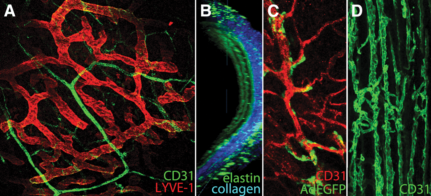
High-resolution imaging of vasculature in small- and large-animal models by 1- and 2-photon microscopy.
Conclusions
Virus vectors are efficient tools for gene transfer applications in CVDs, including myocardial infarction, proangiogenic studies, restenosis, in-stent restenosis, vein graft stenosis, peripheral ischemia, and heart failure. Delivery method, virus vector type, quality of the vector production, and dose all affect the therapeutic outcome. Particularly, development of production and purification methods for all virus vectors in large scale is essential in order to broaden their use in cardiovascular gene transfer. Besides development of vectorology, more knowledge is needed on vascular biology itself in order to find novel molecular targets for treatment of CVDs. This year (2015) we are privileged to organize European Society of Gene and Cell Therapy (ESGCT) meeting in Helsinki. In our program, special emphasis has been put to epigenetics and high-resolution imaging that have begun to transform our knowledge of cell and molecular biology. The findings in these research fields are going to shape our view also on treatment of CVDs by gene transfer. After all, superresolution imaging in heart by nanoscopy is no longer science fiction. 5
Footnotes
Acknowledgments
Drs. Jari Lappalainen, Thomas Theelen, Pyry Toivanen, Tiina Nieminen, and Taina Vuorio are acknowledged for providing tissue samples for imaging. This work was supported by grants from Finnish Academy Center of Excellence, Academy of Finland, Kuopio University Hospital, ERC Advanced Grant, Finnish Cultural Foundation, K. Albin Johansson Foundation, and Ella and Georg Ehrnrooth Foundation.
Author Disclosure
No competing financial interests exist.