
Abstract
In order to pursue a clinical gene therapy for a human neurologic disease, it is often necessary to perform proof-of-concept trials in mouse models of that disease. In order to demonstrate a potential clinical efficacy, one must be able to select an appropriate vector and route of delivery for the appropriate age group in the disease model. Since many diseases require correction early in life, investigators often need to deliver recombinant adeno-associated viral (rAAV) vectors to neonatal mice. Herein, general central nervous system expression patterns of nuclear GFP following delivery of rAAV by three different routes are reported.
A
All animal work was approved by the University of Massachusetts Medical School Institutional Animal Care and Use Committee. All dosing was done in C57Bl6 mice bred in-house and originally obtained from Jackson Labs (Strain #000664). The vector used was a single-stranded rAAV2/9 encoding an H2B-GFP protein under the control of a shortened chicken-beta actin promoter with a cytomegalovirus enhancer. H2B serves to target the GFP protein to the nucleus of the cell. For all delivery routes, the protocols for injection were performed as previously described with only minor modifications. 1,2 All pups were anesthetized using isoflurane anesthesia prior to injections. For the temporal vein and retro-orbital routes of delivery, the mice were administered 4 × 10¹¹ vector genomes diluted in 50 μL total volume of phosphate-buffered saline. For the ICV injection route, mice were administered 5.6 × 10¹⁰ vector genomes per pup in 4 μL of total volume (2 μL per cerebral ventricle). For each injection route, five to six mice were compared. All mice were sacrificed for histology at 8–10 weeks of age. Tissue processing and immunofluorescent staining for GFP were performed as previously described. 3,4
GFP expression was assessed throughout the brain as well as the retina. Brain expression changed based on the route of delivery, even between the two intravenous routes (TV and RO). Overall, brain expression was higher following RO delivery in all areas assessed compared with TV delivery (Fig. 1). The transduction in areas of the brain such as the cortex and hippocampus was comparable between the RO and ICV delivery, while some areas remained higher with ICV delivery, despite the smaller vector dose with that route (Fig. 1). This was particularly apparent in the cerebellum, where modest GFP expression was obtained following ICV delivery but minimal or no expression following either venous delivery methods, although RO did consistently result in low-level expression (Fig. 1).
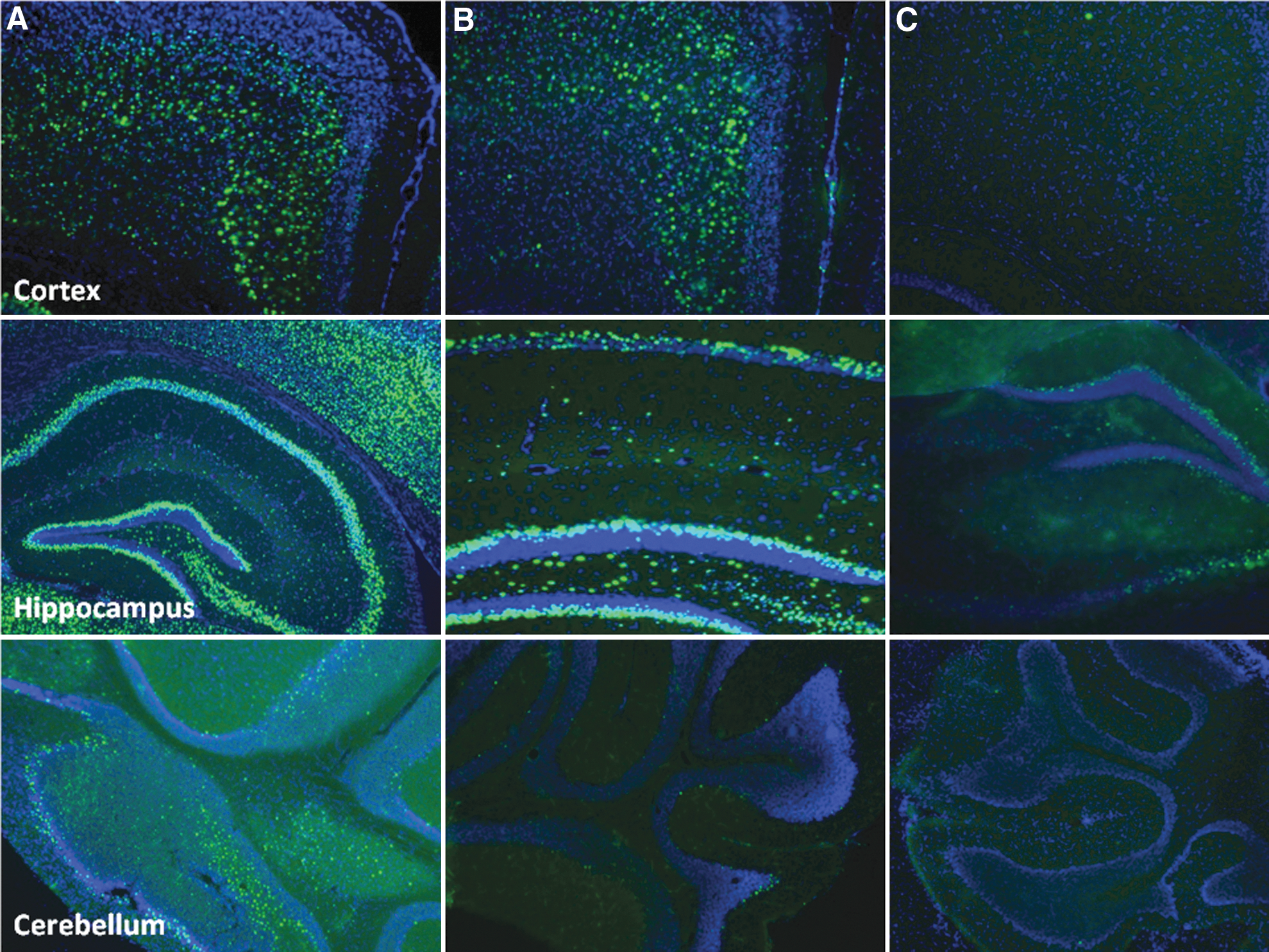
Representative central nervous system GFP expression following rAAV9 delivery by three routes in neonatal mouse pups. This image compares all routes of delivery in the cortex, hippocampus, and cerebellum. (
For retinal expression, both the RO and TV routes resulted in peri-optic nerve expression following P1–P2 delivery (Fig. 2A, B, D, and E). As the retinal vasculature developed, it was found that it was possible to obtain expression further out circumferentially from the optic nerve (Fig. 2C). When retinal cross-sections were examined, it was noted that the intravenous delivery resulted in expression in the innermost layers of the retina (Fig. 2F).

Retinal GFP expression following rAAV9 delivery by two routes in neonatal mouse pups. (
The increased brain transduction following RO delivery could be due to direct venous drainage of the retro-orbital venous sinus intracranially. While the retro-orbital sinus can drain to the external jugular, it can also drain via the cavernous sinus into the basilar venous plexus from the inferior frontal and temporal lobes and brain stem. A higher-volume injection, such as was performed in this study, may allow retrograde flow via this route directly to the brain similar to hydrodynamic tail vein injection in the liver or high-volume isolated limb perfusion.
Based on these data, it is concluded that when using neonatal mice as a model for central nervous system gene therapy, the route of AAV vector delivery is critical in determining expression in a region of interest. Pilot studies with a gene and vector capsid type of interest should be done to determine the optimal delivery route before correction experiments are undertaken. The mouse age at delivery and region of the retina to be targeted should be taken into account if both the brain and retina are target organs using the intravenous route.
Footnotes
Acknowledgments
This work was supported by the Massachusetts Lions Eye Research Fund. We wish to thank Shan Ma, Aditya Venkatesh, Jia Li, Lorelei Stoica, and Sourav Choudhury for their technical training and advice. Thank you to the Horae Gene Therapy Center Vector Core for virus production.
Author Disclosure
No competing financial interests exist.